44
Unusual ocular manifestations
of silent sinus syndrome
Manifestações oculares incomuns na síndrome do seio silencioso
Fabricio Lopes da Fonseca
1,Luciana Mazoti
2,Mariza Polati
31Ophthalmology and Otorhinolaryngology Department, Hospital das Clínicas da Faculdade de Medicina da Universidade de São Paulo - USP – São
Paulo (SP), Brazil.
2Ophthalmology and Otorhinolaryngology Department, Hospital das Clínicas da Faculdade de Medicina da Universidade de São Paulo - USP – São
Paulo (SP), Brazil.
3Head of Strabismus Department. Ophthalmology and Otorhinolaryngology Department, Hospital das Clínicas da Faculdade de Medicina da
Universidade de São Paulo - USP – São Paulo (SP), Brazil.
Study carried out at Strabismus Sector, Ophthalmology and Otorhinolaryngology Department, Hospital das Clínicas da Faculdade de Medicina da Universidade de São Paulo - USP – São Paulo (SP), Brazil.
The authors declare no conflicts of interest
Received for publication: 23/5/2012 - Accepted for publication: 26/9/2012
A
BSTRACTSilent sinus syndrome is an acquired condition in which there is a gradual collapse of the orbital floor and inward retraction of the maxillary sinus (atelectasis of the maxillary sinus). This in turn may cause associated ocular occurrences of enophthalmos and hypotropia. This is a report of an 8 year-old boy with silent sinus syndrome and associated ocular motility disorders. The association between silent sinus syndrome and ocular motility disturbance has been recently described in the literature. However, this is an infrequent association, mainly in childhood.
Keywords: Paranasal sinus diseases; Maxillary sinus; Orbit; Strabismus; Case reports
R
ESUMOA síndrome do seio silencioso é afecção adquirida em que há colapso gradual do assoalho orbital e do seio maxilar (atelectasia do seio maxilar), o que pode acarretar alterações orbitárias e oculares associadas, como enoftalmia e hipotropia. Relatamos o caso de um paciente de 8 anos de idade com síndrome do seio silencioso e distúrbios da motilidade ocular. A associação entre a síndrome do seio silencioso e alterações da motilidade ocular extrínseca tem sido descrita na literatura. No entanto, esta é uma associação pouco frequen-te, principalmente na infância.
Descritores: Doenças dos seios paranasais; Seio maxilar; Órbita; Estrabismo; Relatos de casos
C
ASER
EPORTRev Bras Oftalmol. 2014; 73 (1): 44-6
45
I
NTRODUCTIONThe silent sinus syndrome (SSS) is a rare disorder, first described by Montgomery(1) in 1964 and named by Soparkar et
al.(2) in 1994, characterized more often by enophthalmos and
hypotropia and increased ipsilateral orbital volume secondary to maxillary sinus atelectasis.
The typical syndrome occurs between the third and fifth decades of life3, with equal distribution between genders(3,4) and
unilateral symptoms, most commonly reported as enophthalmos of the ipsilateral eye, proptosis of the contralateral eye or changing of facial appearance(1,5-7). Orbital asymmetry, palpebral
retraction(1,6,7) and deepening of the superior eyelid sulcus(5) are
more specific symptoms. The ocular motility is frequently preserved(8-10), although there are recent articles relating
diplopia(11,12), even in children(13).
The most obvious radiological signs are clouding of the maxillary sinus(10), loss of the maxillary sinus roof convexity and
uprising and thinning of the orbital floor(8,10), nasal septum
deviation at the affected side(2,10) and lateralization of the
ipsilateral middle turbinate(5,10). Increased orbital volume and
atelectasis of the maxillary sinus are required findings for the diagnosis(8). The classical treatment aims to restore normal sinus
aeration and to correct enophthalmos and hypotropia with an orbital implant(4). The disease progression is interrupted with
sinus aeration(10).
Report of case
The case of an 8 year-old boy with exotropia and amblyopia of the left eye since birth. Treatment was patching for 2 years, which was discontinued 3 years ago, when he was started on glasses. There is also no history of trauma or surgery.
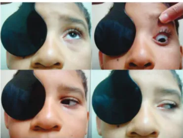
The ophthalmological exam revealed a visual acuity OD 1,0 and OS 0,2 (LH Symbols; Snellen equivalent OD 20/20 and OS 20/100), under cycloplegic refraction of +1.25 -1.50 x 180 and +3.00 -2.00 x 160, respectively; far and near left exotropia and torticollis (tilts and rotates the head to the left) with an angle of turn of 15 degrees. There was a Hertel enophthalmos of 4 mm. The Krimsky test showed an incomitance: at distance, right eye fixating exotropia of 35 prism diopters and left hypotropia of 4 prism diopters; with left eye fixating, an exotropia of 50 prism diopters and left hypotropia of 6 prism diopters. At near
Figure 2. Ductions, showing no adduction deficit
Figure 1. Facial asymmetry, enophthalmos and hypotropia on the left; gaze restriction.
distance, right eye fixating exotropia of 30 prism diopters and left hypotropia of 6 prism diopters; with left eye fixating, an exotropia of 50 prism diopters and left hypotropia of 6 prism diopters.
All the left extraocular muscles had a marked underaction, except for the lateral rectus muscle. It seems an adduction deficit (Figure 1), that is not present in ductions (Figure 2). These findings would be secondary to a large exotropia. The upper eyelid did not follow the left eye when it moved in a downward position. There was also a manifest constant pendular nystagmus, low
Figure 3. T2 image showing the maxillary sinus of reduced size, with elevation of its floor and lowering the orbit floor. It is also observed the ipsilateral middle turbinate laterally displaced
Figure 4. Increased ipsilateral middle meatus due to the lateralization of the uncinate process in T1 image
Figure 5. T1 image showing displacement of left inferior rectus muscle
Unusual ocular manifestations of silent sinus syndrome
46
amplitude and frequency, no null point, observed in all gaze positions, with upper eyelid fasciculation.
In an attempt to open the eyes, they converged.
There was anisocoria of 2 mm, left pupil greater. This condition was absent in the dark. Unfortunately, we do not have measurements in both bright and dim illumination. The left eye was amblyopic. A orbital MRI scan showed an anomalous left maxillary sinus featuring small size, associated with elevation of its floor and lowering of orbital floor, and hyperintense T2 filling content. A slight deviation was also noted of the nasal septum to the affected side, and lateralization of the uncinate process and left middle turbinate (Figures 3 and 4). A coronal section of the MRI showed inferior displacement of extra-ocular muscles positioning, mainly the inferior rectus (Figure 5).
The patient was treated with glasses and referred to Otorrhinolaringology Department for endoscopic maxillary antrostomy. After 12 months of former evaluation, visual acuity and ocular motility were unchanged.
D
ISCUSSIONThe exact pathogenetic mechanism leading to SSS is not clear(14). Initially, the hypothesis that chronic obstructive
sinusitis coexisted with a primary condition of sinus hypoplasia was formulated; however, several authors demonstrated the absolute normality of the maxillary sinus walls before clinical and radiological findings of SSS(14,15) which confirms that it is an
acquired condition. Studies identified infundibular obstruction as a probable pathogenetic event, leading to sinus hypoventilation and development of negative pressure. Persistence of negative pressure seems to produce slow maxillary sinus atelectasis. Furthermore, progressive osteomalacia(14) or probably reduced osteoblastic activity(15) is
thought to contribute to inward bowing of the sinus bone and to orbital floor osteomalacia.
This case presents some unfrequent features associated with silent sinus syndrome. The occurrence of symptoms in this age group is unusual. Although diplopia and gaze restriction are seen in some cases, its presentation is unusual, mainly in this age range. The motility issues is due to a mechanical restriction, as showed by MRI scan and related in previous studies(11-13). The most common findings suggest, with
hypotropia and enophthalmos, as well as the characteristic radiological criteria, the diagnosis of framework compatible with silent sinus syndrome, however, with unusual epidemiological and clinical characteristics. The treatment of SSS typically consists in functional endoscopic surgery to re-move the obstruction and restore positive pressure. Orbital floor lifting and reconstruction may be performed during the same session or at a later date(16). Surgical intervention is
useful to interrupt progression of maxillary and orbital changes and to correct enophthalmos or facial deformity, although it does not seem to produce a significant restoration of orbital muscle function and, consequently, of diplopia(15,17).
Corresponding author:
A/C Fabricio Lopes da Fonseca
Rua Xavier de Almeida, 1135 apto 121 – Ipiranga CEP 04211001 - São Paulo (SP), Brasil.
E-mail: [email protected]
R
EFERENCES1. Montgomery WW. Mucocele of the maxillary sinus causing enoph-thalmos. Eye Ear Nose Throat Mon. 1964;43:41-4.
2. Soparkar CN, Patrinely JR, Cuaycong MJ, Dailey RA, Kersten RC, Rubin PA, et al. The silent sinus syndrome. A cause of spontaneous enophthalmos. Ophthalmology. 1994;101(4):772-8. Comment in:
Langer PD, Patel BC, Anderson RL. Silent sinus syndrome. Ophthal-mology. 1994;101(11):1763-4.
3. Kass ES, Salman S, Rubin PA, Weber AL, Montgomery WW. Chronic maxillary atelectasis. Ann Otol Rhinol Laryngol. 1997;106(2):109-16. 4. Boyd JH, Yafee K, Holds J. Maxillary sinus atelectasis with
enophthal-mos. Ann Otol Rhinol Laryngol. 1998;107(1):34-9.
5. Gillman GS, Schaitkin BM, May M. Asymptomatic enophthalmos: the silent sinus syndrome. Am J Rhinol. 1999;13(6):459-62.
6. Garber PF, Abramson AL, Stallman PT, Wasserman PG. Globe ptosis secondary to maxillary sinus mucocele. Ophthal Plast Reconstr Surg. 1995;11(4):254-60.
7. Wan MK, Francis IC, Carter PR, Griffits R, van Rooijen ML, Coroneo MT. The spectrum of presentation of silent sinus syndrome. J Neuroophthalmol. 2000;20(3):207-12.
8. Buono LM. The silent sinus syndrome: maxillary sinus atelectasis with enophthalmos and hypoglobus. Curr Opin Ophthalmol. 2004;15(6):486-9. 9. Zambarakji HJ, Rose GE. An unusual case of oscillopsia. Br J
Ophthalmol. 2001;85(11):1388.
10. Rose GE, Sandy C, Hallberg L, Moseley I. Clinical and radiologic characteristics of the imploding antrum, or “silent sinus,” syndrome. Ophthalmology. 2003;110(4):811-8. Erratum in Ophthalmology. 2003;110(8):1475.
11. Stevens K, Omer S, Toocaram B, Rich P, Almemar A. The imploding antrum syndrome: an unusual cause of double vision. Pract Neurol. 2010;10(2):101-4.
12. Zhang C, Phamonvaechavan P, Christoff A, Guyton DL. Silent sinus syndrome causing cyclovertical diplopia masquerading as superior oblique paresis in the fellow eye. J AAPOS. 2010;14(5):450-2. 13. Yip CC, McCulley TJ, Kersten RC, Tami TA, Kulwin DR. Silent sinus
syndrome as a cause of diplopia in a child. J Pediatr Ophthalmol Strabismus. 2003;40(5):309-11.
14. Hourany R, Aygun N, Della Santina CC, Zinreich SJ. Silent sinus syn-drome: an acquired condition. AJNR Am J Neuroradiol. 2005;26(9):2390-2.
15. Gaudino S, Di Lella GM, Piludu F, Martucci M, Schiarelli C, Africa E, et al. CT and MRI diagnosis of silent sinus syndrome. Radiol Med. 2012 May 14. [Epub ahead of print].
16. Michielsens A, Herzeel R, Gordts F. [Acquired enophthalmos associ-ated with hypoplasia of the maxillary sinus and asymptomatic chronic maxillary sinusitis]. J Fr Ophtalmol. 1999;22(4):451-5. French. 17. Bossolesi P, Autelitano L, Brusati R, Castelnuovo P. The silent sinus
syndrome: diagnosis and surgical treatment. Rhinology. 2008;46(4):308-16.
Fonseca FL, Mazoti L , Polati M