Immune cell profile of dental pulp tissue treated
with zoledronic acid
P. G. de Barros Silva1 , M. E. Q. L. Verde1, L. A. C. Brizeno2, D. V. T. Wong3, R. C. P. Lima Junior 3, F. B. Sousa1, M. R. L. Mota1& A. P. N. N. Alves1
1Department of Dental Clinic, Division of Oral Pathology, Faculty of Pharmacy, Dentistry and Nursing, Federal University of
Ceara, Fortaleza, Ceara;2Department of Biotechnology, Faculty of Biotechnology, Federal University of Ceara, Fortaleza, Ceara; and3Department of Physiology and Pharmacology, Faculty of Medicine, Federal University of Ceara, Fortaleza, Ceara, Brazil
Abstract
de Barros Silva PG, Verde MEQL, Brizeno LAC, Wong DVT, Lima Junior RCP, Sousa FB, Mota MRL, Alves APNN. Immune cell profile of dental pulp tissue treated with zoledronic acid. International Endodontic
Journal.
Aim To characterize the pulp immune cell profile in the teeth of rats treated with zoledronic acid (ZA).
Methodology Male Wistar rats (n=6 per group) received four intravenous infusions of ZA at doses of 0.04, 0.20 or 1.00 mg kg 1 ZA or saline (control). On the 70th experimental day, they were euthanized. The first right molar was examined microscopically and submitted to toluidine blue reaction and immunohistochemical for CD68, tumour necrosis Fac-tor (TNF)-a, interleukin (IL)-1b, inducible nitric oxide synthase (iNOS), nuclear factor kappa B (NF-kB) and IL-18 binding protein (IL-18 bp). The presence of ectasic/dilated vessels and inflammatory cells was analysed, and mast cells and mononuclear CD68-posi-tive cells were counted along with the intensity of immunostaining (0–3) for inflammatory markers in
odontoblasts and nonodontoblasts pulp cells. The Kruskal–Wallis/Dunn’s test (scores or quantitative data) and the chi-squared test (categorical data) were used (GraphPad Prism 5.0,P<0.05).
Results There was no differences in the number of animals exhibiting dilated/ectasic blood vessels (P=0.242) and inflammatory cells (P=0.489) or in the number of mast cells (P=1.000). However, there was an increase in mononuclear CD68-positive cells (P=0.026), immunostaining of TNF-a (P=0.020), IL-1b (P=0.027) and iNOS (P=0.001) in odonto-blasts, and IL-1b (P=0.013) in nonodontoblast pulp cells dose-dependently. NFkB (nucleus and cytoplasm) and IL-18 bp did not differ between groups.
Conclusion ZA modified the immune cell profile in the dental pulp, increasing the number of macro-phages and expression of pro-inflammatory markers independent of NFkB.
Keywords: acute-phase reaction, dental pulp, inflammation, zoledronic acid.
Received 14 March 2016; accepted 7 December 2016
Introduction
Zoledronic acid (ZA) is a third-generation amino-bisphosphonate. It is an analogue of endogenous pyrophosphate and has an antiresorptive power more than 1000 times greater than that of etidronate, the first bisphosphonate used (Oizumiet al.2009). Due to its potency, high-dose ZA is used for the treatment of diseases such as metastatic cancers of the bone (Sil-verman & Landesberg 2009). However, this drug has Correspondence: Paulo Goberl^anio de Barros Silva, Division of
considerable toxicity and is associated with bisphos-phonate-related osteonecrosis of the jaws (BRONJ).
ZA is directly toxic to several groups of cells, such as epithelial cells, fibroblasts (Scheper et al. 2009), osteoblasts (Naiduet al.2008), macrophages (Scheller et al. 2011), neutrophils (Kuiper et al. 2012) and endothelial cells (Misso et al. 2012), and also impairs the maturation of myeloid cells (Wolf et al. 2006). Nevertheless, little is known about its effect on dental pulp cells.
ZA, like sodium alendronate (Hiraga et al. 2010), can lead to dental teratogenicity, and it alters molar eruption and tooth matrix formation, stimulates odontoclastic resorption and induces denticle and odontoma formation (Massa et al.2006). At low con-centrations, ZA increases collagen type I expression. At high concentrations, it affects phosphatase alkaline synthesis and alters the cellular morphology of odon-toblasts (Bassoet al. 2013). ZA also time-dependently reduces cellular viability, proliferation and protein synthesis in pulp cells (Cviklet al.2011).
Tooth physiology involves several events that depend on complex interactions between inflamma-tory cytokine and protein levels (Rakian et al. 2013). In classical experimental models, amino bisphospho-nates increase levels of TNF-aand IL-1b(Nortonet al. 2012), which are important cytokines of the patho-genesis of pulpitis (ElSalhyet al.2013).
Pulpitis is greatly influenced by variations in cyto-kine levels. Increases in proinflammatory cytocyto-kines (Pezelj-ribaric et al. 2002) are related to the develop-ment of pulpitis and necrosis (Huang et al. 1999, ElSalhy et al. 2013). This is particularly important when there is a systemic proinflammatory stimulus, such as pharmacological treatments or dental caries (Zadiket al.2010).
Odontoblasts and nonodontoblast cells can respond to caries (Horstet al.2011) by increasing the produc-tion of IL-8, TNF (Veerayutthwilai et al. 2007) and matrix metalloproteinase (MMP; Accorsi-Mendoncßa et al. 2013). When this process is added to ZA infusion (Cvikl et al. 2011), there may be further increases in cytokine production, culminating in pulpitis.
Caution is suggested during the endodontic treat-ment of patients prescribed bisphosphonates due to the risk of BRONJ (Moinzadeh et al. 2013). Endodon-tic treatment elevates the risk of BRONJ 5.5-fold (Bar-asch et al. 2011), and there are some case reports of BRONJ induced by pulp and periapical diseases (Katz 2005, Wigler et al. 2013). The major risk factor for
BRONJ is tooth extraction, but ZA infusion generates a paradox: ZA can elevate the risk of developing pul-pitis, which requires endodontic treatment, which is a conservative approach in the prevention of BRONJ. However, endodontic treatment also increases the risk of BRONJ. There are no studies characterizing the immune cell profile in the dental pulp of rats treated with bisphosphonates.
In the light of the role of ZA-dependent cytokine overproduction and ZA’s ability to modulate the immune response in vivo, the objective of this study was to characterize the pulp immune cell profile in the teeth of rats treated with ZA, through a histologi-cal and immunohistochemihistologi-cal study.
Materials and methods
Sample size calculation
Using the research by Cviklet al. (2011) that showed a reduction in the rate of protein synthesis in dental pulp-derived cells treated with ZA 30lmol mL 1 by 24 h (75.2 7.1%) or 48 h (44.1 19.3%), a power of 90% and a confidence of 95% were adopted to define a sample of five animals (t-test). This calcu-lation was based in the hypothesis that ZA chronic infusion modifies the biology of the dental pulp. Due to the possibility of sample loss during the study, a 20% increase in the number of animals was planned. So six animals in each group were used (n=6/ group).
Animals, doses and experimental protocols
Rats (n=6/group) received three consecutive weekly intravascular (penile access) infusions of saline or 0.04, 0.20 or 1.00 mg kg 1of ZA. These doses were calculated by software Dose Calculator provided free by the Food and Drug Administration (http:// www.accessdata.fda.gov). Body weight and surface area were the parameters used for pharmacological conversion of the human dose of ZA for the animals. The mensal dose (4 mg) used to treat multiple mye-loma was calculated to be 0.60 mg kg 1for the Wis-tar rats and divided into three weekly administrations of 0.20 mg kg 1. Then, a dose–response curve was calculated with three values: 0.20 mg kg 1, five times greater (1.00 mg kg 1) and five times less (0.04 mg kg 1; Silvaet al.2015).
3 weeks later (day 70), the animals were sacrificed, and the hemi-mandibles were fixed in 10% neutral buffered formalin (Ethics Protocol: 26/13).
After fixation (24 h), the hemi-mandibles were decalcified (ethylenediaminetetraacetic acid 10%, pH 7.3) for 30 days to prepare the tissue for microscopic slides.
Histological and histochemical assays
Microscopic slides (4lm) were deparaffinized, dehy-drated and cored by the conventional haematoxylin and eosin (H&E) method for histological analysis. Hydrated tissue sections (4lm) were immersed in a 0.1% toluidine blue solution (in 0.9% sodium chlo-ride) for 60 s for histochemical assays.
Immunohistochemical assay
After deparaffinization and rehydration, tissue sections (2.5lm) were used in immunohistochemical assays. Antigenic recuperation was performed by heat in citrate solution (pH 6.0). After reaching room tempera-ture, the slides were blocked in peroxidase with 3% H2O2and diluted in PBS (phosphate-buffered saline) or methanol solution (only for NF-kB) for 30 min.
After blocking with albumin for 1 h, the slides were incubated with the following primary antibodies: CD68 (Dakoâ, Dopenhage, Denmark; 1:500
over-night), Tumor Necrosis Factor (TNF)-a (Abcamâ,
Cambridge, UK; 1 : 50 for 1 h), Interleukin (IL)-1b
(Abcamâ, 1 : 100 for 1 h), Inducible Nitric Oxide
Synthase (iNOS; Abcamâ, 1 : 200 overnight),
Nuclear Factor kappa B (NF-kB; Santa Cruzâ, Finnell
Street Dallas, TX, USA; 1 : 200 overnight) and IL-18 binding protein (IL-18 bp; Santa Cruzâ, 1 : 100
overnight).
Universal Immune-peroxidase Polymer (Histofineâ;
[Nicherei Biosciences Inc., Tokyo, Japan] for Dakoâ
or Abcamâprimary antibodies; 30 min) or secondary
biotinylated anti-rabbit IgG (for primary antibodies Santa Cruzâ; 30 min) plus ABC System (Santa
Cruzâ; 30 min) was used. 5,5-diaminobenzidine tetra
hydrochloride (DAB) was used to identify positive cells (Dakoâ).
Histological, histochemical and immunohistochemical analysis
The right mandibular first molar of each rat was analysed by optic microscopy at 4009magnification
(five microscopic fields per tooth). To characterize the cell profile of the pulp by H&E, the presence of ecta-sic/dilated vessels and inflammatory cells was evalu-ated. The total CD68-positive mononuclear cells (IHC) and mast cells in this tooth were also counted.
To characterize the inflammatory profile of the pulp, the right mandibular first molar was evaluated, and the percentage of odontoblast and nonodonto-blast cells (mesenchymal cells, such as fibrononodonto-blasts and inflammatory cells) with cytoplasmic (and nuclear for NF-kB) expression of each antibody was characterized as (0) no positive cells; (1 – mild) 1–33% of positive cells; (2–moderate) 34–66% of positive cells; and (3 –intense) 67–100% positive cells. The final score was that agreed upon by two observers (kappa=0.921; Etemad-Moghadamet al.2009).
Statistical analysis
Kruskal–Wallis and Dunn’s post-tests were used for scores (Median (Minimum-Maximum)) or mean (meanstandard mean error) analysis; the chi-squared test (absolute and percentage frequency of the animals) was used for categorical analysis in GraphPad Prism 5.0 software (GraphPad Software, Inc., La Jolla, CA, USA;P<0.05).
Power size calculation
Based on the mean number of mononuclear CD68+
positive cells that were found in the dental pulp of the 1.00 mg kg 1 ZA-treated group (2.3
1.7) in rela-tion to the saline group (0.00.0) and considering the sample of six animals per group (n=6), a power of 91.2% to reject the null hypothesis of this study was calculated (t-test).
Results
Effect of ZA in the dental pulp
In the pulp of animals treated with saline or ZA (0.04, 0.20 or 1.00 mg kg 1), there were no signifi-cant differences between the four groups. The number of animals exhibiting dilated and ectasic blood vessels (P=0.242) or inflammatory cells (P=0.489) was similar in all groups (Table 1, Fig. 1).
difference in the number of these cells in the groups treated with 0.04 mg kg 1 (0.7 0.3) or 0.20 mg kg 1 (0.5 0.4) of ZA versus saline. No teeth had mast cells in the pulp (P=1.000; Table 1, Fig. 1).
Effect of ZA in odontoblasts
The groups treated with ZA exhibited high levels of TNF-a expression in the cytoplasm of odontoblasts. The saline group had a median of 0 (0–1) TNF-a -posi-tive cells, but the groups treated with 0.04 mg kg 1 (3, 2–3), 0.20 mg kg 1(3, 2–3) or 1.00 mg kg 1(3, 1–3) ZA exhibited a median of three TNF-a-positive cells, which was significantly higher than the saline group (P=0.020; Table 1, Fig. 2).
The number of IL-1b-positive odontoblastic cells did not differ between the saline (3, 2–3) or 0.04 mg kg 1 ZA groups (3, 2–3). However, the number of IL-1b-positive odontoblasts was signifi-cantly higher in the 0.20 mg kg 1 (3, 3–3) and 1.00 mg kg 1(3, 3–3) ZA groups compared with the saline group (P=0.027; Table 1, Fig. 2).
iNOS immunoexpression was increased in all groups treated with ZA. The number of iNOS-positive
odontoblasts in the saline group (0, 0–1) was signifi-cantly lower than in the 0.04 mg kg 1 (3, 2–3), 0.20 mg kg 1 (3, 3–3) and 1.00 mg kg 1 (3, 3–3) ZA-treated groups (P=0.001; Table 1, Fig. 2).
Odontoblasts showed no immunostaining for IL-18 bp (P=0.572) or nuclear immunostaining for NF-kB (P=1.000). However, the odontoblasts in all groups had 100% cytoplasmic immunostaining for NF-kB (P=0.507; Table 1, Fig. 2).
Chronic treatment with ZA increases IL-1bin nonodontoblast pulp cells
In nonodontoblast pulp cells, the levels of immunos-taining for TNF-a were equal for groups treated with saline (0, 0–2) and 0.04 mg kg 1 (1.5, 0–3), 0.20 mg kg 1(1, 1–2) and 1.00 mg kg 1ZA (2, 1– 3;P=0.162; Table 1, Fig. 2).
The IL-1b-positive nonodontoblast cells did not dif-fer between the saline group (1, 1–2), 0.04 mg.kg 1 ZA-treated group (3, 2–3) or 0.20 mg.kg 1 (3, 2–3) ZA-treated group. However, the median number of these cells was significantly higher in the 1.00 mg kg 1 (3, 3–3) group (P
=0.013; Table 1, Fig. 2).
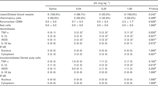
Table 1 Histological, histochemical and immunohistochemical profile of molar dental pulp in rats treated with ZA or saline
ZA (mg kg 1)
Saline 0.04 0.20 1.00 P-Value
Ectasic/Dilated blood vessels 6 (100.0%) 4 (66.7%) 5 (83.3%) 6 (100.0%) 0.242a Inflammatory cells 3 (50.0%) 5 (83.3%) 2 (33.3%) 3 (50.0%) 0.489a Mononuclear CD68+ 0.00.0 0.70.3 0.50.4 2.31.7†
0.026b
Mast cells 0.00.0 0.00.0 0.00.0 0.00.0 1.000b
Odontoblasts
TNF-a 0 (0–1) 3 (2–3)†
3 (2–3)†
3 (1–3)†
0.020b
IL-1b 3 (2–3) 3 (2–3) 3 (3–3)†
3 (3–3)†
0.027b
iNOS 0 (0–1) 3 (2–3)†
3 (3–3)†
3 (3–3)†
0.001b IL-18 bp 0 (0–0) 0 (0–0) 0 (0–0) 0 (0–1) 0.572b NF-kB
Nucleus 0 (0–0) 0 (0–0) 0 (0–0) 0 (0–0) 1.000b
Cytoplasm 3 (2–3) 3 (3–3) 3 (3–3) 3 (3–3) 0.507b Nonodontoblasts Dental pulp cells
TNF-a 0 (0–2) 1.5 (0–3) 1 (1–2) 2 (1–3) 0.162b
IL-1b 2 (1–2) 3 (2–3) 3 (2–3) 3 (3–3)† 0.013b
iNOS 0 (0–1) 0.5 (0–1) 2 (1–3) 1 (1–2) 0.250b
IL-18 bp 0 (0–0) 0 (0–0) 0 (0–0) 0 (0–0) 1.000b NF-kB
Nucleus 0 (0–0) 0 (0–0) 0 (0–0) 0 (0–0) 1.000b
Cytoplasm 0 (0–0) 0 (0–0) 0 (0–0) 0 (0–0) 1.000b
aChi-square (data showed as absolute and percentage frequency),*P<0.05 versus Saline. bKruskal–Wallis/Dunn Test (Median (Minimum-Maximum)).†P
<0.05 versus Saline.
There was no difference in iNOS in nonodontoblast pulp cells in the four groups (P=0.250), and none of these cells was positive for IL-18 bp (P=1.000) or NF-kB (nucleus, P=1.000; cytoplasm, P=1.000; Table 1, Fig. 2).
Discussion
ZA is a toxic drug that is used at high doses for the treatment of bone metastases. ZA can deregu-late the immune system and increase the number of inflammatory cells and levels of cytokines (Ros-sini et al. 2012a,b, Norton et al. 2012, Welton et al. 2013).
Mast cells are poorly visualized in the dental pulp by histochemical methods, and their presence is therefore uncertain. The role of mast cells in pulp diseases is unclear (Bruno et al. 2010), but macro-phages and dendritic cells, which are mononuclear
and CD68 positive, are common cells that appear in the development and normal physiology of the pulp (Iwasaki et al.2011). In this study, there were a sig-nificant number of macrophages in the pulp of rats treated with the highest dose of ZA (1.00 mg kg 1). Macrophages have scavenger receptors (Harre et al. 2012) that recognize apoptotic cells, and they have an important role in defence against caries: macro-phages accumulate in the pulp adjacent to caries-affected regions (Kamal et al. 1997) and phagocyte apoptotic cells (Nishikawa & Sasaki 1999). These cells can serve as antigen-presenting cells (APC) and migrate to the apical region of teeth (Rungvechvut-tivittaya et al. 1998), where they accumulate in response to local overexpression of MCP-1/CCR6 and generate apical granulomas (Liuet al. 2014).
An increase in TNF-a and IL-1b levels was observed. These cytokines are important constitu-tively expressed markers in the pulp, and they can
activate the death domains in pulp cells, stimulating macrophage infiltration and activation (Ohazama et al. 2003, Paula-Silva et al. 2009). Therefore,
these cytokines may play a role in the increased macrophage number in one of the ZA-treated groups.
There was no significant difference in iNOS expres-sion in nonodontoblast pulp cells between treatment groups, but the odontoblasts of ZA-treated rats were associated with iNOS overexpression. iNOS is only present in inflamed pulps and not in healthy pulps (Di Nardo Di Maio et al. 2004). Odontoblasts are impor-tant cells that activate this enzyme in response to car-ies (Veerayutthwilai et al. 2007, Farges et al. 2015). The iNOS staining may reflect hyperaemia or a state of pulpitis (Di Nardo Di Maioet al.2004, Veerayutth-wilai et al. 2007, Farges et al. 2015). TNF-a and IL-1b (increased in this study) may be partially responsible for the modulation of iNOS activity (Bakkeret al.2009).
ZA did not cause direct toxicity to dental pulp cells. High concentrations of this drug are needed to cause this effect, but tissue damage increases with time (Cviklet al.2011).
In the present in vivostudy, the ZA dose was con-verted from human dose to rats. Cvikl et al. (2011) usedin vitroempirical doses of 30 or 100 lmol mL 1 to identify the toxic dose-dependent effect of ZA in dental pulp cells. So, the comparison of these two pro-tocols is not possible due to the difficulty in demon-strating the real concentration of ZA in the pulp cells of rat’s teeth.
The free (not incorporated) ZA is the most associ-ated with ZA toxicity in dental pulp cells (Cviklet al. 2011). In bone, the toxicity of bisphosphonates appears, whilst osteoclasts demineralization releases the incorporated drug (Baronet al. 2011). The odon-toblastic metalloproteinases in inflammatory condi-tions can explain partially this mechanism. The process of dentine matrix degradation and liberation of bisphosphonates by metalloproteinases can be simi-lar in bone, although slower (Chaussain et al. 2013). ZA infusion in peritonitis model leads to IL-1b over-production (Norton et al. 2012). IL-1 is the most important cytokine described in gingival fluid and sal-iva of BRONJ (Bagan et al. 2013, Tsao et al. 2013). Furthermore, IL-1b is an important cytokine involved in pulpitis (Veerayutthwilai et al. 2007), and it was altered in this study. IL-1b can strongly active odon-toblastic metalloproteinases (Hiyamaet al. 2013) and augment bisphosphonate liberation in dental pulps raising IL-1b overexpression (included in nondental pulp cells as shown in this study) perpetuating this process.
Odontoblasts and nonodontoblast pulp cells respond by increasing pro-inflammatory cytokine synthesis, and caries can contribute to the rapid development of
pulpitis in patients who use bisphosphonates (Horst et al.2011).
ZA infusion chronically has been associated with an increase in TNF-a (Cheung et al. 2011), IL-1b
(Bonewald 2004, Tan et al. 2008) and high oxida-tive stress by iNOS activity (Almeida et al. 2010). High levels of these mediators modulate excessive apoptosis and act as a stimulus for the recruitment and activation of macrophages (Kogianni et al. 2008, Muratsu et al. 2013). Thus, ZA may con-tribute to an increase in these proinflammatory markers.
TNF-a and IL-1b are produced by NF-jB activa-tion, but there was no difference in NF-jB levels. NF-jB is constitutively expressed in odontoblasts and is important for the production of collagen type I and dentine sialoproteins. Carious stimuli can modulate the immune response in odontoblasts, leading to overexpression of TNF-a, IL-1b and CCL20 (binder of CCR6; Veerayutthwilai et al. 2007), which participate in macrophage infiltration (Liu et al. 2014).
NF-jB activation is the primary driver of increased TNF-a, IL-1b and iNOS activity, but treatment with ZA did not alter NF-jB immunostaining. However, physiological expression of NF-jB in odontoblasts, accompanied by TNF-a and IL-1b overexpression, leads to pulp cell death (Hozhabriet al. 2015). In the pulp, there are complex ways to regulate these inter-actions, and other proteins can be involved in this process (ABCF1, FOS, IRF3, SP1, STA3, STAT1, FOXO, ERK1, TNFR and many others; Horst et al. 2011). Cytokine production in dental pulp tissue is increased in the presence of lipopolysaccharide (LPS), but a reduction in IjB-a activity, even in the pres-ence of LPS, leads to an increase in TNF-a, IL-1band IL-6 (Muratsu et al. 2013); thus, other mechanisms are responsible for this alteration in the dental pulp of rats treated with ZA.
ZA increased the expression of proinflammatory cytokines, independent from LPS. The addition of car-ies may further increase the production of these medi-ators, modifying the immune profile in the pulp and leading rapidly to an inflammatory disease with pulp damage and irreversible pulpitis and necrosis (ElSalhy et al.2013).
Conclusion
ZA modified the immune cell profile in the dental pulp, increasing the number of macrophages and expression of pro-inflammatory markers (TNF-a, IL-1b
and iNOS) independently of NF-kB immunostaining. This is the first study in vivo showing this relation-ship. However, more studies are needed to investigate the ways that ZA can increase cytokine expression in the dental pulp.
Acknowledgements
This study was partially designed by and is dedicated to Professor Ronaldo Albuquerque Ribeiro (Labo-ratorio de Farmacologia da Inflamacßao e do C~ ^ancer), who is no longer amongst us.
Conflict of Interest
The authors have stated explicitly that there are no conflict of interests in connection with this article.
References
Accorsi-Mendoncßa T, Silva EJ, Marcaccini AMet al.(2013) Evaluation of gelatinases, tissue inhibitor of matrix metal-loproteinase-2, and myeloperoxidase protein in healthy and inflamed human dental pulp tissue. Journal of Endodontics39, 879–82.
Almeida M, Li H, Ambrogini E, Shoshana MB, Manolagas SC (2010) Oxidative stress stimulates apoptosis and activates NF-kappaB in osteoblastic cells via a PKCbeta/p66shc sig-naling cascade: counter regulation by estrogens or andro-gens.Molecular Endocrinology24, 2030–7.
Bagan J, Sheth CC, Soria JM et al.(2013) Bisphosphonates-related osteonecrosis of the jaws: a preliminary study of salivary interleukins.Journal of Oral Pathology and Medicine
42, 405–8.
Bakker AD, Silva VC, Krishnan Ret al.(2009) Tumor necro-sis factor alpha and interleukin-1beta modulate calcium and nitric oxide signaling in mechanically stimulated osteocytes.Arthritis Rheumatism60, 3336–45.
Barasch A, Cunha-Cruz J, Curro FAet al.(2011) Risk factors for osteonecrosis of the jaws: a case-control study from the CONDOR dental PBRN. Journal of Dental Research 90,
439–44.
Baron R, Ferrari S, Russell RG (2011) Denosumab and bis-phosphonates: different mechanisms of action and effects. Bone48, 677–92.
Basso FG, Turrioni PS, Hebling J, de Souza Costa CA (2013) Effects of zoledronic acid on odontoblast-like cells.Archives of Oral Biology58, 467–73.
Bonewald LF (2004) Osteocyte biology: its implications for osteoporosis.Journal of Musculoskeletal and Neuronal Interac-tions4, 101–4.
Bruno KF, Silva JA, Silva TA, Batista AC, Alencar AHG, Estrela C (2010) Characterization of inflammatory cell infiltrate in human dental pulpitis.International Endodontic Journal43, 1013–21.
Chaussain C, Boukpessi T, Khaddam M, Tjaderhane L, George A, Menashi S (2013) Dentin matrix degradation by host matrix metalloproteinases: inhibition and clinical perspectives toward regeneration. Frontiers in Physiology
4, 1–8.
Cheung WY, Liu C, Tonelli-Zasarsky RM, Simmons CA, You L (2011) Osteocyte apoptosis is mechanically regulated and induces angiogenesis in vitro. Journal of Orthopaedic Research29, 523–30.
Cvikl B, Agis H, St€ogerer K, Moritz A, Watzek G, Gruber R (2011) The response of dental pulp-derived cells to zole-dronate depends on the experimental model. International Endodontic Journal44, 33–40.
Di Nardo Di Maio F, Lohinai Z, Darcangelo C et al. (2004) Nitric oxide synthase in healthy and inflamed human den-tal pulp.Journal of Dental Research83, 312–6.
ElSalhy M, Azizieh F, Raghupathy R (2013) Cytokines as diagnostic markers of pulpal inflammation. International Endodontic Journal46, 573–80.
Etemad-Moghadam S, Khalili M, Tirgary F, Alaeddini M (2009) Evaluation of myofibroblasts in oral epithelial dys-plasia and squamous cell carcinoma.Journal of Oral Pathol-ogy and Medicine38, 639–43.
Farges JC, Bellanger A, Ducret Met al.(2015) Human odon-toblast-like cells produce nitric oxide with antibacterial activity upon TLR2 activation. Frontiers in Physiology 6,
1–9.
Harre U, Keppeler H, Ipseiz N et al. (2012) Moonlighting osteoclasts as undertakers of apoptotic cells.Autoimmunity
45, 612–9.
Hiraga T, Ninomiya T, Hosoya A, Nakamura H (2010) Administration of the Bisphosphonate Zoledronic Acid During Tooth Development Inhibits Tooth Eruption and Formation and Induces Dental Abnormalities in Rats. Cal-cified Tissue International86, 502–10.
Hiyama T, Ozeki N, Mogi Met al.(2013) Matrix metallopro-teinase-3 in odontoblastic cells derived from ips cells: unique proliferation response as odontoblastic cells derived from ES cells.PLoS ONE8, e83563.
Horst OV, Horst JA, Samudrala R, Dale BA (2011) Caries induced cytokine network in the odontoblast layer of human teeth.BMC Immunology12, 9.
doi:10.1186/1471-2172-12-9.
Hozhabri NST, Benson MD, Vu MDet al. (2015) Decreasing NF-jB Expression Enhances Odontoblastic Differentiation and Collagen Expression in Dental Pulp Stem Cells Exposed to Inflammatory Cytokines. PLoS ONE 10,
Huang GT, Potente AP, Kim JW, Zhang X (1999) Increased interleukin-8 expression in inflamed human dental pulps. Oral Surgery, Oral Medicine, Oral Pathology, Oral Radiology, and Endodontology88, 214–20.
Iwasaki Y, Otsuka H, Yanagisawa N et al. (2011) In situ proliferation and differentiation of macrophages in dental pulp.Cell Tissue Research346, 99–109.
Kamal MMA, Okiji T, Kawashima N, Suda H (1997) Defense responses of dentin/pulp complex to experimen-tally induced caries in rat molars: an immunohistochem-ical study on kinetics of pulpal la antigen-expressing cells and macrophages. Journal of Endodontics 23, 115–20.
Katz H (2005) Endodontic implications of bisphosphonate-associated osteonecrosis of the jaws: a report of three cases.Journal of Endodontics31, 831–4.
Kogianni G, Mann V, Noble BS (2008) Apoptotic bodies con-vey activity capable of initiating osteoclastogenesis and localized bone destruction. Journal of Bone and Mineral Research23, 915–27.
Kuiper JWP, Forster C, Sun C, Peel S, Glogauer M (2012) Zoledronate and pamidronate depress neutrophil functions and survival in mice. British Journal of Pharmacology16, 532–9.
Liu L, Wang L, Wu Y, Peng B (2014) The expression of MCP-1 and CCR2 in induced rats periapical lesions. Archives of Oral Biology59, 492–9.
Massa LF, Bradaschia-Correa V, Arana-Chavez VE (2006) Immunocytochemical study of amelogenin deposition dur-ing the early odontogenesis of molars in alendronate-trea-ted newborn rats. Journal of Histochemistry and Cytochemistry54, 713–25.
Misso G, Porru M, Stoppacciaro Aet al.(2012) Evaluation of the in vitro and in vivo antiangiogenic effects of deno-sumab and zoledronic acid.Cancer Biology and Therapy13, 1491–500.
Moinzadeh AT, Shemesh H, Neirynck NA, Aubert C, Wes-selink PR (2013) Bisphosphonates and their clinical impli-cations in endodontic therapy. International Endodontic Journal46, 391–8.
Muratsu D, Yoshiga D, Taketomi Tet al. (2013) Zoledronic acid enhances lipopolysaccharide-stimulated proinflamma-tory reactions through controlled expression of SOCS1 in macrophages.PLoS ONE8, e67906.
Naidu A, Dechow PC, Spears R, Wright JM, Kessler HP, Opperman L (2008) The effects of bisphosphonates on osteoblasts in vitro. Oral Surgery, Oral Medicine, Oral Pathology, Oral Radiology, and Endodontology 106,
5–13.
Nishikawa S, Sasaki F (1999) Apoptosis of dental pulp cells and their elimination by macrophages and MHC class II-expressing dendritic cells. Journal of Histochemistry and Cytochemistry47, 303–11.
Norton JT, Hayashi T, Brian C et al. (2012) Cutting Edge: nitrogen bisphosphonate-induced inflammation is
dependent upon mast cells and IL-1.Journal of Immunology
188, 2977–80.
Ohazama A, Courtney JM, Sharpe PT (2003) Expression of TNF-receptor-associated factor genes in murine tooth development.Gene Expression Patterns3, 127–9.
Oizumi T, Yamaguchi K, Funayama Het al.(2009) Necrotic actions of nitrogen-containing bisphosphonates and their inhibition by clodronate, a non-nitrogen-containing bis-phosphonate in mice: potential for utilization of clodronate as a combination drug with a nitrogen-containing bispho-sphonate. Basic and Clinical Pharmacology and Toxicology
104, 384–92.
Paula-Silva FWG, Ghosh A, La Silva B, Kapila YL (2009) TNF-alpha promotes an odontoblastic phenotype in dental pulp cells.Journal of Dental Research88, 339–44.
Pezelj-ribaric S, Anic I, Brekalo I, Miletic I, Simunovic-Soskic M (2002) Detection of tumor necrosis factorain normal and inflamed human dental pulps. Archives of Medical Research33, 482–4.
Rakian A, Yang WC, Gluhak-Heinrich Jet al. (2013) Bone morphogenetic protein-2 gene controls tooth root development in coordination with formation of the periodontium. International Journal of Oral Sciences5, 75– 84.
Rossini M, Adami S, Viapiana O et al. (2012a) Long-term effects of amino-bisphosphonates on circulatingcdT cells. Calcified Tissue International91, 395–9.
Rossini M, Adami S, Viapiana O et al. (2012b) Circulating cdT cells and the risk of acute-phase response after zole-dronic acid administration. Journal of Bone and Mineral Research27, 227–30.
Rungvechvuttivittaya S, Okiji T, Suda H (1998) Responses of macrophage-associated antigen-expressing cells in the den-tal pulp of rat molars to experimenden-tal tooth replantation. Archives of Oral Biology43, 701–10.
Scheller EL, Hankenson KD, Reuben JS, Krebsbach PH (2011) Zoledronic acid inhibits macrophage SOCS3 expres-sion and enhances cytokine production.Journal of Cellular Biochemistry112, 3364–72.
Scheper M, Badros A, Chaisuparat R, Cullen KJ, Meiller TF (2009) Effect of zoledronic acid on oral fibroblasts and epithelial cells: a potential mechanism of bisphosphonate-associated osteonecrosis. British Journal of Haematology
144, 667–76.
Silva PGB, Ferreira Junior AEC, Teofilo CR et al. (2015) Effect of different doses of zoledronic acid in establishing of bisphosphonate-related osteonecrosis.Archives of Oral Biol-ogy60, 1237–45.
Silverman SL, Landesberg R (2009) Osteonecrosis of the jaw and the role of bisphosphonates: a critical review. The American Journal of Medicine122, S33–45.
Tsao C, Darby I, Ebeling PRet al.(2013) Oral health risk fac-tors for bisphosphonate-associated jaw osteonecrosis. Jour-nal of Oral and Maxillofacial Surgery71, 1360–6.
Veerayutthwilai O, Byers MR, Pham TT, Darveau RP, Dale BA (2007) Differential regulation of immune responses by odontoblasts. Oral Microbiology and Immunology 22,
5–13.
Welton JL, Morgan MP, Martı Set al.(2013) Monocytes and cdT cells control the acute-phase response to intravenous zoledronate: insights from a phase IV safety trial.Journal of Bone and Mineral Research28, 464–71.
Wigler R, Steinbock N, Berg T (2013) Oral cutaneous sinus tract, vertical root fracture, and bisphosphonate-related osteonecrosis: a case report. Journal of Endodontics 39, 1088–90.
Wolf AM, Tilg H, Gunsilius E (2006) The effect of zoledronic acid on the function and differentiation of myeloid cells. Haematologica91, 1165–71.
Zadik Y, Vainstein V, Heling I, Neuman T, Drucker S, Elad S (2010) Cytotoxic chemotherapy-induced odontalgia: a dif-ferential diagnosis for dental pain. Journal of Endodontics