vol. 41, n. 3, jul./set., 2005
Fos-like immunoreactivity in central nervous system of mice
simultaneously exposed to the elevated plus-maze and nociception
Karina Santos Gomes1, Cristiane Álvares Garcia2, Cleopatra da Silva Planeta2, Ricardo Luiz Nunes-de-Souza2*
1Departamento de Psicologia e Educação, Faculdade de Filosofia, Ciências e Letras de Ribeirão Preto, Universidade
de São Paulo, 2Laboratório de Farmacologia, Departamento de Princípios Ativos Naturais e Toxicologia,
Faculdade de Ciências Farmacêuticas, Universidade Estadual Paulista/Araraquara
It has been demonstrated that mice exhibit antinociception when they are exposed to the elevated plus-maze (EPM), an animal model of anxiety. To investigate which brain structures are activated during EPM exposure, the present study assessed the immunohistochemical staining for Fos-like immunoreactivity (Fos-LI) in mice intraperitoneally injected with saline or 0.6% acetic acid (which produces nociception) and confined in the open arm (threatening situation) or enclosed arm (control) of the EPM. The following structures were investigated: magnus, dorsal and median raphe nuclei (MR, DR and MnR), periaqueductal gray matter (PAG), dorsal and ventral hippocampus (DH and VH), amygdala (AMY), hypothalamus (HYP) and superior and inferior colliculi (SC and IC). After four days of habituation (habituation was conducted by handling the animals daily for a period of 10 minutes followed by i.p. injection of saline 0.1 mL/10 g), mice received i.p. injection of 0.6% acetic acid or saline and were confined in open or enclosed arms of the EPM. Results showed that open arm confinement increased the number of positive cells for Fos in MnR, PAG and IC, indicating that the fear produced by the threat of the open arms is modulated by these structures. Although statistical analyses did not reveal any effect for nociception factor, (i.e. no effect of acetic acid) the increase in Fos expression was recorded only in animals treated with i.p. acetic acid, suggesting that the simultaneous presence of nociception could be related to an enhanced recruitment of neurons in those midbrains structures.
*Correspondence:
R. L. Nunes-de-Souza Laboratório de Farmacologia Faculdade de Ciências Farmacêuticas Rod. Araraquara-Jaú, km 01 14801-902/Araraquara/ SP/ Brazil Phone: +55 16 3301 6983 FAX: +55 16 222 0073 E-mail: [email protected]
Uniterms
• Fear • Pain • Fos
• Elevated plus-maze • Mice
• Central nervous system
INTRODUCTION
It has been widely demonstrated that animal exposure to learned or innate aversive situations elicits behavioral and
(1980) argued that defensive behaviors and nociceptive responses are incompatible reactions, since the time spent to take care of a body injure could impair the exhibition of an appropriate defensive response. Thus, the antinoci-ception induced by fear events can be considered as part of the defensive reaction (Bolles, Fansellow, 1980; Siegfried
et al., 1990).
The relationship between fear and antinociception has been demonstrated in a range of tests including the elevated plus-maze (EPM) (Lee, Rodgers, 1990; Taukulis, Goggin, 1990; Rodgers et al., 1992), one of the most widely used animal models of anxiety. Briefly, the EPM is an apparatus used to evaluate anxiety responses that rodents (ex. mice and rats) exhibit in an aversive situation, elicited by open arms. The level of anxiety is evaluated by open arm avoidance (% entries and % time in open arms), while general activity is evaluated by the frequency of closed arms entries (Cruz et al., 1994).
The interaction between fear/anxiety and antinoci-ception has also been studied in our laboratory. We have demonstrated that open arm confinement in the EPM produces consistently high magnitude antinociception, as verified by the lower number of abdominal writhes induced by intraperitoneal injection of 0.6% acetic acid (writhing test) recorded in this EPM compartment than exhibited when mice were confined to the enclosed arms (Nunes-de-Souzaet al., 2000).
Although several studies have investigated fear-induced antinociception, the likely brain circuitry involved in the interaction of both responses is not well known. Based on clinical and experimental studies, several brain structures have been widely recognized as important components of the brain defensive systems. Among those are the prefrontal cortex, septum, hippocampus, amygdala, medial hypothalamus, dorsal periaqueductal gray, locus coeruleus, dorsal and median raphe nuclei (for review, see Graeff, 1990; Misslin, 2003; McNaughton, Corr, 2004), and more recently, the deep layers of superior colliculus and inferior colliculus (for review, see Brandão et al, 2003).
The immunochemical detection of Fos, the protein produced by the immediate early-gene c-fos,became a well-established method to map the central nervous system structures involved in functional responses of animals exposed to a variety of environmental stimulus (for review, see Morgan, Curran, 1991; Herdegen, Leah, 1998). In this sense, Fos like-immunoreactivity (Fos-LI) was identified in several limbic structures of animals exposed to EPM (Silveiraet al., 1993; Duncan et al., 1996; Linden et al., 2003; Salomé et al., 2004). Also, Fos-LI induced by noxious thermal, mechanic, chemical and electrical stimuli has been demonstrated in brain structures and spinal cord
of cats, guinea-pigs, rats and mice (for review, see Harris
et al., 1995).
The present study investigated whether concurrent nociception stimulation interferes with Fos expression induced by open arm confinement (fear situation) in the mouse central nervous system. Investigations involving the neurobiology of fear-induced antinociception can contribute to development of new drugs to alleviate pain.
MATERIAL AND METHODS
The expression of the early-gene c-fos in central nervous system (CNS) was assessed in male swiss mice (Paulista State University/UNESP, SP, Brazil), weighing 25-35g and confined to the open arms (fear situation) of the EPM. In order to investigate whether fear situation (open arm confinement) would alter Fos expression induced by nociceptive stimulation some animals were concurrently treated with 0.6% acetic acid i.p. (see below) and exposed to open arm. The effect of nociceptive stimulation on c-fos
expression was also evaluated in animals confined to the enclosed arm (control situation). Saline-treated enclosed arm-confined animals were used as control group. The following brain structures were studied: magnus, dorsal and median raphe nuclei (MR, DR and MnR), periaqueductal gray matter (PAG), dorsal and ventral hippocampus (DH and VH), amygdala (AMY), hypothalamus (HYP) and su-perior and inferior colliculi (SC and IC).
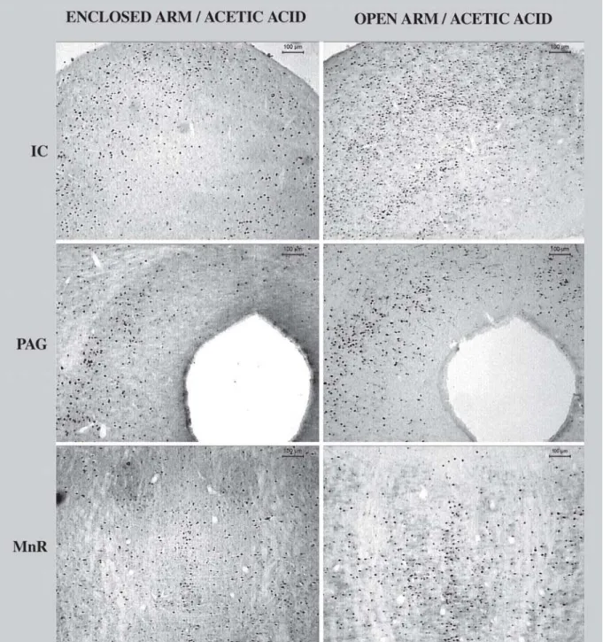
again. Immunoreactivity to Fos was revealed by the addition of the chromogen diaminobenzidin (DAB, 0.02%, Sigma), and hydrogen peroxide (0.04%) and was visualized as a brown insoluble reaction product inside neuronal nucleus. The Fos-LI neurons located through the brain structures were analyzed using a computerized image analysis system. Images (Figure 1) were captured from slides using an optic microscope (Leica DMLB) coupled
with a CCD color camera (JVC) and the Leica Qwin software. To quantify the number of Fos-LI positive cell, the average number from a representative size/area belonging to the proposed structures were measured. In the case of bilateral structure, the measurements were conducted bilaterally. At least two sections per structure per animal were evaluated for all experimental groups. A mean value for cell density in each structure was then calculated.
All structures were analyzed as a whole structure, with the exception of hypothalamus, which was the dorsomedial nucleus the target region for the analysis. The experiments carried out in this study comply with the norms of Brazilian Neuroscience and Behavior Society (SBNeC), based on the US National Institutes of Health Guide for Care and Use of Laboratory Animals.
RESULTS AND DISCUSSION
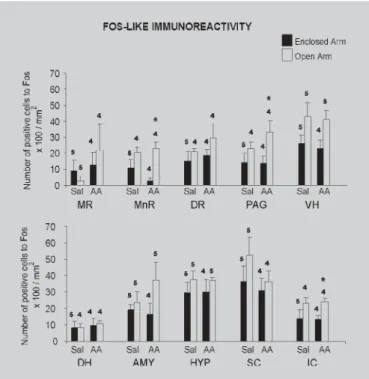
The results obtained through quantitative analyses of Fos-LI in the studied brain regions after exposure to the EPM are shown in Figure 2. All results were initially submitted to Levene’s test for homogeneity of variance and whenF-values were significant, results were analyzed by non-parametric Mann-Whitney’s test. For homogenous samples, data were submitted to two-factor analysis of variance (ANOVA) [Factor 1: treatment (saline or acetic acid); Factor 2: confinement (open or enclosed arm)] followed by Duncan’s multiple comparisons test.
Figure 2 shows the number of positive cells for Fos recorded in several brain structures in mice treated with i.p injection of saline or 0.6% acetic acid and confined to open or enclosed arm of the EPM. Levene’s test revealed significantF-values only for the following structures: DR (F3,13=5.76, p<0.01), HYP (F3,15=4.66, p<0.02) and IC (IC: F3,13=7.38, p<0.01). However, post hoc acceptable between-group (Mann-Whitney test) comparisons revealed significant differences only in the IC, in which the group treated with acetic acid and confined to open arm showed increased Fos-LI when compared to enclosed arm confined group (p<0.02). For homogenous groups (MR, MnR, PAG, DH, VH, AMY and SC), two-way ANOVA (followed by Duncan’s test) revealed significant differences in Fos-LI only for confinement factor and in the following structures: MnR [Factor 1 (treatment): F1,13=0.48, p=0.49; Factor 2 (confinement): F1,13=13.04, p<0.004; treatment x confinement interaction (F1,13=1.54, p=0.23)] and PAG [Factor 1 (treatment): F1,13=0.81, p=0.38; Factor 2 (confinement): F1,13=6.25, p<0.03; treatment x confinement interaction (F1,13=0.36)]. For both structures, post-hoc comparisons for acetic acid treated animals showed an increased Fos-LI in open arm confined group.
The present study demonstrated that open arm confinement tended to increase the number of positive cells for Fos in almost all brain structures investigated. However, the fear situation increased significantly Fos expression only in MnR, PAG and IC in mice receiving acetic acid. Our results corroborate previous studies showing that plus-maze exposure increases c-Fos
expression in limbic structures (Silveira et al., 1993;
Duncanet al., 1996; Linden et al, 2003; Salomé et al., 2004). Interestingly, in these structures ANOVA did not show any difference between the groups exposed to the open arms and treated with acetic acid with the control group (exposed to the same arm and treated with saline). Since these midbrain structures play a role in the brain defensive system (Graeff, 1990; Fanselow, 1991; Carrive
et al., 1997; Brandão et al., 2003; Misslin, 2003) and in the neurobiology of pain (Millan, 2002; Brandão et al., 2003; Behbehani, 1995; Castilho et al., 1999) the present results suggest that both fear (produced by the threat of the open arms) and pain may be modulated by these neural substrates.
However, contrary to expected, i.p. injection of acetic acid did not alter Fos expression in any brain structure investigated in animals confined to the enclosed arms. Being a pain stimulus, it was expected that acetic acid
FIGURE 2 - Means (± S.E.M.) of Fos-positive cells per mm2 X 100 in different brain regions of animals treated with
would increase Fos expression in the central nervous system in animals confined in a non-aversive place (for review, see Harris et al., 1995). This lack of effect impaired one of the purposes of our study, which was to identify the influence of the fear state (i.e. open arm confinement) on Fos expression induced by nociceptive stimulation. In addition, although MnR, PAG and IC are closely involved in modulation of fear reactions (Brandão et al, 2003; Millan, 2002), the other structures (e.g. DR, AMY, HYP and hippocampus) investigated in the present study have also been related to these processes (for review, see Graeff, 1990; Brandão et al., 2003; Millan, 2002). However no significant changes in c-Fos expression was found in these structures, either provoked by open arm exposure or by nociceptive stimulation. One possible explanation for these discrepancies may be related to the small number of subjects (n= 4-5) of the samples used in our study. Small samples usually render a statistical analysis more rigorous in terms of acceptable level of significance. In addition, high levels of Fos-LI were recorded in the control group (saline i.p. + enclosed arm confinement), bringing the means of groups closer and making a between-group difference less probable. In this context, Hinks et al. (1996) also found a high expression of c-Fos mRNA in the brain of control rats confined in the enclosed arms of the EPM. Indeed, it is possible that the animal manipulation per se
has led the number of positive cells for Fos near to the “ceiling effect”. This might explain why the presentation of a more aversive stimulus (e.g. open arm confinement and/ or i.p. injection of acetic acid) produced only mild increases in the Fos-LI in structures like MnR, PAG, IC, DR, AMY, HYP and hippocampus. It is still possible that some potential differences were not detected because the histological analysis of Fos-LI was conducted considering the structures as a whole. Thus, further studies should attempt to control possible experimental variables, such as the duration of handling, the number of exposures to the experimental apparatus and the analysis of Fos-LI in the specific nucleus of the proposed structures. It has been demonstrated that such changes in the experimental protocol facilitate habituation and reduces c-fos baseline in mice (Linden et al., 2003; Ryabinin et al., 1999). Thus, Fos expression in structures like MnR, PAG, IC, DR, VH and AMY in mice exposed to both nociceptive and threatening situations needs further investigation.
ACKNOWLEDGMENTS
The authors thank Elizabete Z. P. Lepera and Rosana F. P. Silva for their efficient technical assistance and Fran-cisco Carlos Rocatelli for photomicrographs preparation.
This work received financial support: Fapesp (00/02790-9, 00/15116-4, 02/03705-0), CNPq (109390/2001-6, 470445/01-7 and 350480/1995-8) and PADC/FCF. K.S. Gomes is recipient of Fapesp scholarship.
RESUMO
Expressão de Fos no sistema nervoso central de camundongos expostos simultaneamente ao labirinto
em cruz elevado e à nocicepção
Recentes evidências têm mostrado que a exposição de ca-mundongos ao labirinto em cruz elevado (LCE), um modelo animal de ansiedade, resulta na exibição de analgesia. Com o objetivo de investigar quais as estruturas que são ativadas durante a exposição ao LCE, no presente estudo foi utiliza-da a técnica de imunocitoquímica para marcação de pro-teína Fos em camundongos que receberam injeção intraperitoneal de salina ou ácido acético 0,6% (estímulo nociceptivo) e que foram confinados no braço aberto (situ-ação ameaçadora) ou fechado (controle) do LCE. As se-guintes estruturas foram estudadas: núcleos magno, dorsal e mediano da rafe (MR, DR e MnR), matéria cinzenta periaquedutal (PAG), hipocampo ventral e dorsal (DH e VH), amigdala (AMY), hipotálamo (HYP) e colículos supe-rior e infesupe-rior (SC e IC). Após quatro dias de manipulação [a manipulação foi conduzida pelo manuseio diário dos animais durante um período de 10 minutos seguidos da injeção i.p. de salina (0,1 mL/10 g)], camundongos recebe-ram injeção i.p. de ácido acético 0,6% ou salina e forecebe-ram confinados nos braços aberto ou fechado do LCE. Os resul-tados revelaram que o confinamento no braço aberto au-mentou o número de células positivas para Fos no MnR, PAG e IC, indicando que o medo produzido pela ameaça dos braços abertos é modulado por essas estruturas. Em-bora a análise não tenha revelado efeito para o fator nocicepção (isto é, nenhum efeito do ácido acético) um incremento na expressão de Fos foi registrado somente em animais tratados com ácido acético i.p., sugerindo que a presença simultânea da nocicepção pode estar relaciona-da ao aumento do recrutamento de neurônios nessas estru-turas mesencefálicas.
UNITERMOS: Medo. Dor. Fos. Labirinto em cruz eleva-do. Camundongos. Sistema Nervoso Central
REFERENCES
BOLLES, R, C.; FANSELOW, M. S. A perceptual-defensive-recuperative model of fear and pain. Behav. Brain Sci.,
Amsterdam, v.3, p. 291-3222, 1980.
BRANDÃO, M. L., TRONCOSO, A. C.; SILVA, M. A. S.; HUSTON, J. P. The relevance of neuronal substrates of defense in the midbrain tectum to anxiety and stress: empirical and conceptual considerations. Eur. J. Pharmacol., Amsterdam, v. 463, p. 225-233, 2003.
CARRIVE, P.; LANG, P.; HARRIS, J. A.; PAXINOS, G. Conditioned fear to context is associated with increased fos expression in the caudal ventrolateral region of the midbrain periaqueductal gray. Neurosci., New York, v. 78, p. 165-177, 1997.
CASTILHO, V. M.;AVANZI, V.; BRANDÃO, M.L. Antinociception elicited by aversive stimulation of the inferior colliculus. Pharm. Bioch. Behav., New York, v. 62, n. 3, p. 425-431, 1999.
CRUZ, A. P. M.; FREI, F.; GRAEFF, F. G. Ethopharmacological analysis of rat behavior on the elevated plus-maze. Pharmacol. Biochem. Behav., New York, v. 49, p. 171-179, 1994.
DEAKIN, J. F. W.; GRAEFF, F. G. 5-HT and mechanisms of defense.J. Psychopharmacol., Berlin, v. 5, n. 4, p. 305-315, 1991
DUNCAN, G. E.; KNAPP, D. J.; BREESE, G. R. Neuroanatomical characterization of Fos induction in rat behavioral models of anxiety. Brain Res., Amsterdam,v. 713, p. 79-91, 1996.
FANSELOW, M. S. The midbrain periaqueductal gray as a coordinator of action in response to fear and anxiety. In: DEPAULIS, A.; BANDLER, R. (eds). The midbrain periaqueductal gray matter. New York: Plenum Press, 1990. p. 151-173.
GRAEFF, F. G. Brain defense systems and anxiety (1990). In: ROTH M.; NOYES, R.; BURROWS, G. D. (eds).
Handbook of anxiety. Amsterdam: Elsevier, 1990, v. 3, p. 307-354.
HARRIS J.A.; WESTBROOK, R. F.; DUFFIELD, T.Q.; BENTIVOGLIO, M. Fos expression in the spinal cord is suppressed in rats displaying conditioned hypoalgesia.
Behav. Neurosci., Washington DC, v. 109, p. 320-328, 1995.
HERDEGEN, T.; LEAH, J. D. Inducible and constitutive transcription factors in the mammalian nervous system: control of gene expresion by Jun, Fos and Krox, and CREB/ATF proteins.Brain Res. Rev., New York, v. 28, n. 3, p. 370-490, 1998.
HINKS, G. L.; BROWN, P.; FIELD, M.; POAT, J. A.; HUGHES, J. The anxiolytics CI-988 and chlordiazepoxide fail to reduce immediate early gene mRNA stimulation following exposure to the rat elevated X-maze.Eur. J. Pharmacol., Amsterdam, v. 312, p.153-161, 1996.
LEE, C.; RODGERS, R.J. Antinociceptive effects of elevated plus-maze exposure: influence of opiate receptor manipulations.Psychopharmacology, Berlin, v. 102, p. 507-513, 1990.
LINDEN, A. M.; BAEZ, M.; BERGERON, M.; SHOEPP, D. D. Increased c-fos expression in the centromedial nucleus of the thalamus in metabotropic glutamate 8 receptor knockout mice following the elevated plus-maze test.Neurosci., New York, v. 121, p.167-178, 2003.
MCNAUGHTON, N.; CORR, P. J. A two-dimmensional neuropsychology of defense: fear/anxiety and defensive distance.Neurosci. Biobehav. Rev., New York, v. 28, p. 285-305, 2004.
MILLAN, M. J. Descending control of pain. Prog. Neurobiol., New York, v. 66, p. 355-474, 2002.
MISSLIN, R. The defense system of fear: behavior and neurocircuitry. Cl. Neuropsysiol., Paris, v. 33, p. 55-66, 2003.
MORGAN, J. L.; CURRAN, T. Stimulus-transcription coupling in the nervous system: involvement of the inducible proto-oncogeneses fos and jun. Anun. Rev. Neurosci., New York, v. 14, p. 421-451, 1991.
RODGERS, R. J, LEE, C.; SHEPHERD, J. K. Effects of diazepam on behavioural and antinociceptive responses to the elevated plus-maze in male mice depend upon treatment regimen and prior maze experience.
Psychopharmacology, Berlin, v. 106, p. 102-110, 1992.
RYABININ, A. E.; WANG, Y.; FINN, D. A. Different levels of fos immunoreactivity after repeated handling and injection stress in two inbread strains of mice. Pharmacol. Biochem. Behav., New York, v. 63, n. 1, p. 143-151, 1999.
SALOMÉ, N.; SALCHNER, O. D.; SEQUEIRA, H.; WIGGER, A.; LANDGRAF, R.; SINGEWALD, N. Neurobiological correlates of high (HAB) versus low anxiety-related behavior (LAB): differential Fos expression in HAB and LAB rats. Biol. Psych., New York,
v. 55, p. 715-723, 2004.
SIEGFRIED, B., FRISCHKNECHT, H. R.; NUNES-DE-SOUZA, R. L. An ethological model for the study of activation and interaction of pain, memory and defensive systems in the attacked mouse. Role of endogenous opioids. Neurosci. Biobehav. Rev., New York, v. 14:, p. 481-490, 1990.
SILVEIRA, M. C. L.; SANDNER, G.; GRAEFF, F.G. Induction of Fos immunoreactivity in brain by exposure to the elevated plus-maze. Behav. Brain Res., Amsterdam,
v. 56, p. 115-118, 1993.
TAUKULIS, H. K.; GOGGIN, C. E. Diazepam-stress interactions in the rat: effects on autoanalgesia and a plus-maze model of anxiety. Behav. Neural Biol., New York, v. 53, p. 205-216, 1990.