TRANSVESICAL ROUTE FOR NOTES UROLOGICAL APPLICATIONS: ADVANCES
& CONTROVERSIES
Estevao Lima
1,2, Carla Rolanda
1,3and Jorge Correia-Pinto
1,4.
1Surgical Sciences Research Domain, Life and Health Sciences Research Institute (ICVS), ICVS/3Bs - PT Government Associate Laboratory. Braga/Guimarães. Portugal. School of Health Sciences. University of Minho. Braga. Portugal. 2Department of Urology, 3Department of Gastroenterology and 4Department of Pediatric Surgery. Hospital de Braga. Braga. Portugal.
Resumen.- La cirugía endoscópica transluminal por orificios naturales (NOTES) ha emergido recientemente en el campo quirúrgico experimental, innovando en el paso de la barrera luminal, la ausencia de cicatrices y la reducción del dolor postoperatorio. Entre los di-ferentes puertos de acceso (transvaginal, transgástrico, transvesical y transcolónico), este artículo es una puesta al día de los avances y controversias del puerto trans-vesical para aplicaciones urológicas del NOTES.
@
CORRESPONDENCE
Estevao Lima, MD, FEBU, PhD
Department of Urology
Hospital of Braga
University of Minho
Campus de Gualtar
4709-057 Braga (Portugal)
[email protected]
Summary.- Natural Orifice Transluminal Endosco-pic Surgery (NOTES) has emerged recently in the ex-perimental surgical field, innovating for the passage of luminal barrier, the absence of scars and reduction of post-operative pain. Among the various ports of access (transvaginal, transgastric, transvesical and transcolonic), this paper is an update on advances & controversies of transvesical port for NOTES Urological applications.Keywords: Endoscopy. Laparoscopy. Surgery. Transvesical surgery. Natural Orifice Transluminal Endoscopic Surgery. NOTES.
INTRODUCTION
In the last decades of the twentieth century, there was a major revolution with the advent of laparoscopy, breaking with the traditional concept of open surgery “large incision, great surgeon” (1). With the initial application of laparoscopy, surgeons have recognized that this approach reduced the post-operative morbidity and was quickly accepted as the gold-standard technique for an increasing number of procedures. Indeed, the laparoscopic small incisions in the abdominal wall reduce post-operative pain by shortening the time of hospitalization and convalescence, with improved aesthetic scar (2). Although the laparoscopic advance, it is a long time since Humans dream of performing surgery without scars. A transvisceral port to achieve the abdominal cavity could be the key to this challenge. Inspired by this possibility, Reddy and Rao, in 2004, in India
in order to overcome the limitations of this port with the first one, performing a pure NOTES cholecystectomy using a combined approach concept (11).
Transvesical port – technique access
In 2006, Lima et al were the first to suggest the bladder as a portal for NOTES (8). Unlike other ports, this organ seemed to be instantaneously approachable with standard urological tools. The placement of the transvesical port is based on the Seldinger principle. Currently, an ureteroscope is introduced through the urethra into the bladder with pneumodistension, emptying urine from the bladder and distending it with CO2. The vesicotomy site is carefully selected on the bladder dome. A mucosal incision is made with scissors introduced through the working channel of the ureteroscope. Subsequently, a 5 Fr open-ended ureteral catheter is pushed forward through the incision into the peritoneal cavity. A 0.035-inch flexible tip guidewire is then inserted into the peritoneal cavity through the lumen of the ureteral catheter. Guided by the flexible tip guidewire, the vesical hole is enlarged with a dilator of a ureteroscope sheath enveloped by a flexible 5.5-mm overtube. A ureteroscope is introduced into the peritoneal cavity through the overtube and allows the creation of a pressure-controlled CO2 pneumoperitoneum.
In 2007 Gettman and Blute described transvesical peritoneoscopy in human attempted before a robot assisted radical prostatectomy using a flexible ureterorenoscope (12). They used the same technical mode of transvesical approach in a porcine model with few modifications (e.g., instead of a ureteral catheter, the authors used a balloon dilator).
Transvesical experimental procedures
The first documented case of surpassing the bladder wall to perform simple intraperitoneal procedures was carried out and reported by Lima et al (8) (Table I). The authors introduced a transurethral and transvesical ureteroscope into the peritoneal cavity, and subsequently, liver biopsy and division of the falciform ligament were performed. Postoperatively, the survival animals were left with a catheter for 4 days, after which necroscopy revealed completely healed cystotomy sites and no evidence of peritoneal complications.
Given the unexpected good results from the first study using transvesical port, Lima et al. felt encouraged to test the possibility to reach even the thoracic cavity (9). An ureteroscope was introduced (3), performed a transgastric appendectomy in
humans and Kalloo et al., in USA (4), described a peritoneoscopy with liver biopsies by a transgastric approach in a porcine model. This was the birth of NOTES (Natural Orifice Transluminal Endoscopic Surgery), a new era in surgery, performing with no scars and therefore without its complications, such as wound infections, adhesions, post-operative ileus, and possibly less post-operative pain (5).
Historical perspective of transluminal ports
Since the last century that surgeries are performed by natural orifices: urologists by an urethral access, gastroenterologists by an oral and anal way, but without exceeding the luminal barrier. It is the transluminal concept that is new in the conceptualization of NOTES, emerging as new approaches the transvaginal, transgastric, transvesical and transcolonic ports. The transvaginal approach was the first to be used as an entrance to the peritoneal cavity, in 1901, by Dimitri Ott who performed a ventroscopy (5). Later, in 1928, Decker & Cherry performed the same procedure, naming it culdoscopy (5). Although the dates go back to early last century and Gynecology has been using a transvaginal port to the pelvic surgery, this access was not explored as a pathway to the abdominal cavity until 2002, when Gettman et al. described a transvaginal nephrectomy in a porcine model (6). Only in 2004, kalloo et al. carried out, by the first time, the transgastric approach into the peritoneal cavity performing liver biopsies and peritoneoscopies in the same model (4). Since this experience and given its relevance to surgery’s history, a large number of research teams have developed and explored the applications of these techniques, leading to that many other procedures were tried out by the transgastric port (7). Given the complications that this approach presents, Lima et al., in 2006, conceived a lower access port, the transvesical pathway, which could solve some of the limitations associated with the isolated transgastric port (8). Thereby, the authors explored the peritoneal cavity by a transvesical port, demonstrating the feasibility of this access. After these experiments and faced with these results, Lima et al., using the same port, acceded the thoracic cavity, expanding the intervention area of NOTES into the thorax (9).
into the peritoneum through the transvesical port and was subsequently advanced into the thoracic cavity. The insufflation was achieved through the ureteroscope, and lung biopsies and inspection of the pleural cavity and lung surface were performed with success. A Foley catheter was left in the bladder for 4 days, and the postmortem examination 15 days after surgery revealed complete healing of the vesical and diaphragmatic incision. Although the authors had been able to perform only limited thoracoscopy and lung biopsies, it definitively extended the intervention field of NOTES from peritoneal to thoracic cavity as well.
Some critics always questioned the feasibility and reproducibility of transvesical port in the human being, particularly regarding the use of rigid instruments. This was the aim for Branco et al. to test the access and feasibility of peritoneoscopy by using a rigid ureteroscope in two human male cadavers. This research group concluded that peritoneoscopy, liver biopsy, and ileocecal appendix manipulation using a rigid ureteroscope through a transvesical port is feasible in a cadaver model (13).
Cholecystectomy is one of the most challenging isolated transgastric approaches. Using two endoscopes, or a single endoscope conjugated with a transabdominal trocar, Park et al. (14) and Swanstrom et al. (15) experienced significant
difficulties performing cholecystectomy using shape-lock technology. These authors reported difficulties related to controlling the pneumoperitoneum and obtaining a stable platform for anatomy exposure, organ retraction, secure grasping, and adequate triangulation of instruments. Rolanda et al. introduced the concept of combined approaches by natural orifice using a combined transgastric and transvesical approach for cholecystectomy (11). This step confirmed the advantage of the transvesical port and initiated the multiple ports of entry concept in NOTES. This concept may provide advantage over the single port, such as monitoring the creation of a second port through the first one, being most likely to successfully close the otomy, and finally but perhaps the main advantage, has increased triangulation and traction.
In a nonsurvival study, Lima et al. expanded the concept of combined transgastric and transvesical approaches performing nephrectomy in female pigs (16). Under ureteroscope visualization through a 5-mm transvesical port, researchers controlled the orally introduced flexible gastroscope by the gastrotomy into the peritoneal cavity. Right or left nephrectomy was carried out using instruments introduced by devices that worked in the renal hilum, alternating intervention on dissection or retraction procedures. In all animals, both kidneys were visualized, and the renal vessels and ureter were reasonably individualized and ligated separately with ultrasonic scissors introduced
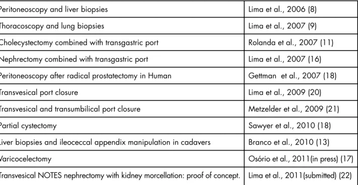
TABLE I. PROCEDURES PERFORMED BY THE VESICAL PORT.
Peritoneoscopy and liver biopsies
Thoracoscopy and lung biopsies
Cholecystectomy combined with transgastric port
Nephrectomy combined with transgastric port
Peritoneoscopy after radical prostatectomy in Human
Transvesical port closure
Transvesical and transumbilical port closure
Partial cystectomy
Liver biopsies and ileoceccal appendix manipulation in cadavers
Varicocelectomy
Transvesical NOTES nephrectomy with kidney morcellation: proof of concept.
Lima et al., 2006 (8)
Lima et al., 2007 (9)
Rolanda et al., 2007 (11)
Lima et al., 2007 (16)
Gettman et al., 2007 (18)
Lima et al., 2009 (20)
Metzelder et al., 2009 (21)
Sawyer et al., 2010 (18)
Branco et al., 2010 (13)
Osório et al., 2011(in press) (17)
through the transvesical port. In two early cases, mild hemorrhage occurred after ultrasonic ligation. Thus, complete renal release and mobilization to the stomach were achieved in all animals, but the gastrotomy site could not be closed. The authors also reported that additional improvements are needed with better devices and instruments.
After these successful upper abdominal procedures the Minho Research Group felt that this approach might be best not only to perform simple procedures in the structure of the upper abdomen and chest, but also to carry out maneuvers in the pelvis and lower abdomen (17). Indeed, they assessed the feasibility and the safety of a bilateral varicocelectomy through a transvesical approach using a combination of flexible cystoscope and thulium laser energy in 6 survival porcine models.
Recently, Sawyer et al. described a partial cystectomy by intravesical transurethral techniques in a porcine model (18). An endoscopic loop device was advanced through one port of the multichannel cystoscope. Through the second port, a flexible toothed grasping device was advanced through the loop to grasp the targeted area of the bladder wall. Then the grasper was slowly withdrawn while maintaining a grip on the “pseudotumor” through the loop. A full-thickness bladder segment was then excised using cutting current. At the end of the procedure, the specimen was removed en bloc with the cystoscope, and the bladder wall defect was reapproximated with endoscopic clips. The authors reported that further investigation in chronic models will be required to determine the potential for safe adaptation to human beings.
In a human case, Gettman et al. performed a transvesical peritoneoscopy using an ureteroscope prior to performing a robot-assisted radical prostatectomy (12). There were nor intraoperative complications nor early postoperative. At discharge and at 2-month follow-up no bowel dysfunction, no pain control problems and no evidence of urine leakage from the bladder were observed.
Safety of the transvesical port
First purpose had been the determination of the safety of the transvesical approach in term of possible peritoneal contamination. Recent works showed elevate risks of infection-related complications in animals undergoing transintestinal (colonic, gastric) incision and peritoneal contamination after transgastric incision in human during laparoscopic procedure (5). Nevertheless, Lima et al. demonstrated that closure
of a 5-mm bladder hole is not absolutely necessary if bladder drainage is assured (8). Anyway, McGee examined the resultant microbial contamination of the human peritoneum after transvesical incision confirming this as a clean portal of entry. This research group examined the resultant microbial contamination of the human peritoneum after transvesical incision during 60 robot-assisted laparoscopic prostatectomy (RALP) procedures (19). With an average time from transvesical incision to vescicourethral anastomosis of 118 minutes peritoneal contamination was seen in 5 of 60 (8.3%) patients. Besides, organism resembled bacteria native of the prostate or seminal fluid because of prostate manipulation and resection during RALP. This study confirmed that transvesical incision would be effectively a clean portal of entry for NOTES.
Transvesical port closure
concluded that these findings provided immediate support for clinical application of this method to close bladder perforations both in management of bladder rupture and transvesical port in NOTES procedures.
More recently, Metzelder et al. described another method for closure bladder perforations in porcine model. In five female piglets underwent right-sided transurethral nephroureterectomy using a hybrid technique with one 15 mm trocar placed umbilically and one 3 mm trocar placed transvesically. Hilar dissection was performed with a 5 mm endoligasure vessel sealing device. After umbilical retrieval of the resected kidney, the urinary bladder was closed by an Endoloop via an umbilical “two in one system” with the assistance of a 2 mm transurethrally placed endoscopic clamp (21).
Transvesical port for abdominal morcellation of specimen
The morcellation was thought as a method to overcome the limitation of the size for removing specimens, regardless of the diameter of the port accessed. One of the limitations of the transvesical port was specimen retrieval, conditioned by the size of the urethra. Recently using this concept, Lima et al reported pure transvesical nephrectomy with kidney morcellation within the abdominal cavity (22). In six pigs, after a pure transvesical nephrectomy, the peritoneal cavity was emptied of CO2 and replaced
with saline solution. Before emptying CO2, the kidney was fixed to the abdominal wall with a fixing needle, created specifically for this purpose. Later, was introduced, through the working channel of the telescope, the morcellator (Piranha-wolf morcellator ®) in the peritoneal cavity and the kidney morcellation began under saline solution. The morcellation proved to be effective, allowing excision of the entire kidney rapidly (median time: 15 minutes, range 10 to 20 minutes). This study showed, for the first time, the feasibility of morcellation of abdominal organs through a natural orifice, in porcine model.
Advantage and limitations of transvesical port in comparison with others ports
Considering the natural orifices that allow NOTES, in order to know what will be the best port or the most suited to a particular procedure, some aspects should be considered: ease of access, ease and safety of the otomy closure, potential infectious complications related to the closure, maximum diameter and type of instruments that the orifices can tolerate and if it allows specimen retrieval (Table II).
The transgastric port has many limitations, such as the lack of sterility of the digestive tract, being a long route, what implicates the use of flexible instruments, and the potential infectious and iatrogenic complications. Some limitations are associated with gastroscopes, particularly the lack of triangulation
Transluminal Port/ Features
Available in both genders
Possibility of using rigid instruments
Access to the abdominal cavity
Sterility
Capability of closure
Limited size of the access
Intact specimen retrieval
Morcellation of specimen
Transvesical
Yes
Yes
Inferior, anterior
Yes
- (a)
Yes
No
Yes
Transvaginal
No
Yes
-No
Easy
No
Yes
-Transcolonic
Yes
Yes
Inferior, posterior
No
- (b)
No
Yes
-Transgastric
Yes
No
Superior
No
- (b)
No
- (c)
No TABLE II. COMPARISON BETWEEN THE TRANSLUMINAL PORTS.
(a) – If otomy up to 5 mm and with adequate bladder drainage is not necessary to close it up. (b) – Lack of an effective closure mechanism.
and traction, the need to work in retroflexion with inverted images and the difficulty in controlling the pneumoperitoneum. The closure of the gastrotomy is also an important limitation (23). In order to overcome these problems, some solutions were launched such as the design of new gastroscopes and transgastric instruments, the combination of transgastric access with a transabdominal port (hybrid NOTES) or with another natural orifice (combined pure NOTES) (24). Despite these attempts, the application in humans depends of an effective gastrotomy closure method that still does not exist.
In the transvaginal approach, the advantages include the possibility of using rigid instruments and direct visualization of upper abdominal structures, without retroflexion and therefore with less difficulty in spatial orientation. Besides, it has an easy method to access the peritoneal cavity through a simple incision at the Douglas pouch. It is considered a good way for specimen retrieval, given its diameter, and the port with the safest otomy closure to date. Nonetheless,
being only available in women is the limiting key factor. Other limitations are related to the patient’s personal history (previous vaginal or pelvic surgery) that may not enable NOTES to be performed. Still without full understanding, but that should be taken into account in future studies, are the effects on sexual function and life’s quality of female patients (24).
Regarding the transcolonic port, this allows the introduction of rigid instruments, direct visualization of the upper abdomen organs, is present in both sexes, allows the use of larger diameter instruments and specimen retrieval due to rectum’s complacency. However, it presents a high risk of infection because it is not sterile and the transvisceral incision closure is not yet effective. It would take a colonic preparation and lumen sterilization to consider its implementation in Humans (10).
The transvesical port broke down some of the most feared complications in Urology: the perforation of the bladder wall that is requested by
ADVANTAGES • Naturally sterile
• Located in most anterior portion of the pelvic cavity allowing peritoneal access above the bowel loops • Providing an en face orientation of the upper abdominal organs allowing better visualization and the ability to work straightforwardly
• Visualization of all the intraperitoneal structures with a direct line of sight
• The possibility of introduce rigid instruments by the working channels of the scopes enhancing the possibility to retract and grasping structures
• The easy and quickly achievement, control and maintaining of the pneumoperitoneum with standard instrument
• The ability to perform in both genders
• Easy endoscopic closure technique of the cystostomy tract although possible a spontaneous healing • Easy spatial orientation
• The possibility of introduce a rigid morcellator into the abdominal cavity above the bowel loops DISADVANTAGE
• The diameter of the urethra limiting the size of the devices and the intact specimen retrieval at the end of the procedures
NOTES to access the abdominal cavity. However, in 2006, Lima et al. saw this port as an advantageous pathway, considering the following statements (Table III): i) the bladder and its content are naturally sterile; ii) the bladder and the lower urinary tract allow the passage of rigid instruments, which facilitates the structure’s retraction; iii) it is a pathway present in both sexes; iv) it is the most anterior anatomical position in the sagittal plane allowing the access to the upper abdominal organs above the bowel loops, reducing the risk of damaging other organs, unlike the transvaginal and transcolonic ports in which the surgeon works through the bowel loops (24). With practice arose other advantages, among which are worth mentioning the simplicity of cystotomy closure and this may not be necessary if the otomy is less than 5 mm with an adequate bladder drainage; the ease and speed which the pneumoperitoneum is achieved and controlled; and the diminished complexity of spatial orientation given the direct visualization of the upper abdomen organs. The disadvantage is related to the diameter of the urethra that limits the size of the instruments used and the intact specimen retrieval.
However recently, Lima et al. reports experimentally, in porcine model, the morcellation as a method for extracting the kidney after nephrectomy by isolated transvesical port.
Potential surgical applications of transvesical port
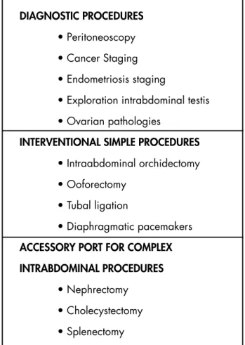
The transvesical port, because of its advantages (in particular, to be naturally sterile and anatomically the most anterior access to the abdominal cavity, allowing the access to the upper abdominal organs above bowel loops), is appealing for both urologic and non-urological procedures. Thus, the transvesical port can be potentially applied to several procedures such as peritoneoscopy, cancer staging, abdominal testes search, intra-abdominal orchidectomy, varicocelectomy, ovarian pathologies treatment, nephrectomy, cholecystectomy, and procedures in the diaphragm (Table IV).
However, the passage of these procedures to Human has been slow and cautious, as it requires rigorous protocol study regarding techniques and materials, but it still points out NOTES as the next evolutionary step of surgery.
DIAGNOSTIC PROCEDURES • Peritoneoscopy • Cancer Staging • Endometriosis staging
• Exploration intrabdominal testis • Ovarian pathologies
INTERVENTIONAL SIMPLE PROCEDURES • Intraabdominal orchidectomy • Ooforectomy
• Tubal ligation
• Diaphragmatic pacemakers ACCESSORY PORT FOR COMPLEX INTRABDOMINAL PROCEDURES
• Nephrectomy • Cholecystectomy • Splenectomy
TABLE IV. POSSIBLE INDICATION FOR TRANSVESICAL PORT.
Harrell AG, Heniford T. Minimally invasive ab-dominal surgery: lux et veritas past, present, and future. Am J Surg 2005;190:239-243.
Autorino R, Cadeddu JA, Desai MM, Gettman M, Gill IS, Kavoussi LR, et al. Laparoendoscopic single-site and natural orifice transluminal endos-copic surgery in urology: a critical analysis of the literature. Eur Urol. 2011;59:26-45.
Rattner D, Kalloo A. ASGE/SAGES Working Group on natural orifice transluminal endoscopic surgery. SAGES/ ASGE Working Group on NO-TES. Surg Endosc 2006;20:329-333.
Kalloo AN, Singh VK, Jagannath SB, Niiyama H, Hill SL, Vaughn CA, et al. Flexible transgastric peritoneoscopy: a novel approach to diagnostic and therapeutic interventions in the peritoneal ca-vity. Gastrointest Endosc 2004;60:114–117. Lima E, Rolanda C, Autorino R, Correia-Pinto J. Experimental foundation for NOTES and Hybrid NOTES. BJUint 2010; 06:913-8.
Gettman MT, Lotan Y, Napper CA, Cadeddu JA. Transvaginal laparoscopic nephrectomy: develop-ment and feasibility in the porcine model. Urolo-gy 2002;59:446-450.
Autorino R, Stein RJ, Lima E, Damiano R, Khanna R, Haber GP, et al. Current status and future pers-*1.
**2.
**3.
4.
**5.
6.
**7.
**8.
9.
10.
11.
12.
**13.
14.
15.
**16.
pectives in laparoendoscopic single-site and natural orifice transluminal endoscopic urological surgery. Int J Urol. 2010;17:410-431.
Lima E, Rolanda C, Pêgo JM, Henriques-Coelho T, Silva D, Carvalho JL, Correia-Pinto J. Trans-vesical endoscopic peritoneoscopy: a novel 5 mm port for intra-abdominal scarless surgery. J Urol 2006;176:802-5.
Lima E, Henriques-Coelho T, Rolanda C, Pêgo JM, Silva D, Carvalho JL, Correia-Pinto J. Trans-vesical thoracoscopy: a natural orifice translu-menal endoscopic approach for thoracic surgery. Surg Endosc 2007;21:854-8.
Pai RD, Fong DG, Bundga ME, Odze RD, Ratt-ner DW, Thompson CC. Transcolonic endoscopic cholecystectomy: a NOTES survival study in a porcine model (with video). Gastrointest Endosc 2006; 64:428–434.
Rolanda C, Lima E, Pêgo JM, Henriques-Coel-ho T, Silva D, Moreira I, et al. Third-generation cholecystectomy by natural orifices: transgastric and transvesical combined approach (with video). Gastrointest Endosc 2007;65:111-7.
Gettman MT, Blute ML. Transvesical peritoneos-copy: initial clinical evaluation of the bladder as a portal for natural orifice translumenal endoscopic surgery. Mayo Clin Proc 2007;82:843-845. Branco F, Pini G, Osório L, Cavadas V, Versos R, Gomes M, et al. Transvesical peritoneoscopy with rigid scope: feasibility study in human male cada-ver. Surg Endosc. 2011;25:2015-9.
Park PO, Bergstrom M, Ikeda K, Fritscher-Ravens A, Swain P. Experimental studies of transgastric gallbladder surgery: cholecystectomy and chole-cystogastric anastomosis (videos). Gastrointest Endosc 2005;61:601–606.
Swanstrom LL, Kozarek R, Pasricha PJ, Gross S, Birkett D, Park PO, et al. Development of a new access device for transgastric surgery. J Gastroin-test Surg 2005;9:1129-1137.
Lima E, Rolanda C, Pêgo JM, Henriques-Coelho
17.
18.
19.
**20.
21.
**22.
23.
*24.
T, Silva D, Osório L, et al. Third-generation ne-phrectomy by natural orifice transluminal endos-copic surgery. J Urol 2007;178:2648-54.
Osório L, Silva D, Autorino R, Damiano R, Co-rreia-Pinto J, Lima E. Pure NOTES transvesical venous ligation: translational animal model of va-ricocelectomy. Urology. 2011;78:1082-6.
Sawyer MD, Cherullo EE, Elmunzer BJ, Scho-misch S, Ponsky LE. Pure natural orifice trans-lumenal endoscopic surgery partial cystectomy: intravesical transurethral and extravesical trans-gastric techniques in a porcine model. Urology 2009;74:1049-53.
McGee SM, Routh JC, Pereira CW, Gettman MT. Minimal contamination of the human peri-toneum after transvesical incision. J Endourol 2009;23:659-63.
Lima E, Rolanda C, Osório L, Pego J, Silva D, Henriques-Coelho T, et al. Endoscopic closure of transmural bladder wall perforations. Eur Urol. 2009;56:151-158.
Metzelder M, Vieten G, Gosemann JH, Ure B, Kuebler JF. Endoloop closure of the urinary bladder is safe and efficient in female piglets un-dergoing transurethral NOTES nephrectomy. Eur J Pediatr Surg 2009;19:362-5.