Alves Filho EF, Costa PFO, Guerra JC. Transanal minimally invasive surgery with single-port (TAMIS) for the management of rectal
neoplasms: a pilot study. J Coloproctol, 2012;32(4): 402-406.
ABSTRACT: Transanal endoscopic microsurgery (TEM) has been used since the 1980’s for the treatment of selected rectal cancers, with clear beneits regarding morbidity and mortality, and good oncological outcomes when compared to radical surgery and conven -tional local resections. The high cost of equipment and the need for long learning curve did not allow the spread of the technique. The aim of this study was to describe the technical characteristics and outcomes of 4 patients operated by this technique, 3 with histologi-cally conirmed adenomas and 1 carcinoid rectal tumor, with no recurrence after an average follow-up of 12 months. The use of single port devices for transanal surgery is a safe method with good oncological results and allows a faster learning curve, by the similarity with conventional laparoscopic procedures and the availability of devices commonly used in laparoscopy.
Keywords: colorectal surgery; rectal neoplasms; TAMIS; laparoscopy.
ReSumo: A microcirurgia endoscópica transanal (TEM) é usada desde a década de 80 para o tratamento de neoplasias retais se-lecionadas, com claros benefícios relacionados à mortalidade e morbidade, e com bons resultados oncológicos, em comparação à cirurgia radical e a ressecções locais convencionais. O custo alto de equipamento de TEM e a necessidade de uma curva de aprendi-zagem longa ainda não permitiram a propagação da técnica. O objetivo deste estudo foi descrever a técnica cirúrgica e os desfechos oncológicos em 4 pacientes operados por esta técnica, 3 com diagnóstico inal de adenomas e 1 de tumor carcinoide, sem recorrência após seguimento médio de 12 meses. A utilização de dispositivos de portal único para a cirurgia transanal é um método seguro e com bons resultados oncológicos, permitindo uma curva de aprendizado mais rápida pela semelhança com os procedimentos laparoscó-picos convencionais e pela disponibilidade de dispositivos comumente utilizados em laparoscopia.
Palavras-chave: cirurgia colorretal; neoplasias retais; TAMIS; laparoscopia.
Transanal minimally invasive surgery with single-port (TAmIS)
for the management of rectal neoplasms: a pilot study
Eduardo Fonseca Alves Filho1, Paulo Frederico de Oliveira Costa2, João Cláudio Guerra3
1Titular Member of Sociedade Brasileira de Coloproctologia (SBCP) – Rio de Janeiro (RJ), Brazil. 2Afilieted Member of
SBCP – Rio de Janeiro (RJ), Brazil. 3Associate Member of SBCP – Rio de Janeiro (RJ), Brazil.
Studiy carried out at the Coloproctology Service of Hospital Português da Bahia – Salvador (BA), Brazil. Financing source: none.
Conlict of interest: nothing to declare.
Submitted on: 11/06/2012 Approved on: 02/08/2012
INTRoDuCTIoN
Since the introduction of transanal endoscopic mi-crosurgery (TEM) by Buess in 1985, there was a change in the paradigms for the treatment of several rectal neo-plasms. This technique has proven to be effective and safe in treating early rectal tumors and polyps. When compared with traditional techniques as local excision, TEM has the advantages of better visualization of the
rectum, access to higher lesions and better ability to ob-tain clear margins with the possibility of excision with-out fragmentation of the surgical specimen1-13.
From the development of surgical techniques, such as Natural Oriice Transluminal Endoscop -ic Surgery (NOTES), and the use of single portal devices for laparoscopic surgery, a new alternative for resection of rectal lesions by transanal surgery called Transanal endoscopic microsurgery per-formed by single port (TAMIS) was introduced1-3, combining the TEM traditional technique with other instruments commonly used in laparoscopic surgery.
The aim of this study was to describe a case se-ries of the irst four patients operated by the TAMIS technique in our service, with emphasis on technical details and surgical oncological outcomes.
meTHoDS
This is a retrospective description (case series) of four patients submitted to rectal neoplasms resection by the TAMIS technique. All patients were operated with the SILS® device (Covidien, USA) (Figure 1), an equipment primarily developed for single-port lapa-roscopic surgery, made of lexible synthetic materi -al, with three openings for the introduction of 5 and 12 mm trocars and a CO2 connection for insuflation and pneumorectum achievement.
All patients underwent colonoscopy and biopsy previously. As a preparation for the operations, they underwent mechanical anterograde bowel prepara-tion, antibiotic prophylaxis and were operated on lithotomy position under general anesthesia, with uniform technique. The device was introduced into the anal canal after its lubriication (Figure 2). Lat
-eral, anterior and posterior stitches were performed to maintain proper placement. Rectal insuflation with carbon dioxide was performed, with an aver-age pressure of 12 to 15 mmHg. Three trocars were then introduced on the SILS device. Superiorly, an optics with 5 mm and 30 degrees was introduced, and inferiorly one ordinary laparoscopic forceps of 5 mm was placed. On the third port, a hook, scis-sor or sealing clamp was placed, according to the surgical needs. A conventional monopolar cautery was connected to these previous laparoscopic in-struments, as needed.
After its identification, the lesions were bounded and the disection was started (Figure 2). One crutial step of the procedures was to outline partial or total commitment of the rectal wall. De-pending on the size of the lesion, it was removed along with the device withdrawal, which could again be introduced and fixed. Then, the wound could be reviewed and its primary closure could be performed in cases of full thickness resection of the rectal wall.
ReSuLTS
Four patients were treated with this method and represented our initial experience, three women and one man. The average age was 55 years old. Regard-ing the preoperative diagnosis, three rectal adenomas with low grade dysplasia were identiied and one rec -tal submucosal tumor whose chromogranin A serum level conirmed the diagnosis of carcinoid tumor.
Figure 2. Introdução do dispositivo SILS
Three patients underwent magnetic resonance imag-ing (MRI) of the pelvis preoperatively, and there was no evidence of invasion of the rectal wall.
The average size of the lesions was 1.5 cm, and the median distance from the anal margin was 6.5 cm. The mean duration of the procedures was 110 minutes. All operations were carried out successfully, without conversion for conventional transanal resection. There were no complications and no need for new interven-tions. There were two submucosal resection and two full-thickness resection with primary closure of the wound with separate stitches.
In all cases, the lateral and deep margins were clear, and the postoperative diagnosis were three tubule-villous adenomas with low grade displasia and one carcinoid tumor measuring 1 cm. No ad-ditional therapy was needed for all cases. With an average follow-up of 12 months, no recurrences were detected.
DISCuSSIoN
TEM has been accepted as a safe alternative when colonoscopic rectal adenomas resections or conventional transanal local resections are not fea-sible or appropriate from the oncologic standpoint. Colonoscopic resection may be associated with high rates of recurrence (21 to 33%) for polyps whose resection margins are positive or less than 1 mm. This is most likely to happen in polyps larg-er than 2 cm and in piecemeal resections14. TEM provides larger resections with adequate margins and encompassing the whole rectal wall. This pro-vides a recurrence rate of aproximately 5%, with conversion rates of 5.7% and complications in 3 to 7% of the cases14. Recurrence is particularly in-creased in patients who previously underwent a re-section by both TEM and colonoscopy15,16. In all cases of our initial experience, no recurrence and no morbidity was demonstrated, possibly due to patient selection.
Recent meta-analysis that evaluated the re-sults of TEM in the treatment of T1 and T2 rec-tal tumors demonstrated that when compared with radical surgery, TEM has higher chances of posi-tive margins, higher rates of local recurrence and lower disease-free survival rates15. On the other
hand, the method also proved to have lower mor-bidity and no difference in overall survival rates. This can be explained by the fact that patients with recurrent T1 tumors after TEM are often referred to radical surgery and chemoradiation. When com-pared to local resection, TEM had better results in achieving negative margins and disease-free survival, however no differences in complication rates and overall recurrence were demonstrat-ed15,16. TEM is also described as inadequate for the treatment of T2 rectal tumors15.
The submucosal invasion and tumor size are considered the most important predictors of local recurrence. Tumors smaller than 3 cm and without submucosal invasion have recurrence rates of 7%, as compared to up to 38% in lesions larger than 3 cm with invasion of the submucosal layer17.
In selected cases of early rectal adenocarcino-mas with certain features such as superficial inva-sion of submucosa (pT1 SM1), histologically well differentiated, with <3 cm of diameter and without lymphatic or vascular invasion, a local recurrence rate of less than 5% can be demonstrated when treated by TEM. In these cases, TEM had similar recurrence and disease-free survival, with fewer rates of mortality and morbidity, when compared to radical surgery18.
In patients with suspected partial response af-ter neoadjuvant treatment, where any lump or ir-regularity in the rectal wall can be identified, re-section by TEM of these suspicious lesions with adequate lateral and deep margins, can confirm a complete pathologic response (ypCR), allowing the inclusion of these patients in Watch and Wait protocols19.
TEM can also be used to resect submuco-sal rectal neoplasms such as carcinoids tumors20. These must be smaller than 2 cm without any evi-dence of muscular invasion to be considered a good indication for the method. When these lesions are removed by colonoscopy, usually they result in positive margins, compromising the oncological radical profile of the procedure. Our patient with the carcinoid tumor presented with this features and had no complications on the follow-up.
TEM. Decreased resting pressures and contraction can be presented until one year after these proce-dures, causing temporary incontinence. The main risk factor for this complication was the duration of the procedure1. It was demonstrated by endorec-tal ultrasound that partial lesions of the internal anal sphincter can occur in up to 29% of patients1. Other studies have shown no significant changes in incontinence scores or in quality of life ques-tionnaires in the long term evaluation21,22. Despite the fact that our patients were not submitted to sphincter evaluation with ultrasound, no anal in-continence was referred in this initial experience.
Unlike what is done in TEM and local resec-tions, the positioning of the patients in TAMIS is independent of the location of the lesion. The li-thotomy position is suitable for most tumor resec-tions, even in lesions of the anterior rectal wall1,3,4. The attachment of an external arm to the surgical table is also not needed. The instruments needed are the same commonly used in laparoscopic pro-cedures, like cholecystectomies and appendecto-mies. The costs of the TEM surgical rectoscope and its instruments is estimated by US$ 85,000 in Europe, while the single port devices cost around US$ 500. This makes TAMIS a feasible alterna-tive in any center with regular laparoscopic equip-ments and experienced professionals in colorectal laparoscopic surgery14.
The technical limitations of TAMIS are simi-lar to those of laparoscopic surgery. There is a ten-dency when using conventional electric cautery to produce smoke that impairs the view of the opera-tive field. Depending on the mobilization of the
in-struments, a partial loss of insufflation (pneumor-ectum) can also occur. The visibility of the rectum is generally excellent. Lower lesions, nearby the dentate line, are not generally resectable by both TEM and TAMIS, being conventional local re-section the most appropriate method indicated7. TAMIS, when compared to TEM, does not allow access to higher rectal lesions (15 to 20 cm ver-sus 18 to 25 cm). Currently, more than one single portal device have been tested for the resection of rectal lesions, and it seems to be no difference among the different equipments tested13.
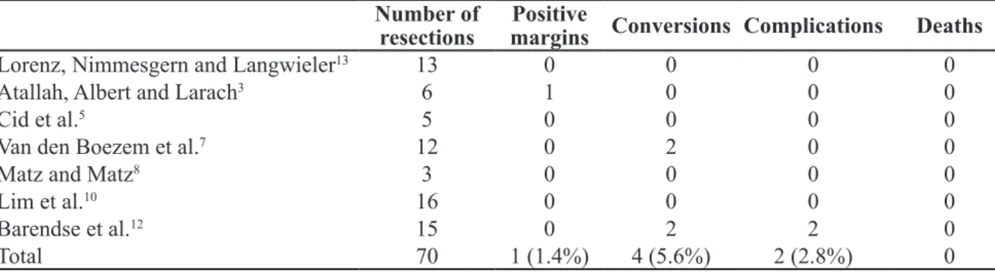
The main question that exists with TAMIS is if this new adapted technique will have simi-lar results as compared to TEM. TAMIS is not the first modification of the TEM method. Other ap-paratus as TEO, using 2D optical systems, in con-trast to the three-dimensional view of TEM, are also used with similar results to TEM23. Based on reports in the literature, we could identify more than 70 patients treated by TAMIS (Table 1), with results comparable to those of TEM for negative margins, conversion and complication rates. There were no reported deaths related to the procedure. Long term results regarding recurrence and over-all survival rates may answer these questions in a near future.
The initial experience of this pilot study, in accordance to the literature, suggests that TAMIS, as a new adaptation of TEM, can be safely per-formed with similar results. Due to its lower costs, and the possible shorter learning curve, this adapt-ed technique can contribute to the dissemination of minimally invasive treatment for rectal lesions.
Table 1. Resections by TAMIS.
Number of resections
Positive
margins Conversions Complications Deaths
Lorenz, Nimmesgern and Langwieler13 13 0 0 0 0
Atallah, Albert and Larach3 6 1 0 0 0
Cid et al.5 5 0 0 0 0
Van den Boezem et al.7 12 0 2 0 0
Matz and Matz8 3 0 0 0 0
Lim et al.10 16 0 0 0 0
Barendse et al.12 15 0 2 2 0
ReFeReNCeS
1. Lorenz C, Nimmesgern T, Back M, Langwieler TE. Transanal single port microsurgery (TSPM) as a modiied technique of transanal endoscopic microsurgery (TEM). Surg Innov 2010;17(2):160-3.
2. Khoo RE. Transanal excision of a rectal adenoma using single-access laparoscopic port. Dis Colon Rectum 2010;53(7):1078-9.
3. Atallah S, Albert M, Larach S. Transanal minimally invasive surgery: a giant leap forward. Surg Endosc 2010;24(9):2200-5. 4. Ragupathi M, Haas EM. Transanal endoscopic video-assisted excision: application of single-port access. JSLS 2011;15(1):53-8.
5. Cid RC, Pérez JC, Elosua TG, Pinto FL, Alegre JM, Martín R, et al. [Transanal resection using a single port trocar: a new approach to NOTES]. Cir Esp 2011;89(1):20-3.
6. Dardamanis D, Theodorou D, Theodoropoulos G, Larentzakis A, Natoudi M, Doulami G, et al. Transanal polypectomy using single incision laparoscopic instruments. World J Gastrointest Surg 2011;3(4):56-8.
7. Van den Boezem PB, Kruyt PM, Stommel MW, Tobon Morales R, Cuesta MA, Sietses C. Transanal single-port surgery for the resection of large polyps. Dig Surg 2011;28(5-6):412-6.
8. Matz J, Matz A. Use of a SILS port in transanal endoscopic microsurgery in the setting of a community hospital. J Laparoendosc Adv Surg Tech A 2012;22(1):93-6.
9. Demirbas S, Cetiner S, Ozer TM, Oztas M, Duran E. The use of single port surgery for polyps located in the rectum.Turk J Gastroenterol 2012;23(1):66-71.
10. Lim SB, Seo SI, Lee JL, Kwak JY, Jang TY, Kim CW, et al. Feasibility of transanal minimally invasive surgery for mid-rectal lesions. Surg Endosc 2012. [Epub ahead of print] 11. Smith RA, Anaya DA, Albo D, Artinyan A. A stepwise
approach to transanal endoscopic microsurgery for rectal cancer using a single-incision laparoscopic port. Ann Surg Oncol 2012. [Epub ahead of print]
12. Barendse RM, Verlaan T, Bemelman WA, Fockens P, Dekker E, Nonner J, et al. Transanal single port surgery: selecting a suitable access port in a porcine model. Surg Innov 2011. [Epub ahead of print]
13. Lorenz C, Nimmesgern T, Langwieler TE. Transanal endoscopic surgery using different single-port devices. Surg Technol Int 2012;XXI:107-111. [Epub ahead of print]
14. Mulsow J, Winter DC. Sphincter preservation for distal rectal cancer – a goal worth achieving at all costs? World J Gastroenterol 2011;17(7):855-61
15. Middleton PF, Sutherland LM, Maddern GJ. Transanal endoscopic microsurgery: a systematic review. Dis Colon Rectum 2005;48(2):270-84.
16. Nahas SC, Nahas CSR, Marques CFS, Dias AR, Pollara WM, Cecconello I. Transanal endoscopic microsurgery (TEM): a minimally invasive procedure for treatment of selected rectal neoplasms. ABCD 2010;23(1):35-9.
17. Sgourakis, Lanitis S, Gockel I, Kontovounisios C, Karaliotas C, Tsiftsi K, et al. Transanal endoscopic microsurgery for T1 and T2 rectal cancers: a meta-analysis and meta-regression analysis of outcomes. Am Surg 2011;77(6):761-72.
18. Doornebosch PG, Zeestraten E, de Graaf EJ, Hermsen P, Dawson I, Tollenaar RA, et al. Transanal endoscopic microsurgery for T1 rectal cancer: size matters! Surg Endosc 2012;26(2): 551-7.
19. Habr-Gama A, Perez R, Proscurchin I, Gamarodriguez J. Complete clinical response after neoadjuvant chemoradiation for distal rectal cancer. Surg Oncol Clin N Am 2010;19:829-45. 20. Moraes RS, Malafaia OT, Queiroz JE, Trippia MA, Buess
GF, Coelho JCU. Transanal endoscopic microsurgery in the treatment of rectal tumors: a prospective study in 50 patients. Arq Gastroenterol 2008;45(4):268-74
21. Doornebosch PG, Gosselink MP, Neijenhuis PA, Schouten WR, Tollenaar RA, de Graaf EJ. Impact of transanal endoscopic microsurgery on functional outcome and quality of life. Int J Colorectal Dis 2008;23(7):709-13.
22. Allaix ME, Rebecchi F, Giaccone C, Mistrangelo M, Morino M. Long-term functional results and quality of life after transanal endoscopic microsurgery. Br J Surg 2011;98(11):1635-43.
23. Rocha JJR, Féres O. Transanal endoscopic operation: a new proposal. Arq Gastroenterol 2008;45(4):268-74.
Correspondence to: Eduardo Fonseca Alves Filho Centro Médico do Hospital Português
Avenida Princesa Isabel, 914, sala 208, Barra Avenida CEP: 40144900 – Salvador (BA), Brazil