Relationship Between Internal Derangement of
Temporomandibular Joint and Changes in Body Posture
Master’s Dissertation in Physiotherapy
Tiago Miguel Rodrigues Rocha
Coimbra
Relationship Between Internal Derangement of
Temporomandibular Joint and Changes in Body Posture
Dissertation submitted by Tiago Miguel Rodrigues Rocha to the
Escola Superior de Tecnologia da Saúde de Coimbra do Instituto
Politécnico de Coimbra for comply the necessary requisites to
obtain the Master degree in Physiotherapy – Human Movement
Specialization, performed under the scientific guidance of PhD.
Maria António Castro, Associate Professor of the Escola Superior
de Tecnologia da Saúde de Coimbra and co-orientation of PhD.
Daniele Manfredini, Associate Professor of the University of
reflective tape and to my friend Juliano Soares for giving me the contact;
To Mr. Alberto Pinheiro, representing the company Flexicel, sited in Braga, Portugal, for having offered the coating to the referential;
To my friend Rui Neto for the brainstormings and idealization of the referential;
To my friend Pedro Magalhães who was always there and had a key role in the design of the schemes;
To my friend Carlos Torres also for the idealization but especially by the laborious construction of the referential;
To my friend Pedro Seromenho who with his countless talent, made the drawings of the temporomandibular joint;
To my friend Hugo Costa for the graphical support whenever necessary;
To my friend Clara Rebelo who beyond itself be a volunteer for the study, captivated many other volunteers;
To my friend Manuel Almeida who was always an anchor in the course of the study, sharing his technical expertise;
To my friend Tiago Ferrete for always being there and available to help, providing a help particularly fundamental at statistical level;
To my “flaminguistas” friends for all the support given;
To D. Fernanda, borrowed grandmother, for all the help provided;
To MSc. Márcio Vieira and PhD. Andrea Ribeiro for sharing their scientific knowledge, giving very good advices;
To the authors PhD. Boudewijn Stegenga, PhD. Shirish Ingawale, PhD. Emily Hoelscher, PhD. Stefan Rues, PhD. Jeffrey Nickel, PhD. Luigi-Maria Gallo, PhD. Laura Iwasaki, PhD. Machiel Naeije, PhD. Aart Visser, PhD. Giacomo Chessa and PhD. Tommaso Castroflorio, whom with their sympathy and scientific goodwill, shared with me their articles;
Particularly to the authors PhD. Maurizio Bergamini and PhD. Barry Cooper, whom besides sharing with me their articles, offered their help to dissipate any doubt, invinting me to know their studies;
To Instituto Superior de Saúde do Alto Ave (ISAVE), in the person of its President PhD. Jónatas Pego, for the spirit of open mind and acceptance of the study. To all officials of this institution whom were tireless in logistical support. To the department of Physiotherapy, with a very special gratitude to MSc. Nuno Soares and MSc. Márcio Vieira, whom since the beginning were available for the study and provided an essencial help for its development;
To the department of Audiology of the ESTESC, in the persons of MSc. Inês Araújo and MSc. Margarida Serrano, whom with all their sympathy and goodwill provided us the use of the balance platform whenever requested, exemplifying its operation and clarifying any questions;
To my friend and external supervisor, PhD. Daniele Manfredini, for all uncountable and valuable help, for the acceptance and personal knowledge sharing in its professional and scientific aspects, since the idealization of the project until its completion;
To my friend and internal supervisor, PhD. Maria António Castro, for having been present in data collections, for always being available when a question arose, but especially for making this study easier with her good mood and positive spirit;
To the memory of PhD. João Gil, who with much regret left us without seeing a small part of his work completed. He was always a companion trying to solve all our problems in the best way possible as if it him own problems. For his dedication and professionalism always inside the group, and for his straightforwardness and friendliness always present;
To my girlfriend Ana Luísa Dias, for everything you had to endure during the frustrations of a less good day, for my lack of time and patience, always staring these things positively, giving me in exchange a opened smile and a friendly word, which always gave me an extra strength. Thanks for being who you are and how you are, thanks for be always by my side because without your support everything would be much more difficult. I am very proud to have you in my life. “You are a lighthouse when I’m lost at sea” Hope – Shawn McDonald;
"A nossa vida é toda ela feita de acasos.
Mas é o que em nós há de necessário que lhes há-de dar um sentido."
VIII | Escola Superior de Tecnologia da Saúde de Coimbra
Key-Words: Temporomandibular joint, internal derangement of temporomandibular
joint, temporomandibular joint dysfunction, body posture.
Background: The presence of body posture changes among patients with
temporomandibular disorders (TMD) has been a controversial issue in the literature, in which it supporters point out the muscular origin as the main etiological factors, mainly associated with postural changes in head. Due to this controversy, it is pertinent to check whether this relationship exists on the most common etiology of TMD, the disk displacement, which translates a biomechanical internal disorder of the temporomandibular joint (TMJ).
Objectives: Assess body posture changes in subjects with internal derangement of the
TMJ when compared to subjects without this biomechanical dysfunction, characterize the patterns of the jaw movements and assess to the muscle activation during jaw movements.
Methods: 21 subjects with TMJ disc displacement (DD) (test group) and 21 subjects
without any TMD (control group) was assessed for body posture changes through evaluation of several body segments by posturography and also was evaluated the postural balance reactions through the center of mass during jaw movements using a balance platform. For the characterization of the jaw movement patterns it was done a kinematic analysis during jaw movements (active ROM and path of the jaw). For the muscle activation during jaw movements it was evaluated the masseter, sternocleidomastoid and spinae erector muscles by surface electromyography (EMG).
Results Discussion: Both groups show forward head posture and extension of the
cervical spine, not noticing any other significant body posture changes in subjects with DD, and if we had to see in detail, in general, subjects without TMD shows more body posture changes than subjects with DD. The pattern of jaw movements is similar in both groups, but in subjects with DD the closing movements are more instable than the opening movements, related to a less effective movement control to counteract the force of gravity and the disk displacement. The bilateral muscle activation during jaw movements is higher in subjects with DD, likely related to a less stable pattern of movement which leads in a higher muscle activation to guide the movement and ensure the best as possible articular stability.
Conclusion: The disk displacement with reduction should be viewed as part of a set of
Escola Superior de Tecnologia da Saúde de Coimbra | IX
Abstract ... VIII
1. Introduction ... 14
2. Literature Review ... 19
2.1 Temporomandibular Joint (TMJ) ... 19
2.1.1 TMJ Anatomy and Biomechanics ... 19
2.1.2 Biomechanics of Mandibular Movements ... 20
2.2 Etiology of Temporomandibular Disorders (TMD) ... 22
2.2.1 TMJ Internal Derangement ... 24
2.3 The use of electronic devices in assessment of TMD ... 28
2.3.1 Surface Electromyography ... 28
2.3.2 Kinematic Analysis ... 30
2.3.3 Posturography ... 30
2.4 Relationship Between Body Posture and TMD ... 31
3. Methodology ... 34
3.1 Experimental Setup ... 35
3.2 Instruments ... 36
3.2.1 Balance Platform ... 36
3.2.2 Surface EMG ... 37
3.2.3 Digital Cameras ... 37
3.2.4 Referential ... 38
3.3 Procedures ... 41
3.4 Analysis Procedures ... 45
3.4.1 Kinematic Analysis ... 45
3.4.2 Postural Analysis ... 47
3.4.3 COG Sway Velocity and Path ... 50
3.4.4 EMG Analysis ... 50
3.5 Statistics ... 51
4. Results ... 52
4.1 Sample Characterization ... 52
4.2 Measurements Repeatability ... 54
X | Escola Superior de Tecnologia da Saúde de Coimbra
4.4 Postural Analysis ... 60
4.4.1 Posture Parameters: ... 61
4.5 COG Sway Velocity and Path Analysis ... 70
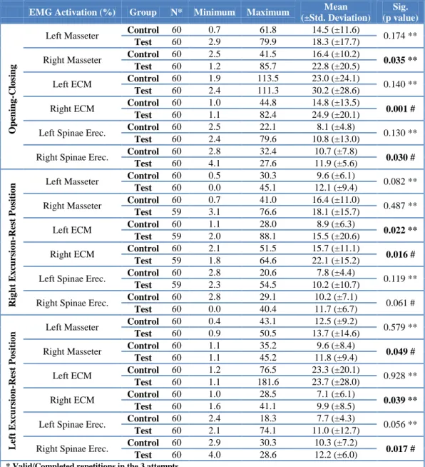
4.6 EMG Analysis ... 71
4.7 Maximum ROM and Vertical Displacement ... 74
5. Discussion ... 75
5.1 Sample Characterization ... 75
5.2 Kinematic Analysis ... 75
5.2.1 Active Jaw ROM ... 75
5.2.2 Jaw Path ... 76
5.3 Postural Analysis ... 77
5.3.1 Posture Parameters ... 77
5.4 COG Sway Velocity and Path ... 79
5.5 EMG Analysis ... 79
5.6 Maximum ROM and Vertical Displacement ... 80
5.7 Implications for the Physiotherapy and Study Limitations ... 81
6. Conclusion ... 82
Escola Superior de Tecnologia da Saúde de Coimbra | XI
IMAGE 1:TEMPOROMANDIBULAR JOINT MOVEMENT DURING MOUTH OPENING. ... 19
IMAGE 2:NORMAL TMJ, DISK ADAPTATIONS DURING MOUTH OPENING. ... 25
IMAGE 3:TMJ DISK DISPLACEMENT WITH REDUCTION, DISK ADAPTATIONS DURING MOUTH OPENING. ... 25
IMAGE 4:TMJ DISK DISPLACEMENT WITHOUT REDUCTION, DISK ADAPTATIONS DURING MOUTH OPENING. ... 26
IMAGE 5:EXPERIMENTAL SETUP SCHEME. ... 35
IMAGE 6:BALANCE PLATFORM COMPONENTS. ... 36
IMAGE 7:SURFACE EMG COMPONENTS. ... 37
IMAGE 8:REFERENTIAL SCHEME. ... 38
IMAGE 9:REFERENTIAL FRONT. ... 39
IMAGE 10:REFERENTIAL HEADREST. ... 40
IMAGE 11:HEADREST FUNCTION. ... 40
IMAGE 12:REFERENTIAL BACK. ... 41
IMAGE 13:STERNOCLEIDOMASTOID MVC PROCEDURE. ... 42
IMAGE 14:SPINAE ERECTORS MVC PROCEDURE. ... 43
IMAGE 15:PASSIVE ROUNDNESS MARKERS. ... 43
IMAGE 16:PASSIVE ROUNDNESS MARKERS PLACED IN THE SUBJECT. ... 44
IMAGE 17:PASSIVE SPHERIC MARKERS PLACED IN THE SUBJECT. ... 45
IMAGE 18:MAXIMUM ROM ANALYSIS. ... 45
IMAGE 19:JAW DISPLACEMENT ANALYSIS. ... 46
IMAGE 20:ASYMMETRICAL ELEVATIONS ANALYSIS. ... 47
IMAGE 21:ANTERIORIZATION OF THE HEAD AND FLEXION/EXTENSION OF THE HEAD ANALYSIS. ... 48
IMAGE 22:BILATERAL DISTANCES ANALYSIS. ... 48
XII | Escola Superior de Tecnologia da Saúde de Coimbra
TABLE 1:DIAGNOSTIC CRITERIA FOR DISK DISPLACEMENT ACCORDING TO THE RDC/TMD(DWORKIN E LERESCHE,1992). 27
TABLE 2:DIAGNOSTIC CRITERIA FOR DISK DISPLACEMENT ACCORDING TO THE AAOP. ... 28
TABLE 3:ELECTROMYOGRAPHY CONFIGURATION. ... 42
TABLE 4:POSTURE PARAMETERS DESCRIPTION. ... 49
TABLE 5:MALE AND FEMALE SAMPLE DISTRIBUTION ON CONTROL AND TEST GROUPS. ... 52
TABLE 6:AGE, HEIGHT AND WEIGHT SAMPLE CHARACTERIZATION MEASUREMENTS. ... 52
TABLE 7:DISK DISPLACEMENT DIAGNOSTIC DISTRIBUTION. ... 52
TABLE 8:STUDY VARIABLES MEASUREMENTS REPEATABILITY. ... 54
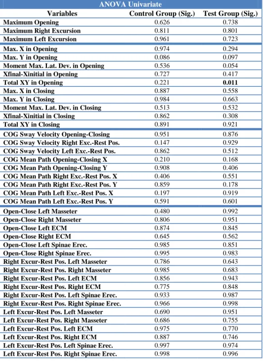
TABLE 9:MEAN AND STATISTICAL SIGNIFICANCE OF THE ACTIVE ROM ANALYSIS. ... 55
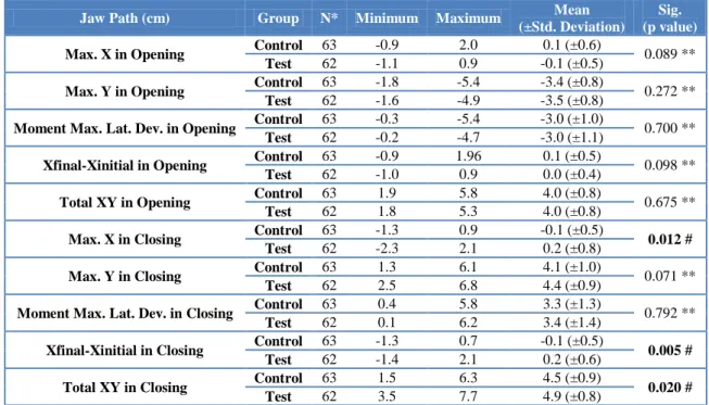
TABLE 10:MEAN AND STATISTICAL SIGNIFICANCE OF THE JAW PATH ANALYSIS. ... 55
TABLE 11:MEAN AND STATISTICAL SIGNIFICANCE OF THE POSTURE SEGMENTS ANALYSIS. ... 60
TABLE 12:MEAN AND STATISTICAL SIGNIFICANCE OF THE COG SWAY VELOCITY AND COG PATH ANALYSIS. ... 70
Escola Superior de Tecnologia da Saúde de Coimbra | XIII
GRAPH 1:DISK DISPLACEMENT DIAGNOSTIC PERCENTAGE... 53
GRAPH 2:HORIZONTAL JAW DISPLACEMENT (CM). ... 56
GRAPH 3:VERTICAL JAW DISPLACEMENT (CM). ... 57
GRAPH 4:MOMENT OF MAXIMUM JAW DISPLACEMENT (CM). ... 57
GRAPH 5:HORIZONTAL JAW DISPLACEMENT DIFFERENCE FROM THE END TO THE START POINT (CM). ... 58
GRAPH 6:TOTAL OF VERTICAL AND HORIZONTAL JAW DISPLACEMENT (CM)... 59
GRAPH 7:POSTURE PARAMETER 1... 61
GRAPH 8:POSTURE PARAMETER 2... 61
GRAPH 9:POSTURE PARAMETERS 3 AND 4. ... 62
GRAPH 10:POSTURE PARAMETER 5... 62
GRAPH 11:POSTURE PARAMETERS 6 AND 7. ... 63
GRAPH 12:POSTURE PARAMETER 8... 63
GRAPH 13:POSTURE PARAMETER 9... 64
GRAPH 14:POSTURE PARAMETER 10... 64
GRAPH 15:POSTURE PARAMETER 11... 65
GRAPH 16:POSTURE PARAMETER 12... 65
GRAPH 17:POSTURE PARAMETER 13... 66
GRAPH 18:POSTURE PARAMETER 14... 66
GRAPH 19: POSTURE PARAMETERS 15 AND 17. ... 67
GRAPH 20:POSTURE PARAMETERS 16 AND 18. ... 67
GRAPH 21:POSTURE PARAMETERS 19 AND 21. ... 68
GRAPH 22:POSTURE PARAMETERS 20 AND 22. ... 68
GRAPH 23:POSTURE PARAMETERS 23 AND 24. ... 69
GRAPH 24:EMG MUSCLE ACTIVATION DURING OPENING-CLOSING MOVEMENTS (%). ... 72
GRAPH 25:EMG MUSCLE ACTIVATION DURING RIGHT EXCURSION-REST POSITION MOVEMENTS (%). ... 72
GRAPH 26:EMG MUSCLE ACTIVATION DURING LEFT EXCURSION-REST POSITION MOVEMENTS (%). ... 73
GRAPH 27:RELATIONSHIP BETWEEN MAXIMUM ACTIVE ROM OPENING AND MAXIMUM VERTICAL JAW DISPLACEMENT DURING OPENING MOVEMENT (MM). ... 74
14 | Escola Superior de Tecnologia da Saúde de Coimbra
1.
The TMJ moves approximately two thousand times per day, during movements inherent to speech, mastication, swallowing, yawning, etc., therefore, this it is the more used joint in the body (Arellano, 2002). It is part of the stomatognathic system, formed by several internal and external structures able to perform complex movements. Chewing, swallowing, phonation and posture depends on the function, health and stability of this joint to work properly (Quinto, 2000). When there is any disorder or derangement in this joint, temporomandibular disorders (TMD) may occur, defined as a heterogeneous group of pathological conditions affecting the TMJ, masticatory muscles and adjacent related structures (McNeill, 1997; Egermark, Carlsson and Magnusson, 2001; McNeely, Olivo and Magee, 2006; Manfredini, 2010; Jerolimov, 2009).
TMD are a set of joint and muscle disorders affecting the temporomandibular region, characterized by signs and symptoms such as pain and/or tenderness in the preauricular area and/or muscles of mastication, reduction and/or changes in mandibular range of motion, joint sounds like click and/or crepitus during jaw movements (Manfredini, 2010), decreased TMJ function, pain or muscle tenderness on palpation, pain during mandibular movement, facial pain and headache, classified as a subgroup of musculoskeletal disorders and rheumatologic in general (McNeill, 1997, Iwasaki et al. 2010; Moreno, Young, Yanaze and Cunali, 2002). Temporomandibular disorders are considered the most common orofacial pain condition of nondental origin (Manfredini et al. 2011). The frequent concurrent symptoms as sore throats, hoarseness, balance loss (Cooper and Kleinberg, 2007), earache, headache, neuralgia and tooth pain makes the assessment of TMD a complex issue (Leresche, 1997). From a psychosocial viewpoint, patients with chronic TMD report depressive symptoms, poor sleep quality, low energy and interference in social activity (Carlson et al. 1998; Manfredini et al. 2009; Manfredini et al. 2010; Tjakkes et al. 2010). Such aspects are worthy to be investigated by dedicated instruments, viz., the so-called axis II assessment, because of their importance in the clinical setting to predict treatment effectiveness.
Escola Superior de Tecnologia da Saúde de Coimbra | 15
The body posture is defined as the relationship between a segment or body part with the adjacent segments, as well as the interconnections between all segments which compose the human body (Gonzales and Manns, 1996). A combination of all support structures to obtain maximum efficiency on static and dynamic balance with minimal overload and energy expense, is considered the ideal posture. Poor posture is seen as a faulty relationship between the different body segments, provoking an increased demand for adaptation to support structures and decreased equilibrium efficiency (Gagey e Weber, 2000).
Good posture is essential to get the appropriate sensory feedback necessary to know the spatial orientation of body segments (kinesthetic position) and to maintain body balance. Poor posture may be viewed as a risk factor for muscle and joint pain, particularly at the neck, shoulders, spine and knees due to the shortening of muscle fibers resulting from an imbalance in the musculoskeletal system. This may overload the joints and be related with degenerative changes of the articular surfaces, such as osteoarthritis (Swann, 2009).
Posture has a major role also to explain how an individual reacts to emotions and to identify some personality traits. People who have an erect posture are seen as more confident and outgoing, whereas a more bending posture indicates insecurity and depression (Swann, 2009).
The human body is a system of rigid segments and it is possible to determine their position through its location, orientation and articular configuration (Zatsiorsky, 1998). The posture of each individual is determined by muscle chains, fascias, ligaments and bone structures, interdependent each other, and covering the whole body (Marques, 2000). Posture is an effectiveness indicator of the biomechanics, balance and neuromuscular coordination (Magee, 2002).
Posture and balance cannot be separated because if posture is the relationship between a segment or part of the body with the adjacent segments, the balance represents the relationship between all the forces that acts in the human body, contributing in a major way for the posture and for the mechanisms of posture control (Montgomery and Connoly, 2003). The main mechanisms of posture control are the oculomotor apparatus (motion planning and avoid obstacles in your path), the vestibular system (detects linear and angular accelerations) and the proprioceptive system (composed of several types of receptors that determine the position and velocity of the body segments in space) (Winter, 1995; Simoneau, Ulbrecht, Derr and Cavanagh, 1995; Chessa, Capobianco and Lai, 2002). The posture control is a complex function involving the central nervous system for control (command), the afferent fibers of the peripheral nervous system for regulation and the musculoskeletal system to perform (effector) (Missaoui, Portero, Bendaya, Hanktie and Thoumie, 2008).
16 | Escola Superior de Tecnologia da Saúde de Coimbra
balance according to Newton’s second law (Knudson, 2007). The body posture is dependent on the perfect relationship between mobility and stability which determines the equilibrium state. This relationship, in turn, depends on the efficient weight distribution in the base of support, highly determined by the body segments characteristics (lenght and weight), which is usually referred to as “postural balance” (Knudson, 2007).
The postural balance is a condition in which all the forces acting on the body are combined into a single point called center of mass, and its vertical projection to the ground is commonly known as center of gravity (Winter, 1995). The uncoordinated action of external forces and internal forces can move the center of mass, requiring the action of the postural control mechanisms to restore balance and posture (Montgomery and Connolly, 2003).
The presence of postural changes among TMD patients has been occasionally described in the literature, with particular regard to postural changes in the head posture associated with pain in the head and/or in the cervical region, related with some peculiar facial morphologies.
Whereas the muscle action is responsible for the surface contacts of the TMJ, changes in head and body position may alter the response patterns of the muscles responsible for the jaw movement (Kimmel, 1994).
Some hypothesis attribute a major role to the posture in the onset of TMD symptoms, and claimed that factors such as an anterior position of the head (Munhoz, Marques and Siqueira, 2004; Janda, 1981) or premature contacts of the dental arch with subsequent potential asymmetric muscle functioning may be viewed as risk factors (Christensen and Rassouli, 1995).
However, the mainstream literature showed that occlusal features play a minor role in the etiology of TMD, so there is still some controversies on the issue, also because of the many healthcare professionals managing with TMD patients.
Currently in Portugal, due to lack of knowledge and training in the area, TMD is still seen as a purely dental problem, opting for treatments often very expensive, which do not solve the problem. Therefore, the first aim of this study is to inform and raise awareness among health care practitioners, about this condition. Often and depending on the patient's impairments, if they do not present referred or local pain, loss of function, altered sensitivity, social and/or psychological conditions, the most important treatment is information about its condition and counseling about some oral and leisure habits.
Escola Superior de Tecnologia da Saúde de Coimbra | 17
of the Physiotherapy when compared to other treatments (Toledo, Silva, Toledo and Salgado, 2012; McNeelly, Olivo and Magee, 2006; Carmeli, Sheklow and Bloomenfeld, 2001; Sato and Kawamura, 2008; Kalamir, Pollard, Vitiello e Bonello, 2007), and should be considered the treatment option, with a cost-effect relationship, more suitable.
The scientific aim of this study is to assess postural changes in subjects with internal derangement of the TMJ when compared to subjects without this biomechanical dysfunction. For this purpose will be evaluated several body segments by posturography and also will be evaluated the postural balance reactions through the center of mass during jaw movements using a balance platform.
Will also be an objective, characterize the patterns of the jaw movements. For that objective, a kinematic analysis of these movements (ROM and path of the jaw) will be performed and the activation of the masseter muscles, sternocleidomastoid muscles and spinae erector muscles, during jaw movements, will be evaluated by surface electromyography.
With this objectives and with the interest to find the answers to the questions/hypothesis: Subjects with disc displacement (DD) show active ROM in opening and lateral excursion movements, significantly different from individuals without DD?; Are there differences in the pattern of depression and elevation of the jaw in subjects with DD when compared to subjects without DD?; Is the total of displacement (in axes X and Y) for the movements of depression and elevation of the jaw, higher in subjects with DD?; Are there significant body posture changes at head level in subjects with DD when compared to subjects without DD?; Are there significant body posture changes in the generality of the body in subjects with DD when compared to subjects without DD?; Are there significant body posture changes between the right hemibody and the left hemibody in subjects with DD?; Are there significant body posture changes between the right hemibody and the left hemibody in subjects without DD?; Does the displacement velocity of the center of mass during the jaw movements, show significant differences between subjects with DD and subjects without DD?; Is the average path of COG during jaw movements, significantly different between subjects with DD and subjects without DD?; Are there significant differences in bilateral muscle activation of the Masseter muscles, Sternocleidosmatoid muscles and Spinae Erector muscles during jaw movements in subjects with DD?; Are there significant differences in bilateral muscle activation of the Masseter muscles, Sternocleidosmatoid muscles and Spinae Erector muscles during jaw movements in subjects without DD?; we set out to conduct this study, with the desire to provide the necessary information to health care providers who deal with TMD, so that they can more adequately develop their treatment targets, adapting their methods.
18 | Escola Superior de Tecnologia da Saúde de Coimbra
Escola Superior de Tecnologia da Saúde de Coimbra | 19
2.1 Temporomandibular Joint (TMJ)
The TMJ is formed by the temporal bone and the mandible, more specifically by the fossa and articular eminence of the temporal bone and the mandibular condyle. Between these two articular surfaces, a biconcave fibrocartilaginous disk adapts itself to provide the joint structures with stability during mandibular movements (Hlináková et al. 2010; Siéssere et al. 2008; Ingawalé and Goswami, 2009; Sommer et al. 2003).
Image 1: Temporomandibular joint movement during mouth opening.
Because the presence of this disk, the intra-articular space is divided into two cavities, the superior and the inferior compartments, also featuring the bilaminar zone (tissue with elastic fibers in the posterior region of the disk), the synovial membrane, the articular cartilage and the joint capsule (Hlináková et al. 2010; Siéssere et al. 2008; Ingawalé and Goswami, 2009). The extra-articular zone is composed by ligaments and muscles responsible for coordinating mandible movements along the different axes of motion. The main muscles acting on the TMJ are those of the mastication (temporalis, masseter, medial pterygoid and lateral pterygoid), and the digastric (Xu et al. 2008; Hannam at al. 2008; Siéssere et al. 2008). An equilibrium between the agonist and antagonist contractions of those muscles allows the best intra-articular stability, from which mandibular movements begin (Hlináková et al. 2010; Siéssere et al. 2008).
2.1.1 TMJ Anatomy and Biomechanics
20 | Escola Superior de Tecnologia da Saúde de Coimbra
joint capsule involving superiorly the fossa and articular eminence, and inserting inferiorly in the periosteum of the condyle branch (Hlináková et al. 2010; Siéssere et al. 2008; Ingawalé and Goswami, 2009). It also presents a peculiar feature, that is related with the articular surfaces are covered with fibrocartilage instead of hyaline cartilage, as is common in this type of joints (Hlináková et al. 2010; Ingawalé and Goswami, 2009). Typically, the fibrocartilage, because of its load-resistance properties to degenerative changes and best regenerating qualities, was mostly found in structures with high impact and repeated loadings (Norkin e Levangie, 2001).
The presence of the disk provides stability and consistency to the articular surfaces, distributes the forces over a larger area and spreads the synovial fluid, providing the lubrification and nutrition needed to the articular structures. The joint disk is composed of water, collagen, proteoglycans, elastin, fibrocartilage and chondral cells (Chin, Aker and Zarrinnia, 1996). The anterior part of the disk attaches to the articular eminence, to the condyle head and to the joint capsule, while the posterior part, which is highly vascularized and composed mainly of loose connective tissue, attaches to the bilaminar zone and to the joint capsule. In the lateral and medial levels the disk is closely attached to the joint capsule and to the condyle head, whilst the anteromedial plan it attaches to the superior part of the lateral pterygoid muscle (Willard, Arzi and Athanasiou, 2011; Sommer et al. 2003; Siéssere et al. 2008).
In the posterior region of the disk, attached superiorly to the retrodiscal space of the mandibular fossa and inferiorly to the condyle, is located the bilaminar zone, also generically known as “retrodiscal tissues”, composed of collagen, elastic fibers, numerous blood vessels and nerves (Siéssere et al. 2008; Tanaka et al. 2002). The characteristics of the bilaminar loose tissue allow a wide range of motion to the disk, and prevent those tissues’ dislocation during mouth opening (Sommer et al. 2003). With the jaw in the closed mouth position the retrodiscal tissue is organized into a dense network behind the condyle; with progressive jaw opening, this elastic tissue expands in all directions (Tanaka et al. 2002).
2.1.2 Biomechanics of Mandibular Movements
The functional dynamics of the jaw is obtained through a complex combination of intra-articular movements (condyle-disk; disk-eminence; attachments of the disk) with extra-articular movements (muscles and ligaments). Such coordinated actions result in mouth opening (jaw depression), mouth closing (jaw elevation), projection of the chin forward (jaw protrusion), slide the jaw backwards (jaw retropulsion) and sliding jaw on both sides (right and left lateral excursions) (Norkin and Levangie, 2001; Baskan and Zengingul, 2006). The normal range of motion for jaw movements is usually set between 40 and 55 mm for mouth opening and at least 7 mm for lateral excursions and protrusion (De Leeuw, 2008).
Escola Superior de Tecnologia da Saúde de Coimbra | 21
necessarily influence the movement in the contralateral side (Palomar and Doblaré, 2006); if the movements are identical in the two joints, the jaw movement is considered symmetric (Kang, Updike and Salathe, 1993).
With the jaw in the resting position, the condyle rests on the articular fossa of the temporal bone. For the mouth opening to occur it is necessary that a complex combination of rotation in the inferior space (condyle-disk) and sliding in the superior space (disk-fossa) happens (Gallo, Brasi, Ernst and Palla, 2006). In an healthy joint, mouth opening is determined mainly by the condyle rotation. The rotation in the inferior space involves a movement of the condyle with the disk attached to its bone surface. The disk follows the movement sliding relative to the superior space, and only a continuous condylar rotation can allow complete mouth opening (Ferrario, Sforza, Lovecchio and Mian, 2005). If load occurs in the mouth closed position, the disk deformation happens mainly in the central area, but if there is a sliding of the condyle forward (protrusion or begining of the mouth opening), the disk deformation tends to be in the lateral region, suggesting that certain regions of the disk suffer more loads than others (Beek, Koolstra and van Eijden, 2003).
In the case of the opening-closing movement, the condylar movements of the two sides are ideally symmetrical, whilst for the lateral excursions the condyles should perform asymmetrical movements. When the jaw makes a lateral excursion to the right side, the contralateral condyle (left) performs forward sliding until the articular eminence, while the ipsilateral condyle (right) remains in the articular fossa only performing a slight sliding movement to the same side of excursion (Koolstra and van Eijden, 1999).
22 | Escola Superior de Tecnologia da Saúde de Coimbra
The primary muscle responsible for mandibular depression is the digastric. Electromyographic recordings showed a minor role of the lower portion of the lateral pterygoid too. The main muscles involved in jaw closing are the masseter, temporalis and medial pterygoid, even if the upper portion of the lateral pterygoid is also important because of its stabilizing action of the disk over the condyle. The masseter, medial pterygoid and lateral pterygoid are responsible for mandibular protrusion and the posterior fibers of the temporalis are responsible for retropulsion, also featuring a mild intervention of the digastric and suprahyoid muscles. The lateral excursions, unlike previous movements, are the only movements during which muscle contraction is not bilateral or symmetrical: an isolated contractions of the medial pterygoid or lateral pterygoid muscles may perform this movement, and if there is a contraction in ipsilateral muscle synergy of the lateral pterygoid and temporalis, it is also possible obtain an effective lateral excursion (Norkin and Levangie, 2001; Xu et al. 2008). A study of Koolstra and van Eidjen stated that the jaw opening muscles (digastric and lateral pterygoid) are not able to make this move over 3.3cm of distance between the incisors. In this position these muscles appears to be overly shortened and no longer with remaining strength to counteract the passive forces of the jaw closing muscles. The muscles responsible for jaw closing (masseter, temporalis and medial pterygoid) are much stronger than the jaw opening muscles and furthermore, the jaw opening muscles cannot produce passive forces able to counteract the jaw closing muscles (Koolstra and van Eidjen, 1997).
The ligaments composing the TMJ are the triangular, the sphenomandibular and the stylomandibular. The triangular ligament has an external portion which inserts in the condyle branch and an internal portion which inserts in the lateral region of the condyle and in the posterior region of the disk. This ligament acts as a strong lateral stabilizer inhibiting the posterior sliding of the mandible (Norkin and Levangie, 2001; Siéssere et al. 2008).
2.2 Etiology of Temporomandibular Disorders (TMD)
Escola Superior de Tecnologia da Saúde de Coimbra | 23
Over the past decades, several purported malocclusions (skeletal class malocclusion I, II, III, posterior crossbite, anterior openbite, horizontal overlap, vertical overlap) were considered as a major cause of TMD based on the hypothesis that they can determine muscle hyperactivity that can exitate in TMD . Actually, evidence-based data suggested that the role of occlusal features as risk factors in the development of TMD is less important than believed in the past (Goldstein, 1999; Mohlin et al. 2007; Bonjardim et al. 2009; Manfredini and Lobbezoo, 2010).
The biopsychosocial theory is now the most widely accepted framework to assess temporomandibular disorders, and it postulates that the TMJ intra-articular and extra-articular etiology is complex and multifactorial, being directely dependent on predisposing, precipitating, perpetuating and contributing factors (Laskin, 1969; Greene, 1995; Suvinen et al. 2005).
Based on these concepts, it is likely that knowledge on TMD etiology will be strongly improved in the near future thanks to researches focusing on the triangle of factors composed by bruxism, pain, and psychosocial factors. On that purpose, several recent works by Manfredini and colleagues contributed a lot to get deeper into the issue (Manfredini and Lobbezoo, 2009; Manfredini, Cantini, Romagnoli and Bosco, 2003; Manfredini, Landi, Fantoni, Segù and Bosco, 2005; Manfredini and Lobbezoo, 2010; Manfredini, Peretta, Guarda-Nardini and Ferronato, 2010; Manfredini, Fabbri, Peretta and Guarda-Nardini, 2011).
24 | Escola Superior de Tecnologia da Saúde de Coimbra
since in some patients it may be viewed as an attempt to restore the physiological airway patency and avoid apnea episodes.
2.2.1 TMJ Internal Derangement
Internal derangement of the TMJ is defined as an abnormal mechanical relationship of the disk in relation to the condyle and mandibular fossa, with interference in the normal (functional) movement of the jaw (Maydana et al. 2010; Molinari et al. 2007). This abnormal relationship is clinically characterized as disk displacement (Tanaka et al, 2008; Manfredini, 2009; Nitzan 2001; Maydana et al. 2010; Palomar and Doblaré, 2006), causing joint pain, joint sounds, muscle tenderness and limitation of mouth opening (Nitzan, 2001; Tanaka et al. 2008). According to Truelove and colleagues, the TMJ internal derangement can be classified into three types: internal derangement type I (disk displacement with reduction); internal derangement type II (disk displacement with reduction and blocking episodes); internal derangement type III (disk displacement without reduction) (Truelove, Sommers, LeResche, Dworkin and vonKorff, 1992).
The etiological or risk factors for internal derangement, such as traumatic events, joint hypermobility (ligamentous laxity), degenerative diseases, occlusal factors, anatomy of the articular eminence and the role of the lateral pterygoid muscle (Isberg and Westesson, 1998; Gokalp, Turkkahraman and Bzeizi, 2001; Loughner et al. 1996; Molinari et al. 2007; Manfredini, 2009; Nitzan, 2001), have been controversial. Current theories suggested that the mechanisms which cause friction and repetitive loads (mandibular condyle) causing defects in joint lubrification, represent the role major in disk displacements (Manfredini, Basso, Salmaso e Guarda-Nardini, 2008; Tanaka et al. 2008; Nitzan, 2001; Manfredini, Basso, Arboretti and Guarda-Nardini, 2009; Gallo, 2005; Palomar and Doblaré, 2007; Spilker, Nickel and Iwasaki, 2009; Gallo, Nickel, Iwasaki and Palla, 2000; Tymofiyeva et al. 2007; Harper and Schneiderman, 1996). In the healthy TMJ the friction coefficient is around 0.0145-0.0239 (Tanaka et al. 2008), and if there is an excessive increase of this ratio, it may reduce the fluency of mandibular movements and facilitate the processes leading to changes in the condyle-disk relationship (Manfredini, 2009).
Escola Superior de Tecnologia da Saúde de Coimbra | 25
Disk displacements may be with reduction or without reduction (McNeill, 1997; Okeson, 1997; Schiffman, Anderson, Fricton and Lindgren, 1992; Dworkin and LeResche, 1992).
Disk displacement with reduction is characterized by a temporary displacement, where the disk tries to regain its relationship with the condyle during condylar movement, thus resulting in a joint sound (click) on opening. During closing a reciprocal click usually occurs, although this is of lesser magnitude, corresponding to the anterior or anteromedial displacement of the disk. In the disk displacement without reduction the relationship of the disk with the condyle is permanently disturbed, since the disk remains in an anterior position with respect to the condyle throughout the whole condylar traslation. This condition is not usually accompanied by any joint sounds, but a limitation in the jaw range of motion with a deflection toward the affected side is clinically detectable (McNeill, 1997; Palomar and Doblaré, 2007). However, the displacement of the disk without further limitating conditions for the subject (referred or local pain, loss of function, altered sensitivity, social and/or psychological conditions, etc.), is not considered a pathological marker by itself (Manfredini, 2010), even if it represents only a risk factor for further degeneration of the joint surfaces. As the movements of the condyle are dynamic and continue to transmit loads, friction and shear forces induced on the disk will increase, leading to a worsening state of the internal derangement, the disk can begin to deform, adopting a biconvex form, becoming more long and thin, until may even suffer cracks and be perfurated (Palomar and Doblaré, 2007; Molinari et al. 2007). The evolution of disk displacement appears to be related with poor load distribution and biomechanical failure of the TMJ (Palomar and Doblaré, 2007).
Image 2: Normal TMJ, disk adaptations during mouth opening.
26 | Escola Superior de Tecnologia da Saúde de Coimbra
Image 4: TMJ disk displacement without reduction, disk adaptations during mouth opening.
A study of Timofiyeva and colleagues aimed to determine the condyle movement in healthy TMJ and in TMJ with disk displacement and joint sounds (click). The findings suggested that the asymptomatic joints have a characteristic pattern of movement (in the first degrees of jaw opening the condyle performs rotation and as the range increases, the condyle will be moving to forward of the fossa until the articular eminence). On the other hand, the symptomatic TMJ have shown a different pattern of movement (in the opening, the condyle almost does not perform the rotation movement, and in a advanced range of motion, the condyle seems to jump to a position of higher protrusion, reducing the joint space, when compared with the previous positions) (Tymofiyeva et al. 2007).
Escola Superior de Tecnologia da Saúde de Coimbra | 27
In the literature, two main guidelines for the diagnosis of disk displacement are found, viz., the Research Diagnostic Criteria for Temporomandibular Disorders (RDC/TMD) proposed by Dworkin and LeResche in 1992 and the Clinical Classification System of the American Academy of Orofacial Pain (AAOP). Patients with TMD should be classified according to widespread schemes such as RDC/TMD for epidemiological and research purposes and AAOP guidelines for classifications in clinical practice.
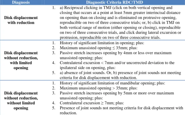
Table 1: Diagnostic Criteria for disk displacement according to the RDC/TMD (Dworkin e LeResche, 1992).
Diagnosis Diagnostic Criteria RDC/TMD
Disk displacement with reduction
1. a) Reciprocal clicking in TMJ (click on both vertical opening and closing that occurs at a point at least 5mm greater interincisal distance on opening than on closing and is eliminated on protrusive opening, reproducible on two of three consecutive trials; or, b) click in TMJ on both vertical range of motion (either opening or closing), reproducible on two of three consecutive trials, and click during lateral excursion or protrusion, reproducible on two of three consecutive trials.
Disk displacement without reduction,
with limited opening
1. History of significant limitation in opening; plus: 2. Maximum unassisted opening ≤ 35mm; plus:
3. Passive stretch increases opening by 4mm or less over maximum unassisted opening; plus:
4. Contralateral excursion < 7mm and/or uncorrected deviation to the ipsilateral side on opening, plus:
5. a) absence of joint sounds. Or, b) presence of joint sounds not meeting criteria for disk displacement with reduction.
Disk displacement without reduction, without limited
opening
1. History of significant limitation of mandibular opening; plus: 2. Maximum unassisted opening > 35mm; plus:
3. Passive stretch increases opening by 5mm or more over maximum unassisted opening; plus:
4. Contralateral excursion ≥ 7mm; plus:
28 | Escola Superior de Tecnologia da Saúde de Coimbra
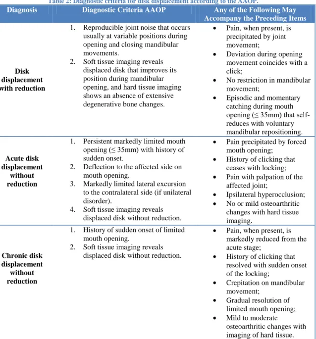
Table 2: Diagnostic criteria for disk displacement according to the AAOP.
Diagnosis Diagnostic Criteria AAOP Any of the Following May Accompany the Preceding Items
Disk displacement with reduction
1. Reproducible joint noise that occurs usually at variable positions during opening and closing mandibular movements.
2. Soft tissue imaging reveals displaced disk that improves its position during mandibular opening, and hard tissue imaging shows an absence of extensive degenerative bone changes.
Pain, when present, is precipitated by joint movement;
Deviation during opening movement coincides with a click;
No restriction in mandibular movement;
Episodic and momentary catching during mouth
opening (≤ 35mm) that self -reduces with voluntary mandibular repositioning.
Acute disk displacement
without reduction
1. Persistent markedly limited mouth
opening (≤ 35mm) with history of sudden onset.
2. Deflection to the affected side on mouth opening.
3. Markedly limited lateral excursion to the contralateral side (if unilateral disorder).
4. Soft tissue imaging reveals displaced disk without reduction.
Pain precipitated by forced mouth opening;
History of clicking that ceases with locking; Pain with palpation of the affected joint;
Ipsilateral hyperocclusion; No or mild osteoarthritic changes with hard tissue imaging.
Chronic disk displacement
without reduction
1. History of sudden onset of limited mouth opening.
2. Soft tissue imaging reveals displaced disk without reduction.
Pain, when present, is markedly reduced from the acute stage;
History of clicking that resolved with sudden onset of the locking;
Crepitation on mandibular movement;
Gradual resolution of limited mouth opening; Mild to moderate
osteoarthritic changes with imaging of hard tissue.
2.3 The use of electronic devices in assessment of TMD
Several electronic devices, such as surface electromyography, kinesiographic records and posturography, have been described as reliable and accurate in assessment and evaluation of TMD, however its role is far to be consensual.
2.3.1 Surface Electromyography
Escola Superior de Tecnologia da Saúde de Coimbra | 29
Some authors proposed that quantitative methods as the sEMG may be used as a complement in the diagnosis of TMD and to monitorize the effectiveness of some treatments (Pedroni, Borini and Bérzin, 2004; Tartaglia et al. 2011; Widmalm, Lee and McKay, 2007; Cooper, 2011); however, it seems that much is yet to be done before defining the real specific indications of such devices.
Some studies claimed that sEMG may provide objective records of the function and dysfunction of the masticatory muscles, but it must be pointed out that most studies came from the research setting (Koyano, Kim and Clark, 1995; Castroflorio et al. 2005; Ferrario et al. 2007) and did not authorize the uncontrolled use of commercial sEMG in the clinical setting, as proposed by others (Widmalm, Lee and McKay, 2007). Electromyographic investigations performed in patients with TMD showed that the muscles of mastication may have altered firing features (Tosato and Caria, 2007; Koyano, Kim and Clark, 1995; Ardizone et al. 2010; Ferrario et al. 2007) and present higher silent periods (McCall, Uthman and Mohl, 1978) than subjects without TMD. In any case, the main sEMG finding associated with the presence of pain is the reduced activity during maximum clenching, in line with the pain adaptation model (Visser, McCarroll and Naeije, 1992, Manfredini et al. 2011).
The belief that the clinical applicability of surface EMG may be justified for the diagnosis and treatments of TMD is based on the assumption that the various pathological or dysfunctional conditions can be revealed by electromyographic records of muscle activity of the masticatory muscles. In any case, it must be borne in mind that current evidence-based concepts suggest that several biological factors (physiologic variation, age, sex, skeletal morphology, psychological factors, density of the skin, weight) and technical factors (position of the electrodes, position and interelectrode distance, cross-talk, head or body movements, existence of painful conditions, facial expressions, history of bruxism) may influence the reliability, validity, sensitivity and specificity of surface EMG as a diagnostic and treatment procedure (Klasser and Okeson, 2006).
Considering that, some systematic reviews suggests that the role of bioelectronic devices, in particular surface EMG, is less important than believed in the past, since they seem to be able, at best, to provide ancillary information (Lund, Widmer and Feine, 1995; Klasser and Okeson, 2006; Suvinen and Kemppainen, 2007; Manfredini et al. 2011).
30 | Escola Superior de Tecnologia da Saúde de Coimbra
Manfredini et al. 2011), featuring true-positive rates below 60% and false-positives between 44-89% (Manfredini et al. 2010).
Also at rest, comparisons of the painful side with the non-painful side on the same patient with unilateral facial pain showed that myoelectric activity levels did not differ significantly between the two sides evaluated. A clear and consistent relationship between the painful state of a muscle and its level of electric activity by surface EMG, has not been established yet (Baba et al. 2001).
In summary, the surface EMG seems to have some potential as a complementary tool in the investigation of the masticatory function (Suvinen and Kemppainen, 2007), but not as a tool for symptoms diagnosis (Lund, Widmer and Feine, 1995; Baba et al. 2001; Klasser and Okeson, 2006; Suvinen and Kemppainen, 2007; Manfredini et al. 2011).
2.3.2 Kinematic Analysis
The kinematic analysis it is a variation of kinesiographic recordings and describes the motion using linear (meters) or angular (degrees) measurements (Knudson, 2007). The most common methods to obtain kinematic data are high speed video systems that record the position of body segments with respect to time, typically using passive markers placed in the segments under consideration. Subsequently, the images are analyzed by specific software, some of free access such as Kinovea, using a reference system that allows collect coordinates in space (Hamill and Knutzen, 2009). Kinematic analysis of human movement through the aforementioned system is widely described in the literature (Decker et al. 2003; Wu et al. 2005; Medved, 2001), however, this analysis for active jaw movements was not found in related literature.
2.3.3 Posturography
The use of photographic techniques to analyze the body posture is widely described in literature (Santos, Silva, Sanada and Alves, 2009; Watson, 1998; Iunes et al. 2005; Cuccia and Carola, 2009; Burke et al. 2010; Ayub, Way and Kraus, 1984; Miranda et al. 2010; Raine and Twomey, 1994) and there are several studies that attempt to relate the body posture with TMD, by the use of balance platforms, generically known as posturography (Wakano et al. 2011; Chessa, Capobianco and Lai, 2002; Ferrario, Sforza, Schmitz and Taroni, 1996; Perinetti, 2007; Perinetti and Contardo, 2009; Manfredini, Castroflorio, Perinetti and Guarda-Nardini, 2012). As in the case of sEMG, also in the field of posturography, clinicians’ beliefs about its potential usefulness in the clinical setting clash with findings from the research setting.
Escola Superior de Tecnologia da Saúde de Coimbra | 31
Several studies using the posturography to establish a relationship between body posture and TMD, are focused on the correlation between the stomatognathic system and the cervical region, which cannot be representative of the global body posture (Perinetti and Contardo, 2009). Based on quantitative data obtained from two literature reviews, the several postugraphic devices and methods appears to be similar regarding to the high variability in records, resulting in low accuracy on clinical diagnosis (Perinetti and Contardo, 2009; Manfredini, Castroflorio, Perinetti and Guarda-Nardini, 2012).
2.4 Relationship Between Body Posture and TMD
The position of the head is an important center of balance for the body and its movements depends on the positioning and stability of the skull on the cervical region (Maciel, 2003). This region has the main function to maintain the centered position of the head on the spine and optimize their mobility (Sachse and Schildt-Rudloff, 2003).
A study of Tosato and colleagues indicates that women with cervicalgia showed more signs and symptoms of TMD when compared to a group of women with low back pain (Tosato et al. 2007).
Another study aiming to verify if the head posture affects the mandibular kinematics showed that different mandibular postures influence the intra-articular space of the TMJ and, therefore, the movement of the mandibular condyle. In the military posture of the head, the opening movement path of the incisal point is shifted anteriorly relative to its path in a natural head posture, whereas in a forward head posture this path is shifted posteriorly (Visscher, Slater, Lobbezoo and Naeije, 2000).
In the head there are two of the three mechanisms for posture control, viz., the oculomotor and the vestibular systems, which together with the function of the cervical spine determines the body position assumed by the subject. The TMJ is the link which shares neuromuscular structures between the jaw with the skull and cervical spine; when pain in the TMJ or jaw muscles is present it can trigger body postural changes (Wakano et al. 2011).
32 | Escola Superior de Tecnologia da Saúde de Coimbra
Relationship between agonist and antagonist muscles can contribute to postural changes (Marques, 2000). The hyperactivity of masticatory muscles (antagonist) interferes with the activity of the posterior muscular chain (agonist), causing changes on stretching-shortening relationship, which promotes exaggerated muscle tension that can lead to postural changes (Gagey and Weber, 2000).
One study suggests that in patients with TMD, postural changes and an abnormal muscle function are more common when compared to individuals without TMD, thus shows the influence of the cranio-mandibular system on body posture (Nikolakis et al. 2000).
Several studies have shown that the electromyographic activity of masticatory muscles can modify the electrical activity of postural muscles, especially the posterior cervical muscles (Lous, Sheik-Ol-Eslam and Moller, 1970; Ehrlich, Garlick and Ninio, 1999; Bergamini, Pierleoni, Gizdulich and Bergamini, 2008; Monaco, Spadaro, Cattaneo and Giannoni, 2010).
Many studies have attributed a relationship between TMD and posture, with the TMD of myofascial origin the most described, especially related to a Forward Head Posture (Ayub, Glasheen-Way and Kraus, 1984; Friedman and Weisberg, 1982; Janda, 1981; Goldstein, Kraus, Williams and Glasheen-Way, 1984; Urbanowicz, 1991; Gonzalez and Manns, 1996; Miranda et al. 2010).
Other studies indicate that postural changes such as unlevel shoulders (Clark, Green, Dorman and Flack, 1987; Fuents, Freesmeyer and Henriquez, 1999; Rocabado and Tapia, 1987), cervical lordosis increased (Clark, Green, Dorman and Flack, 1987; Darling, Kraus and Glasheen-Way, 1984; Munhoz, Marques and Siqueira, 2005; Neto et al. 2010) and rotation and/or head inclination (Farias, Alves and Gandelman, 2001), are also associated with TMD patients.
Another study shows that patients with intra-articular (internal derangement) TMD have postural deviations in the head, spine, shoulders, pelvis and hip joint. However, the major postural changes were found in structures adjacent to the TMJ. The body posture does not change randomly, but following a cranio-caudal standard. This suggest that the postural changes are a consequence of TMD and not the contrary (Munhoz and Marques, 2009).
Conversely, studies of Perinetti and Iunes, found that there were no significant postural changes in TMD patients (Perinetti, 2007; Iunes et al. 2009).
Escola Superior de Tecnologia da Saúde de Coimbra | 33
personality characteristics (psychosocial factors), a need for studies on the physiology of such relationship in the absence of pain symptoms is strongly recommended.
34 | Escola Superior de Tecnologia da Saúde de Coimbra
3.
This research study represents a cross-sectional quasi-experimental design.
To perform the study, subjects from two institutions of higher education and from a temporomandibular rehabilitation clinic were evaluated. All subjects underwent physical evaluation of joint range of motion and joint noises during jaw opening and lateral excursions active movements according to the protocols described by Dworkin and Leresche (RDC/TMD) and the American Academy of Orofacial Pain (AAOP). The study took place at the facilities of Instituto Superior de Saúde do Alto Ave (days 11, 12 and 18 May 2012) and Escola Superior de Tecnologia da Saúde de Coimbra (days 24 and 25 May 2012).
All subjects in the study were properly informed about the procedures to be carried out and about the objectives of the study with their rights to privacy and confidentiality assured. Participants were also informed that they could withdraw at any time and that from the ongoing investigation does not result any consequences to their physical integrity. They were allowed to place any issue that was not properly clear and finally, it was asked the voluntarily signing of the informed consent according to the Helsinki Declaration.
For this study, subjects were selected according to the following criteria:
- Aged between 15 and 40 years;
- Presence of joint sounds during jaw movements reproducible in 2 of 3 repeated trials and/or history of joint sound currently evolved to blocking or marked limitation in range of mandibular movements (Test Group).
- Absence of joint sounds during jaw movements and no limitation in range of mandibular movements (Control Group).
Exclusion Criteria:
- Degenerative diseases such as osteoarthritis or osteoarthrosis;
- Systemic diseases such as rheumatoid arthritis, systemic lupus erythematosus or collagen disease;
- History of trauma to the neck and/or in facial region;
- History of changes in the balance (frequent falls) or pain symptoms influenced by orthostatic position;
- Orthopedic or dentistry surgery with impact on the mobility of the TMJ.
Escola Superior de Tecnologia da Saúde de Coimbra | 35
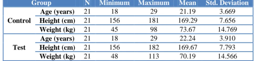
female and 4 are male, the mean age stands at 22.2 (±3.9) years, the mean height is 169.7 (±7.8) cm and mean weight is 70.2 (±14.6) kg.
3.1 Experimental Setup
The room was prepared according to the space required, taking into account the luminosity and the inside temperature. The placement of the referential (described later in section 3.2.4), constituted the reference for the placement of the remaining instruments, whose distance was measured from the plywood board. The NeuroCom ® platform was placed 0.28 meters [m] from the anterior part of the referential, laterally centered therewith, the center of the tripod supporting the camera Casio® EX-FH20 for kinematic analysis was at a distance of 2.76 m from the anterior part of the referential. The center of the tripod supporting the camera Sony® HX-100V for the postural analysis was 3.43 m from the posterior part of the referential, as illustrated in the image 5.
Image 5: Experimental setup scheme.
36 | Escola Superior de Tecnologia da Saúde de Coimbra
3.2 Instruments
3.2.1 Balance Platform
Data from subjects balance was collected with the Basic Balance Master® platform from the company NeuroCom® International, Inc., headquartered in Clackamas, United States of America (USA).
This instrument consists of a force plate linked to a computer with the software provided by the manufacturer (Image 6). It was developed to support the assessment and treatment skills related to balance and mobility in patients with disabilities and/or functional limitations resulting from the orthopedic, neurological, geriatric or vestibular diagnostics (Basic
Balance Master® System Operator’s
Manual, 2003).
The software used was the Clinical Test for the Sensory Interaction on Balance (CTSIB). Each test consists of three trials of 10 seconds each, in which are the initial alignment of the center of gravity (COG), the oscillation speed of the COG and the path of the COG.
In the first test three trials of opening-closing were performed, in the second test three trials of right lateral excursion and return to the midline while in the third test three trials of left lateral excursion and return to the midline. In the fourth test only one trial was done without any movement with the subject looking straight and focusing on a fixed point – it was done for comparison purposes only. The data which was taken into account was the speed of oscillation of the COG (degrees/second) [º/s] and the mean of displacement of the COG (degrees) [º]. All values were collected at an acquisition rate of 100 Hz.
Escola Superior de Tecnologia da Saúde de Coimbra | 37
mass (COM) are equivalent points in space where gravity is the only force taken into account (Basic Balance Master® System Operator’s Manual, 2003).
Balance Master® platform measures the speed of oscillation of the COG as the ratio of distance traveled by COG (degrees) versus the time of repetition (seconds) indicating the amount of oscillation showed for the subject (Basic Balance Master ® System Operator's Manual, 2003). The ability to control the COG in the base of support in various external conditions (different surfaces, forces acting on the body, visual feedback, etc.), is the main function of balance, where low oscillation values indicates little movements of the body, meeting the preservation of this ability.
3.2.2 Surface EMG
The bioPLUXresearch® device from
the company Plux – Engenharia de
Biosensores, Ltd., based in Covilha, Portugal was used to collect the electromyographic signal. The software supplied by the
manufacturer, enables collecting the
electromyographic signal at a sampling rate of 1000 Hz (bioPLUXresearchUser Manual, 2010).
Ambu® Blue Sensor electrodes,
reference N-00-S, from the company Ambu A/S based in Ballerup, Denmark are used. These sensors are silver/silver chloride with
wet gel conduction system
(http://www.ambu.com/corp/products/patient_monitoring_and_diagnostics.aspx/product
.aspx?ProductID=PROD844, seen in 22/08/2012).
3.2.3 Digital Cameras
Two digital cameras were used, one for jaw movements video recording (kinematic analysis) and the other to the photographic records to perform the postural analysis. The camera used for the video recording was the Casio EX-FH20® from the company Casio Computer CO., LTD., Ltd. Tokyo, Japan. To make the photographic records was used the camera Sony® HX-100V, Sony Corporation, Tokyo, Japan. The tripods used were Hama® “Star 63”, Hama Lda. Basingstoke, United Kingdom.
38 | Escola Superior de Tecnologia da Saúde de Coimbra
3.2.4 Referential
A referential was constructed to facilitate the analysis of the collected data (Image 8). Supporting structure was built of galvanized steel with a board of plywood in between, previously prepared with a grid chart for postural evaluation, scaled in centimeters. The basis of this structure was formed by two lateral bars with 1.0 m each and a central bar to connect the two lateral bars with 0.86 m. At the bottom of the two side bars, one wheel was placed at each end which raised the referential structure at the platform height (0.06 m) (Image 9). In the central part of the two side bars, leaving two vertical bars with 2.0 m where two rails fit through the plywood board. The central part of the central bar also leaves a vertical bar with previously prepared 2.0 m measuring tape. On the top of this bar, a height adjustable headrest was placed to provide proprioceptive information for subjects not to move the head upwards and sideways during mandibular motion (Image 10). As the purpose of this instrument was not restraining subjects’ normal movement, it just landed on top of the hair, not making any pressure on the head (Image 11). This head support is built with a steel bar with 0.56 m which engages on the vertical bar and the flat part is constructed with a plywood board with 0.25 m long by 0.26 m wide and lined with a ethylene-propylene-diene rubber (EPDM). Passive markers were placed on the grid chart, translating specific measures in order to minimize the measurement errors in the analysis (Image 12).
Escola Superior de Tecnologia da Saúde de Coimbra | 39
40 | Escola Superior de Tecnologia da Saúde de Coimbra Image 10: Referential headrest.
Escola Superior de Tecnologia da Saúde de Coimbra | 41
Image 12: Referential back.
3.3 Procedures
Procedures started by informing the subjects about the study, allowing them to clear any question and asking them permission to carry on with the study.
Then, some sample characterization questions were asked and height measurement was performed using the referential. In the test group the temporomandibular joints were also evaluated to define the diagnosis of disc displacement to the right, left or bilaterally.
Next step was skin preparation for placement of the electrodes surfaces (Ambu® Blue Sensor N-00-S) on the muscles used on the study. With this purpose, the skin on the muscles area was shaved if necessary, dead cells removed with Omnitape® adhesive from the company Hartmann, Heidenheim, Germany, and skin cleaning with alcohol soaked wipes from the company Romed®, Wilnis, Netherlands.
42 | Escola Superior de Tecnologia da Saúde de Coimbra
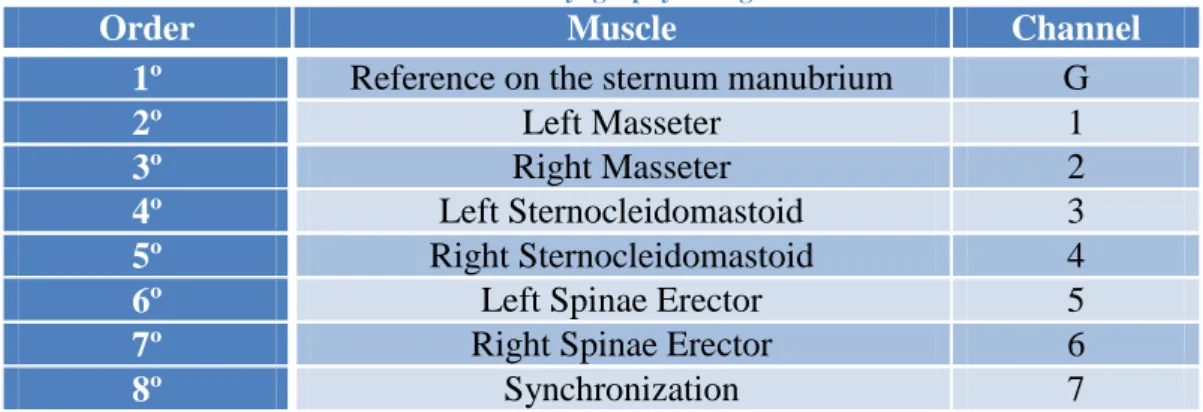
the sternum manubrium. The inter-electrode distance (center to center) was 2 cm and their placement had the following configuration:
Table 3: Electromyography configuration.
Order Muscle Channel
1º Reference on the sternum manubrium G
2º Left Masseter 1
3º Right Masseter 2
4º Left Sternocleidomastoid 3
5º Right Sternocleidomastoid 4
6º Left Spinae Erector 5
7º Right Spinae Erector 6
8º Synchronization 7
Then electromyographic recording of the maximum voluntary contraction (MVC) of the muscles under evaluation for the purposes of normalization in intensity was made, and two followed contractions have been requested to the subject:
Masseters: with the subject in a standing position, 2 dental cotton rolls from the
brand Celluron® Hartmann, Heidenheim, Germany were placed in the region of the molars and premolars between the upper and lower dental arch (1 in the right side and 1 in the left side). The subject was asked to bite the rolls with maximum force for 3 seconds;
Sternocleidomastoid: with the subject in
the standing position and the evaluator also standing, looking forward to a left profile view of the subject, the right arm of the evaluator stabilizes the right shoulder of the subject, the subject is asked for a maximum contraction during 3 seconds in the position of cervical rotation to the right and inclination to the left, while the evaluator resists the movement with his left hand on the left temporal region of the volunteer. This
process evaluates the left
sternocleidomastoid muscle, and it was repeated for the right sternocleidomastoid muscle, reversing the evaluator and subject positions.
Image 13: Sternocleidomastoid MVC
Escola Superior de Tecnologia da Saúde de Coimbra | 43 Spinae Erectors: with the
subject in a standing position and the feet slightly apart from the couch, he was asked to lean the pelvis against the couch and then make a
maximum contraction of
extension movement of the trunk for 3 seconds with the evaluator behind the subject to carry out manual resistance in the scapular region.
Passive roundness markers made of reflective material previously glued to parchment paper to improve the dental adherence (Image 15) were placed between the upper and lower central incisors, with 5 mm of diameter (Image 16). Dental cotton rolls were also placed for clearance of the lips and better visualization of the passive markers.
Image 15: Passive roundness markers.
44 | Escola Superior de Tecnologia da Saúde de Coimbra Image 16: Passive roundness markers placed in the subject.
The subjects were asked to put on top of the balance platform and the headrest was set to their height, just resting lightly on his head. The synchronization between instruments was performed by a switch that sends simultaneously a light signal captured on the video and an electrical signal (5V) recorded by the BioPlux device. Subjects were then requested to perform 3 maximum openings, 3 right excursions and 3 left excursions. During these movements the kinematics of the mandible, the path and the sway velocity of the COG and also the EMG were recorded.
At the end of this procedure, the passive markers and all the electromyographic apparatus were removed and skin was cleaned with alcohol soaked wipes to remove any gel from the removal of detection surfaces.