Case 12396
Primary Carcinoma of the Rectovaginal Septum
Mónica Vieira , Teresa Margarida Cunha , Jorge São Martinho 1 2 3 Hospital José Joaquim Fernandes
Genital (Female) Imaging Section:
2015, Jan. 8 Published:
61 year(s), female Patient:
Authors' Institution
1. Hospital José Joaquim Fernandes, Unidade Local de Saúde do Baixo Alentejo, Imagiologia; R. Dr. António Fernando José Covas Lima 7801-849 Beja, Portugal; Email:vieira@med.up.pt 2. Department of Radiology, Instituto Português de Oncologia de Lisboa Francisco Gentil EPE, Lisbon, Portugal
3. Clinica Ecorad, Vila Franca de Xira, Portugal
Clinical History
A 61-years-old Caucasian female presented with pelvic pain and left inguinal lymphadenopathy. Her gynecological history was not contributory.
Gynecological and digital rectal exams revealed a slight bulge in the upper part of the posterior vaginal wall without mucosal lesion and a compact fixed palpable mass on the anterior rectal wall.
Imaging Findings
Sigmoidoscopy showed two lesions, one infiltrating the lower rectum (Fig.1A) and another producing extrinsic compression in the sigmoid (Fig.1B).
Computed tomography (CT) revealed a heterogeneous, solid mass with 5 cm in the pouch of Douglas, presenting invasion of the parametrium, uterus and anterior rectal wall. Inguinal, pelvic and upper abdominal lymphadenopathies were also present (Fig.2).
lobulated contour and heterogeneous enhancement affecting the posterior aspect of the cervix and vagina, and the anterior wall of the rectum (Fig.3). There were no signs of endometriosis, no ascites, and no ovarian or endometrial pathology.
Cervicovaginal cytology excluded malignancy.
Histological and immunohistochemical stain analysis of the inguinal lymphadenopathy suggested a poorly differentiated carcinoma of Müllerian origin (Fig.4).
Due to the absence of resectability criteria, neoadjuvant chemotherapy was completed, but the patient presented residual disease, for which surgical resection was impossible.
Discussion
Malignant tumors of the rectovaginal septum, although uncommon, are thought to arise either from adjacent organs. Less frequently they develop in the tissue of the rectovaginal space.
Primary carcinoma of the rectovaginal septum (PCRS) is very rare and arising in most cases from endometriosis [1-9].
These tumors manifest as a well or ill-defined large mass in the upper third of the rectovaginal septum of solid or mixed heterogeneous appearance and spherical or irregular shape. It frequently involves the cervix, uterus, ovaries, rectal wall (usually preserving the mucosa) and lymph nodes [1-6, 9]. Our case was the first to describe MRI findings.
In our case the following facts should be considered:
(1) Due to the involvement of the rectal mucosa observed on endoscopy, it could be hypothesized the rectal origin, but that does not justifies the mass seen on CT and MRI, of which the major component was outside the bowel wall (Fig.2D and Fig.3F). Rectal carcinomas are mucosal lesions and may be associated with peri-rectal lymphadenopathy. Rectal gastrointestinal stromal tumors tend to manifest as a focal well-circumscribed exophytic submucosal mass, usually in the absence of adenopathies. Finally, immunohistochemical stain results excluded that origin;
(2) Cervical carcinomas are usually centered at the level of the cervix and are seen disrupting the low-signal-intensity fibrous stroma on MRI. In our case, apart from cervicovaginal cytology results, MRI also ruled out this hypothesis (Fig.3B). Cervical lymphomas are also very rare and may preserve the inner layer of the cervical fibrous stroma, but tend to be well-defined. Histological results excluded lymphoma;
(3) Even though the histological results allowed this hypothesis, on imaging studies, unremarkable ovaries were identified separately from the tumor (Fig.3G and Fig.3H);
(4) Primary vaginal tumors are rare and can appear as an ulcerating, fungating or annular constricting lesion. The carcinoma is the most common and arises characteristically from the posterior wall of the upper third of the vagina. In our case, the tumor was outside the vaginal wall (Fig.3A) and the gynecological exam excluded vaginal origin;
(5) Two cases of mesothelioma of the rectovaginal septum were reported [10]. Localized peritoneal malignant mesothelioma may have similar appearance, but nodal metastases are uncommon, and its presence should suggest another etiology. Loculated ascitic fluid may be present. In our case, the histological features also excluded this hypothesis;
(6) The hypothesis of mesenchymal tumors of the extraperitoneal space [11] was also excluded by histology.
Primary carcinoma of the rectovaginal septum
Differential Diagnosis List
Rectal cancer , Ovarian carcinoma, Cervical carcinoma, Vaginal carcinoma, Mesothelioma , Mesenchymal tumors of the extraperitoneal space
Figures
Figure 1 Sigmoidoscopy
Sigmoidoscopy shows (A) bulging of the sigmoid wall (asterix) about 25% of the
circumference at 12 cm from anal canal, and (B) vegetative lesion with about 3cm (arrow) at
6 cm from the anal canal.
© Instituto Português de Oncologia de Lisboa Francisco Gentil EPE, Lisbon, Portugal
Area of Interest: Genital / Reproductive system female;
Imaging Technique: RIS;
Procedure: Diagnostic procedure;
Special Focus: Neoplasia;
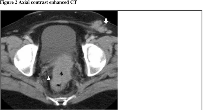
Figure 2 Axial contrast enhanced CT
of parametrium (arrowhead), with no cleavage plane with rectum or cervix.There are
enlarged left inguinal nodes.
© Ecorad Clinic, Vila Franca de Xira, Portugal
Area of Interest: Foetal imaging;
Imaging Technique: CT;
Procedure: Diagnostic procedure;
Special Focus: Neoplasia;
There are enlarged lymph nodes in left external iliac (asterix) and pre-sacral (white arrow)
lymphatic chains.
© Ecorad Clinic, Vila Franca de Xira, Portugal
Area of Interest: Genital / Reproductive system female;
Imaging Technique: CT;
Procedure: Diagnostic procedure;
Special Focus: Neoplasia;
There are enlarged lymph nodes in celiac (asterix) and retrocrural (black arrow) lymphatic
chains.
© Ecorad Clinic, Vila Franca de Xira, Portugal
Imaging Technique: CT;
Procedure: Diagnostic procedure;
Special Focus: Neoplasia;
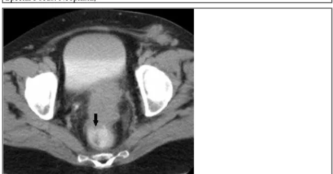
Image acquisition in the excretory phase with rectal administration of contrast helps to better
define the involvement of rectum wall (black arrow).
© Ecorad Clinic, Vila Franca de Xira, Portugal
Area of Interest: Genital / Reproductive system female;
Imaging Technique: CT;
Procedure: Diagnostic procedure;
Special Focus: Neoplasia;
Figure 3 MRI
Sagittal FSE T2-WI demonstrates a large mass (asterix) in the pouch of Douglas /
rectovaginal septum with intermediate/high signal intensity, that extends to the uterine cervix
and myometrium, compressing the sigmoid colon (arrowhead).
Area of Interest: Eyes;
Imaging Technique: MR;
Procedure: Diagnostic procedure;
Special Focus: Neoplasia;
The low signal of the inner layer of fibromuscular cervical stroma is preserved (circle).
© Ecorad Clinic, Vila Franca de Xira, Portugal
Area of Interest: Genital / Reproductive system female;
Imaging Technique: MR;
Procedure: Diagnostic procedure;
Special Focus: Neoplasia;
The mass was inseparable from rectum wall (arrow, see the correlation with sigmoidoscopy
in Fig. 1A).
Area of Interest: Genital / Reproductive system female;
Imaging Technique: MR;
Procedure: Diagnostic procedure;
Special Focus: Neoplasia;
There were multiples enlarged lymph nodes; some are shown (circles).
© Ecorad Clinic, Vila Franca de Xira, Portugal
Area of Interest: Genital / Reproductive system female;
Imaging Technique: MR;
Procedure: Diagnostic procedure;
Special Focus: Neoplasia;
On axial SE T1-WI, a mass with intermediate signal intensity is seen between the uterine
cervix and rectum.
© Ecorad Clinic, Vila Franca de Xira, Portugal
Procedure: Diagnostic procedure;
Special Focus: Neoplasia;
Sagittal contrast-enhanced fat-saturated T1-WI shows heterogeneous enhancement with
slight area of necrosis. The invasion of the anterior rectal wall is better seen (arrow).
© Ecorad Clinic, Vila Franca de Xira, Portugal
Area of Interest: Genital / Reproductive system female;
Imaging Technique: MR;
Procedure: Diagnostic procedure;
Special Focus: Neoplasia;
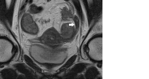
Coronal FSE T2-WI shows unremarkable right ovary separately identified from the mass
(arrow).
© Ecorad Clinic, Vila Franca de Xira, Portugal
Area of Interest: Genital / Reproductive system female;
Imaging Technique: MR;
Coronal FSE T2-WI shows unremarkable left ovary separately identified from the mass
(arrow).
© Ecorad Clinic, Vila Franca de Xira, Portugal
Area of Interest: Genital / Reproductive system female;
Imaging Technique: MR;
Procedure: Diagnostic procedure;
Special Focus: Neoplasia;
Figure 4 Histologic and immunohistochemical features
(A, B) Hematoxylin and eosin stain section of biopsy showing solid tumor growth and
marked cytologic atypia. Tumor showing positive staining for CAM5.2 (C) and CK7 (D). (E)
Tumor showing nuclear positive stain for WT1.
© Instituto Português de Oncologia de Lisboa Francisco Gentil EPE, Lisbon, Portugal
Area of Interest: Genital / Reproductive system female;
Imaging Technique: Percutaneous;
Procedure: Biopsy;
Special Focus: Neoplasia;
References
[2] Davis JM (1967) Carcinoma in the rectovaginal septum Proc R Soc Med 60:502
[3] Leteurtre E, Boman F, Farine MO, Querleu D, Gosselin B (1999) Serous papillary
cystadenocarcinoma of the recto-vaginal septum: a rare primary localization Ann Pathol 19:42-55
[4] Berger A, Rouzier R, Carnot F, et al (2001) Primary adenocarcinoma of the rectovaginal septum: a report and literature review Eur J Obstet Gynecol Reprod Biol 95:111-3
[5] Papacharalabous EN, Awad SA, Edwards JL (2004) A malignant tumour of the rectovaginal septum not arising from endometriosis, presenting a diagnostic enigma J Obstet Gynaecol 24:599-600
[6] Nelson GS, Ghatage P, Mainprize TC, Duggan MA, Buie D, Nation JG (2005) Primary carcinoma of the rectovaginal septum diagnosed as uterine prolapse J Obstet Gynaecol Can 27:1027-30
[7] Guiou M, Hall WH, Konia T, Scudder S, Leiserowitz G, Ryu JK (2008) Primary clear cell adenocarcinoma of the rectovaginal septum treated with concurrent chemoradiation therapy: a case report Int J Gynecol Cancer 18:1118-21
[8] Langmár Z, Harsányi L, Székely E, Járay B, Csömör S, Kazy Z (2008) Primary adenocarcinoma of the rectovaginal septum without associated endometriosis Orv Hetil 149:2251-3
[9] Giordano G, Bersiga A, Marchetti G, Melpignano M (2010) Primary adenocarcinoma of the rectovaginal septum arising in pregnancy in the absence of endometriosis Eur J Gynaecol Oncol 31:211-3
[10] Kengsakul K, Kor-Anantakul O, Pinjareon S, Sukthomya C, Suthipinthawong C. J (1987) Primary tumor of the rectovaginal septum: mesothelioma or papillary serous cystadenocarcinoma. A case report with transmission electron microscopy Med Assoc Thai 70:410-5
[11] Nishimura H1, Zhang Y, Ohkuma K, Uchida M, Hayabuchi N, Sun S (2001) MR imaging of soft-tissue masses of the extraperitoneal spaces Radiographics 5:1141-54
Citation
Mónica Vieira , Teresa Margarida Cunha , Jorge São Martinho (2015, Jan. 8) 1 2 3 Primary Carcinoma of the Rectovaginal Septum {Online}