Melissa MascherettiI Ciléa H TenganI Helena Keiko SatoI Akemi SuzukiII
Renato Pereira de SouzaII Marina MaedaII
Roosecelis BrasilII Mariza PereiraIII Rosa Maria TubakiIII Dalva M V WanderleyIII Carlos Magno Castelo Branco FortalezaIV
Ana Freitas RibeiroI Yellow Fever Group*
I Centro de Vigilância Epidemiológica.
Secretaria de Estado da Saúde de São Paulo. São Paulo, SP, Brasil
II Instituto Adolfo Lutz. Secretaria de Estado
da Saúde de São Paulo. São Paulo, SP, Brasil
III Superintendência de Controle de Endemias.
Secretaria de Estado da Saúde de São Paulo. São Paulo, SP, Brasil
IV Universidade Estadual Paulista “Julio de
Mesquita Filho”. Botucatu, SP, Brasil * The list of group members is in the
acknowledgements section. Correspondence:
Melissa Mascheretti
Rua Cardoso de Almeida, 1006/13 05013-001 São Paulo, SP, Brasil E-mail: [email protected] Received: 5/3/2012
Approved: 6/11/2013
Article available from: www.scielo.br/rsp
Yellow fever: reemerging in the
state of Sao Paulo, Brazil, 2009
ABSTRACT
OBJECTIVE:To describe the investigation of a sylvatic yellow fever outbreak
in the state of Sao Paulo and the main control measures undertaken.
METHODS: This is a descriptive study of a sylvatic yellow fever outbreak in
the Southwestern region of the state from February to April 2009. Suspected and conirmed cases in humans and in non-human primates were evaluated. Entomological investigation in sylvatic environment involved capture at ground level and in the tree canopy to identify species and detect natural infections. Control measures were performed in urban areas to control Aedes aegypti. Vaccination was directed at residents living in areas with conirmed viral circulation and also at nearby cities according to national recommendation.
RESULTS: Twenty-eight human cases were conirmed (39.3% case fatality rate) in rural areas of Sarutaiá, Piraju, Tejupá, Avaré and Buri. The deaths of 56 non-human primates were also reported, 91.4% were Allouatta sp. Epizootics was conirmed in two non-human primates in the cities of Itapetininga and Buri. A total of 1,782 mosquitoes were collected, including Haemagogus leucocelaenus, Hg. janthinomys/capricornii, and Sabethes chloropterus, Sa. purpureus and Sa. undosus. Yellow fever virus was isolated from a group of Hg. Leucocelaenus from Buri. Vaccination was carried out in 49 cities, with a total of 1,018,705 doses. Nine serious post-vaccination adverse events were reported.
CONCLUSIONS: The cases occurred between February and April 2009 in
areas with no recorded yellow fever virus circulation in over 60 years. The outbreak region occurred outside the original recommended vaccination area with a high percentage of susceptible population. The fast adoption of control measures interrupted the human transmission within a month and the conirmation of viral circulation in humans, monkeys and mosquitoes. The results allowed the identiication of new areas of viral circulation but further studies are required to clarify the dynamics of the spread of this disease.
DESCRIPTORS: Yellow Fever, epidemiology. Disease Outbreaks.
Yellow fever (YF) is an infectious disease, endemic in the rainforests of Latin Americaa and Africa, provoking
outbreaks and epidemics which impact on public health.18 The disease is caused by a virus from the
Flaviviridae family, genus Flavivírus, and transmitted by the bite of bloodsucking insects of the Culicidae family, genus Haemagogus and Aedes.11 Urban and
sylvatic cycles are how it is classically described. Sylvatic yellow fever (SYF) occurs in forested areas, where it is transmitted by mosquitoes to non-human primates (NHP), amplifying hosts of the virus. Occasionally, susceptible humans who enter in contact with forested areas become infected.2 The clinical mani
-festation of the disease can be asymptomatic, or with mild or moderate symptoms, or serious and malignant. Mortality varies between 5.0% and 10.0%, reaching 60.0% in the most severe forms.23
According to the World Health Organization, YF is endemic in 33 countries in Africa, with 23 at high risk of epidemic.18 Although a highly effective vaccine is
available, epidemiological data show a resurgence of areas in which the virus circulates in West Africa and the Americas over the last 20 years.3
The last report of urban transmission in Brazil occurred in the state of Acre, Northern, in 1942.5 From this time
onwards, only sporadic sylvatic transmission occur in endemic area, with records of cyclical epidemics at regular intervals of ive to seven years, alternating with small numbers of cases, a pattern observed until 1997.23
From 1998 onwards, a change in this pattern was noted, with increased number of cases and expansion in the disease transmission area, cases in regions that had been previously unaffected and outbreaks in the states of Pará and Tocantins and in the Chapada dos Veadeiros National Park in Goiás, Northern Brazil, in 2000. Since then, there have been reports of transmission in the states of Acre, Amazonas, Pará, Tocantins, in region Northern Brazil, and Bahia, Northeastern Brazil, Minas Gerais, Southeastern Brazil, Mato Grosso, and Goiás, and in the Federal District, in Region Midwest Brazil.24
Occurrence of sylvatic yellow fever (SYF) increased in Brazil in 2008, based on records of deaths in non-human primates and cases in non-humans in the states of Sao Paulo17 and Rio Grande do Sul.21
The irst reports in Sao Paulo date from 1935 in the municipalities of Rifaina, Ribeirao Preto, Viradouro, INTRODUCTION
Penapolis, Tanabi, Mirassol and Monte Aprazível. In 1936, there was a record of epidemics in municipalities in the Alta Sorocabana region, especially in the cities of Avaré, Piraju, Assis and Itapeva, with the last recorded outbreak occurring in 1953.19,b After almost 50 years
with no reported epidemics, two autochthonous cases of SYF were conirmed in the municipalities of Santa Albertina and Ouroeste, in 2000, and a suspected epizooty in Miguelopólis, in 2003.c In 2008, virus
circulation was conirmed in the state, with an epizooty in the municipalities of Mendonça, Nova Aliança and Urupês and two autochthonous human cases in Luiz Antonio and Sao Carlos.17,d The spread of the virus
resulted in a widening of the areas in which vaccination is recommended for residents and visitors.
In February 2009, SYF transmission was detected in the region of Botucatu, a non-endemic area where vaccination has not been recommended. The Ministry of Health considered the event to be a public health emergency of national importance.
The aim of this study was to describe the investigation of an outbreak of sylvatic yellow fever in the state of Sao Paulo and the principle control measures utilized.
METHODS
A descriptive study of an outbreak of SYF in 2009, covering the southeast of the state of Sao Paulo, encompassing 49 municipalities, with an estimated population of 1,174,142 inhabitants. The majority of the municipalities in this region (more than 70.0%) belong to the Alto Paranapanema basin, with an area of 28,447 km2.
The study took place between February and April 2009.
For case deinition, the Ministry of Health recommenda-tions in the epidemiological vigilance guidelines were used. Individuals who had not been vaccinated, who had a high fever accompanied by jaundice and/or hemorrhage, resident or having come from an SYF risk area in the preceding 15 days were considered to be suspected cases.e
After the virus circulation has been conirmed, the most sensitive case deinition was employed: individuals who had not been vaccinated, with a high fever (up to seven days) accompanied by at least two symptoms (headache, myalgia, nausea or vomiting), resident or having come from an area with conirmed SYF cases in the preceding 15 days. The search for cases included
a Organização Mundial de Saúde. Yellow Fever, key facts. Geneva; 2013 [cited 2013 Sept 16]. (Fact sheet,100). b Franco O. Historia da febre amarela no Brasil. Rio de Janeiro: Ministério da Saúde; 1976.
c Secretaria do Estado de São Paulo. Centro de Vigilância Epidemiológica “Prof. Alexandre Vranjac”. Informe técnico sobre febre amarela. São
Paulo; 2006 [cited 2010 Apr 23]. Available from: http://www.cve.saude.sp.gov.br/htm/zoo/FA_INFORME.htm
d Secretaria do Estado de São Paulo. Centro de Vigilância Epidemiológica “Prof. Alexandre Vranjac”. Casos de febre amarela silvestre em
residentes do Estado de São Paulo, 2007-2008. Bol Epidemiol Paul. 2008;5(55):12-5.
retrospective assessment of deaths from unknown causes and/or from ictero-hemorrhagic syndrome.d
Suspected cases that had reactive results in at least one diagnostic method (detection of IgM antibodies using the MAC-ELISA technique, virus isolation, detection of viral genome or viral antigen using the immunohis -tochemistry test) and with a clinical-epidemiological link (suspected death without samples being collected in the area of transmission during the period) were considered conirmed cases.d
A serological survey was applied between March 16 and 19 in the municipality of Sarutaiá, where there were no reports of human cases or epizooty (not considered at risk of the disease) and where the irst conirmed case resided. The individual had not been vaccinated for yellow fever and the only place he visited was a leisure area within the municipality. The aim of the survey was to detect circulation of the virus in the municipality and identify other cases. To calculate the sample size, an expected disease frequency of 50.0% was assumed, with level of signiicance of 95.0% and acceptable error of 10.0%. For a population of 2,966 inhabitants, with 996 residences divided into 9 sectors covered by the Family Health Care Program, the houses were randomly selected and 93 individuals were included in the study. Blood samples from 86 asymptomatic participants were collected for serology (Mac Elisa IgM).
Deaths of NHP with laboratory evidence of the virus infection in at least one animal was considered to be epizooty of yellow fever.d
The data on humans and NHP were taken from Information System for Notiiable Diseases (SINAN) forms and stan-dardized spreadsheets. Notiications were made to the State Epidemiological Health Department by telephone, fax or electronically. Probable locations of infections were identiied and georeferenced using Garmin model Etrex (GPS). The geographical coordinates were obtained and referred to the Datum SAD 69. The municipalities were mapped using the Brazilian Institute of Geography and Statistics (IBGE) 2007 Digital Municipal Grid and the Mapinfo program, version 7.0, Mapinfo Corporation.
The blood, serum and cerebrospinal luid samples were processed using the Mac-Elisa test, following the protocol described by Kuno et al.13
Samples of human blood and serum, tissue material suspensions obtained from NHP autopsies and mosqui-toes were inoculated in 1 to 3-day-old Swiss mice. The brains of animals that showed signs of the disease underwent the following steps.1,f The samples were
inoculated in cell cultures of mosquitos Ae. albopictus, clone C6/36.10 Identification of the isolates was
f Travassos da Rosa APA, Travassos da Rosa ES, Travassos da Rosa JFS, Degallier N, Vasconcelos PF, Rodrigues SG, et al. Os Arbovírus no Brasil:
generalidades, métodos e técnicas de estudo. Belém: Instituto Evandro Chagas; 1994. (Documento técnico, 2).
proceeded using the indirect immunoluorescence tech-nique standardized by Gubler et al,8 using a polyclonal
anti-lavivirus. Positive samples were typed by indirect immunoluorescence with monoclonal antibodies for the yellow fever virus YFV (Biomanguinhos – RJ).
Polymerase chain reaction: The extraction was performed from total RNA from tissue samples, serum or suspension prepared from mice previously inoculated with the use of speciic commercial kits: QIAamp®
RNA Blood for tissues and QIAamp® Viral RNA Kit
for serum (Qiagen Inc., Ontário, Canadá), following the manufacturer’s instructions. Ampliication of viral RNA was performed by single-step reverse transcriptase (one step RT-PCR) followed by a second ampliication (semi-nested).6 The ampliied products were visualized
by electrophoresis in 1.5% agarose gel stained with ethidium bromide to verify band size.
DNA sequencing: ABI-377 sequencer was used to sequence positive samples and Chromas software version 1.45 and EditSeq (Lasergene DNASTAR Inc.) for editing nucleotide sequence.
Histopathology and immunohistochemistry: Samples of brain, heart, lung, liver, spleen and kidney, ixed in formaldehyde and embedded in parafin were processed and examined after completion of histolog -ical sections and staining with hematoxylin and eosin. Slices of liver with 0.3 µm were placed on slides with silane and subjected to immunohistochemistry with polyclonal anti-yellow fever virus, diluted 1/2,000 and detection system using mouse and rabbit anti-immunoglobulins combined with peroxidase or alkaline phosphatase (Envision®, Dako Cytomation, EUA).9
Entomological investigation activities were triggered by notiication of the death of a NHP and suspected and/ or conirmed cases of yellow fever in humans.
Capture took place at ground level and in the tree canopy, on the edges and within the forests at loca -tions of probable infection (LPI). The mosquitoes were collected using protected humans with mouth aspirators and nets at 15 to 20 minute intervals between 9:00 am and 4:00 pm, for four days in each area. Center for Disease Control and Prevention (CDC) style dry ice traps were also used.7 In order to try and record natural
infection the mosquitoes were stored in cryo-resistant tubes transported in liquid nitrogen and then frozen and stored at -70°C for later identiication and grouped into batches and processed for virus isolation.
resident or had visited, according to the current tech -nological norms.g Before these actions came into force,
adult mosquitoes were captured for virus isolation using aspirators and traps.
Entomological monitoring was intensiied in municipalities not infested with Ae. aegypti to research the larvae, elimi -nate potential breeding sites and ind adult mosquitoes.
Vaccination for yellow fever, 17DD Biomanguinhos, was recommended according to the Brazilian Ministry of Health epidemiological vigilance guidelines,d which, in case of epidemics, include individuals aged over six months resident in municipalities with conirmed circulation of the virus and neighboring municipalities. Vaccination was given house to house in the rural areas and in health care centers in the urban areas.
Cases of serious adverse events after vaccination were classiied as acute viscerotropic disease (AVD) and acute neurotropic disease according to CDC deinitions.4
RESULTS
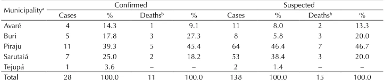
There were 138 reported suspected human cases in the region studied between February and April 2009. Among them, 110 were excluded and 28 were conirmed, of which 11 resulted in death (mortality 39.3%). The probable locations of infection in these conirmed cases were rural areas in the municipalities of Sarutaiá, Piraju, Tejupá, Avaré and Buri (Table 1). Ten cases were in rural workers who worked in the forest and 18 in individuals who undertook some kind of leisure activity outdoors.
Eighteen of the 28 conirmed cases were male (64.3%). Age varied between three days old and 52 years old (mean 29 years). Four cases occurred in children aged between three days and 16 years. The conirmed cases had mild, moderate and serious symptoms; of the conirmed cases,
50.0% fulilled the criteria of fever, jaundice and/or hemor-rhage, and 71.0% were hospitalized. With regards to the laboratory diagnosis, 42.9% of cases were conirmed using serology and 50.0% by more than one method (Table 2).
No yellow fever antibodies were detected in a serological survey carried out on an asymptomatic sample of the population in the municipality of Sarutaiá.
The irst conirmed case occurred in the municipality of Sarutaiá, with symptoms beginning on February 22 and the last case was in the municipality of Buri on April 1 (Figure 1).
Histopathological indings, identiied in ive cases, consisted of predominantly mediozona lesions, extending to the hepatic parenchyma, the presence of apoptosis and necrotic foci, micro and macrogoticular steatosis, hyperplasia and hypertrophy of Kupffer cells and portal spaces with slight lymphoid iniltration, with no signs of lesion to the interface. There was positive immunostaining for YF antigens in hepatocytes and Kupffer cells in immunohistochemistry.
There were 56 reported NHP deaths, followed up until August, distributed throughout seven municipalities in the region, and 91.4% of the NHP were Alouatta sp genus. The highest number of NHP deaths (77.5% of the total) were reported in the municipality of Buri. Material for laboratory diagnosis was collected from 7.0% of the animals, with viscera, serum/blood or brains collected. Laboratory epizooty was conirmed in two NHP of the Alouatta sp genus; one in the municipality of Buri using the RT-PCR technique on serum and the other was in Itapetininga using immunohistochemical technique on the viscera. The Histopathological ind-ings were similar to those of the humans.
Entomological activities began in Sarutaiá, Piraju and Itatinga immediately after conirmation of the irst cases, and later in Avaré, Buri and Itapetininga. There
g Superintendência de Controle de Endemias. Normas e recomendações técnicas para a vigilância e controle de Aedes aegypti no Estado de
São Paulo - NORTE. São Paulo; 2005.
Table 1. Distribution of confirmed and suspected cases of sylvatic yellow fever in humans, according to probable municipality
of infection. Sao Paulo State, February to April, 2009.
Municipalitya Confirmed Suspected
Cases % Deathsb % Cases % Deathsb %
Avaré 4 14.3 1 9.1 11 8.0 2 13.3
Buri 5 17.8 3 27.3 8 5.8 3 20.0
Piraju 11 39.3 5 45.4 64 46.4 7 46.7
Sarutaiá 7 25.0 2 18.2 53 38.4 3 20.0
Tejupá 1 3.6 – – 2 1.4 – –
Total 28 100.0 11 100.0 138 100.0 15 100.0
Sources: Sistema de Informação de Agravos de Notificação. Divisão de Zoonoses do Centro de Vigilância Epidemiológica.
Coordenação de Controle de Doenças. Secretaria de Estado da Saúde de São Paulo.
were infestations of Ae. aegypti in homes in Piraju and Avaré, while no infestations were found in Sarutaiá, Tejupá, Itatinga or Buri.
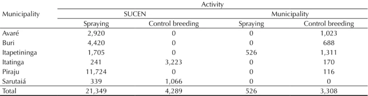
Activities to control the immature and adult forms of Ae. aegypti were performed in infested municipalities. Entomological control measures were carried out in all homes in Piraju, where the highest concentration of suspected and conirmed cases occurred, spraying homes with an adulticide over a ten day period (Table 3). No investigation was conducted in Tejupá, which was included as a transmission area after a retrospective assessment.
There were 1,782 female specimens from 58 species of mosquitoes collected in the forest during the ento -mological monitoring activities. Among the groups identiied, the following stood out: Ae. serratus group (32.2%), Psorophora ferox (22.4%), Hg. leucocelaenus (5.5%), Ochlerotatus scapularis (3.0%), Hg. janthi-nomys/capricornii (2.2%) and Ae. albopictus (0.9%). Specimens from the Sabethini (11.9%) genus were also found: Sa. purpureus, Sa. chloropterus, Sa. undosus, Sa. intermedius, Sa. albiprivus and Sa. tridentatus.
Among the mosquitoes captured for virus isolation, 1,782 were processed in 281 batches, constituted according to species, location and time of capture, with 58 species identiied. There were 1,210 specimens, 26 batches, captured in the municipality of Buri during the irst fortnight of April. The YF virus was isolated in mice and in cells, identiied using RT-PCR, from a batch of the Hg. leucocelaenus species, composed of six specimens.
Table 2. Confirmatory criteria of the human cases of sylvatic
yellow fever. Sao Paulo State, 2009.
Confirmatory criteria
Diagnostic method no. of confirmed
cases %
Laboratorial Serology 12 42.8
Serology and RT-PCR 8 28.6
Serology, RT-PCR and
viral isolation 2 7.1
Serology, RT-PCR, viral isolation and Immunohistochemistry
2 7.1
RT-PCR, viral isolation and Immunohistochemistry
1 3.6
RT-PCR and Immunohistochemistry
1 3.6
Immunohistochemistry 1 3.6
Clinical
epidemiological Not done 1 3.6
Total 28 100.0
Source: Divisão de Zoonoses do Centro de Vigilância Epidemiológica e do Instituto Adolfo Lutz. Coordenação de Controle de Doenças/ Secretaria de Estado da Saúde de São Paulo.
RT-PCR: polymerase chain reaction
20-F
eb
21-F
eb
22-F
eb
23-F
eb
24-F
eb
25-F
eb
26-F
eb
27-F
eb
28-F
eb
1-Mar 2-Mar 3-Mar 4-Mar 5-Mar 6-Mar 7-Mar 8-Mar 9-Mar 10-Mar 11-Mar 12-Mar 13-Mar 14-Mar 15-Mar 16-Mar 17-Mar 18-Mar 19-Mar 20-Mar 21-Mar 22-Mar 23-Mar 24-Mar 25-Mar 26-Mar 27-Mar 28-Mar 29-Mar 30-Mar 31-Mar 1-Apr 2-Apr
0 1 2
Cases
3
Onset symptons date
Sarutaiá Avaré Piraju Buri Tejuba
Source: Sistema de Informação de Agravo de Notificação. Divisão de Zoonoses do Centro de Vigilância Epidemiológica. Coorde-nação de Controle de Doenças. Secretaria de Estado da Saúde de São Paulo.
Figure 1. Temporal distribution of human cases of yellow fever, according to municipality probable date of infection and onset
Figure 2 shows the YF transmission areas conirmed in laboratory in humans, NHP and mosquitoes.
Vaccination was initiated immediately following conirmation of the irst case, in Sarutaiá, and was rolled out into areas of probable viral circulation. There were 49 municipalities where vaccination was recom -mended, with a general population of 1,174,142. A total of 1,018,705 doses of vaccine were given between March and April, with vaccination coverage of 86.8%. There were three conirmed cases of acute neurotropic disease, one case of immediate hypersensitivity, all of whom recovered, and ive cases of acute viscerotropic
disease, who died. The recent circulation of the yellow fever virus in Avaré, Buri, Itapetiniga, Piraju, Sarutaiá and Tejupá in the state of Sao Paulo lead to the recom -mended area of vaccination being expanded.
DISCUSSION
Conirmed cases of SYF occurred between February and April 2009 in the Southeast of the state, in areas where there had been no virus circulation recorded in more than 60 years and, therefore, not recommended for yellow fever vaccine and with a susceptible human popula -tion. The region is mountainous with a large number of
Source: Sistema de Informação de Agravo de Notificação. Divisão de Zoonoses do Centro de Vigilância Epidemiológica. Coorde-nação de Controle de Doenças. Secretaria de Estado da Saúde de São Paulo.
Figure 2. Map of distribution of confirmed cases of sylvatic yellow fever in humans, nonhuman primates and vectors by
probable municipality of infection and municipalities whose epidemiological monitoring actions were expanded. Sao Paulo State, February-April, 2009.
Vaccination area NUGEO/NIVE-CVE
Opromolla & Vieira 2010
Human cases Confirmed monkeys
Confimed Yellow fever in human, monkeys and vector
Ribeirão Branco
Guapiara Nova
Campina Ribeirão Grande Capão Bonito
São Miguel Arcanjo
Pilar do Sul Sarapui Alambari
Riversul Itaporanga Barão de Antonina Fartura Timburí
Sarutaiá
Coronel Macedo Taquarituba
Taguaí Itaí
Paranapanema Angatuba Guareí Quadra
Cesario Lange
Tatuí Itatinga
Avaré Pratânia
Botucatu
Pardinho Bofete
Tejupá Piraju Ipaussu
Bernardino de Campos Óleo
Manduri Cerqueira
César Águas de Santa Bárbara
Iaras
Arandu
Itapetininga Campina do
Monte Alegre
Taquarivaí Buri
Itapeva Itaberá
Itararé
Sao Paulo State Brazil
km km
N N
N km
-50° -60°
-70° -50° -40° -30°
-60° -70° -50°
-50°
-50° -40° -30°
-20°
-30
°-20°
-10
°0
°
-30
°-20°
-10
°0
°
-20°
-50° 0250 500 1 1,5
050 100 200 300 400 0 10 20 40 60 80
Table 3. Distribution of the number of properties covered in the control of Aedes aegypti in municipalities with circulation of
the Yellow Fever virus, according to type of activity and who carried it out. Sao Paulo State, March-April, 2009.
Municipality
Activity
SUCEN Municipality
Spraying Control breeding Spraying Control breeding
Avaré 2,920 0 0 1,023
Buri 4,420 0 0 688
Itapetininga 1,705 0 526 1,311
Itatinga 241 3,223 0 170
Piraju 11,724 0 0 116
Sarutaiá 339 1,066 0 0
Total 21,349 4,289 526 3,308
rivers and fast lowing streams. The vegetation consists of small, broken up forest formations interspersed with plantations, pasture and areas of reforestation, with favorable conditions for viral circulation.
The cases either lived or travelled through the forested area for leisure activities or work in the municipalities of Sarutaiá, Piraju, Tejupá, Avaré and Buri.
The swiftness of the joint and integrated activities carried out by the diverse organizations was essential in conirming viral circulation and adopting appropriate control measures, interrupting human transmission within a month. The vaccination campaign was started in 49 municipalities immediately after conirmation of the irst human case. More than a million individuals were vaccinated in three months. The vaccination activities were expanded gradually in the municipalities according to the LPI of conirmed cases: irst, in every house in the rural area and later in health facilities in urban area. Vaccination was recommended for travelers heading to the affected area and no imported cases were detected in this period.
The investigation of suspected cases by monitoring acute icterohemorrhagic febrile syndrome and deaths from unknown causes was an important instrument in identifying the irst cases. Using the more sensitive dei-nition for suspected cases throughout the work allowed mild and moderate cases to be identiied.
Human cases without early detection and reporting epizooty shows the need to intensify epizootic vigilance so that it constitutes a sentinel event of the virus circu -lation. Even after the active investigation, no relevant monkey deaths were veriied, except in Buri, in contrast to Rio Grande do Sul.h In Buri, it was possible to
iden-tify the virus in humans, NHP and in mosquitoes, which made it possible to complete the epidemiological chain associated with this form of the disease.20
The hypothesis of urban transmission of YF was raised in Piraju due to the large number of human cases in an area with Ae. aegypti. The epidemiological investiga -tion, however, conirmed that those cases had conducted activities in the forested area near their homes where entomological research confirmed the presence of sylvatic vectors. This made the possibility of urban transmission less likely and conirmed the disease’s sylvatic transmission.
Identiication of transmission in forested areas high-lights the risk of infection for the human population living close by or travelling through these areas, or other similar areas in the state.
In most investigated locations, Hg. leucocelaenus was more common than Hg. janthinomys/capricornii. This created a doubt as to their role as the primary or secondary vector in the transmission of sylvatic yellow fever. Hg. leucocelaenus was described as an abundant species in the South of the country.12 Recent epizooties
in Rio Grande do Sul have raised the hypothesis that this species may be a primary vector.23Sa. chloropterus is also considered a secondary vector, as isolation has been done in naturally infected specimens.22 Among the
mosquitoes captured, Oc. scapularis and Ps. ferox were the species with proven experimental transmission and positive for viral isolation.5,14-16 In the state of Sao Paulo, the virus was only isolated in Hg. leucocelaenus in Buri.
In addition to conirming circulation of the yellow fever virus in this area, it is important that investigation continues in order to understand the dynamics of viral transmission. Initial questioning raises the doubt as to whether the disease could have been circulating in this area, undetected, for years. The possibility should also be considered that the virus was recently introduced. Possible routes include from the state of Paraná, with records of NHP in the border area the previous year, or introduction into the state of Sao Paulo by humans due to population movement in the viremic period, traficked HNP or some other vector.24 Other possible
routes should not be ruled out. The use of molecular techniques such as sequencing and phylogenetic analysis of the virus may contribute to the knowledge of circulation and viral origin in this region.
ACKNOWLEDGEMENTS
The authors received collaboration from the following investigators of the Yellow Fever Group Center of the Centro de Vigilância Epidemiológica of the Secretaria de Estado da Saúde de São Paulo:
Ana Lívia Geremias, Roberta Spinola, Paula Opromolla, Pedro Antônio Vieira (in memoriam), Ricardo Mangabeira Albernaz, Maria Gomes Valente, Dulce Junqueira, Maria Teresa Jahnel, Elizabete Maria Nunes (Centro de Vigilância Epidemiológica. Secretaria de Estado da Saúde de São Paulo); Iray Maria Rocco, Ivani Bisordi, Selma Marina C. N. Petrella, Luiz Eloy Pereira, Terezinha Lisieux M. Coimbra, Vivian Regina Silveira, Adriana Yurika Maeda, Fernanda Giselle da Silva, Felipe Scassi Salvador, Cristina Takami Kanamura, Suely Nonogaki, Marina Suehuko Oyafuso, Yara de Menezes, Gislene Mitsue N. Nishina, Amanda Aguiar(Instituto Adolfo Lutz. Secretaria de Estado da Saúde de São Paulo); Eduardo Sterlino Bergo, Simone Luchetta Reginato, Sueli Yasumaro Diaz
h Ministério da Saúde. Secretaria de Vigilância em Saúde. Febre Amarela Silvestre, Brasil, 2009. Boletim de atualização de dezembro de
(Superintendência de Controle de Endemias. Secretaria de Estado da Saúde de São Paulo); Regiane Menezes Tironi, Luis Filipe Mucci (Superintendência de Controle de Endemias. Secretaria de Estado da Saúde de São Paulo); Ricardo Augusto Monteiro de Barros Almeida, Edna Maria de Souza Carvalho (Universidade Estadual Paulista “Julio de Mesquita Filho”); Beatriz Yuko Kitagawa, Eduardo S. Moreno (XI Programa de Treinamento em Epidemiologia Aplicada aos Serviços do SUS do Centro de Vigilância Epidemiológica. Secretaria de Estado da Saúde de São Paulo); Maria Saleti Carli (Grupo de Vigilância Epidemiológica de Botucatu do Centro de Vigilância Epidemiológica. Secretaria de Estado da Saúde de São Paulo); Simone Cristina B. N. S. Neves (Vigilância Epidemiológica do município de Sarutaiá); Yaeko Kawata (Vigilância Epidemiológica do município de Pirajú. Sociedade de
1. Beaty B, Calisher CH, Shope RE. Arboviruses. In: Schmidt NJ, Emmons RW, editors. Diagnostic procedures for viral, rickettsial and chlamydial infections. 6. ed. Washington (DC): American Public Health Association; 1989. p. 797-855.
2. Bicca-Marques JC, Freitas DS. The role of monkeys, mosquitoes and human in the occurrence of a yellow fever outbreak in a fragmented landscape in south Brazil: protecting howler monkeys is a matter of public health. Trop Conserv Sci. 2010;3(1):78-89.
3. Bryant JE, Holmes EC, Barrett ADT. Out of Africa: A Molecular Perspective on the Introduction of Yellow Fever Virus into the Americas. PLoS Pathog.
2007;3(5):e75. DOI:10.1371/journal.ppat.0030075 4. Staples JE, Gershman M, Fischer M; Centers for
Disease Control and Prevention (CDC). Yellow fever vaccine. Recommendations of the advisory committee on immunization practices (ACIP). MMWR Recomm Rep. 2010;59(RR-7):1-27.
5. Degállier N, Hervé JP, Rosa APAT, Vasconcelos P, Rosa JFST, Sá Filho G. A ecologia dos arbovírus na Amazônia: pesquisas atuais e perspectiva. Rev Fund
SESP. 1986;31(2):127-30.
6. Deubel V, Huerre M, Cathomas G, Drouet MT, Wuscher N, LE Guenno B, et al. Molecular detection and characterization of yellow fever virus in blood and liver specimens of a non-vaccinated fatal human case. J Med
Virol. 1997; 53(3):212-7.
DOI:10.1002/(SICI)1096-9071(199711)53:3<212::AID-JMV5>3.0.CO;2-B 7. Forattini OP, Gomes AC, Galati EAB, Rabello EX,
Iverson LB. Estudos ecológicos sobre mosquitos Culicidae no Sistema da Serra do Mar, Brasil. 1-Observações no ambiente extradomiciliar.
Rev Saude Publica. 1978;12(4):297-325.
DOI:10.1590/S0034-89101978000400008 8. Gubler DJ, Kuno G, Sather GE, Velez M, Oliver
A. Mosquito cell culture and specific monoclonal antibodies in surveillance for dengue viruses. Am J
Trop Med Hyg. 1984;33(1):158-65.
9. Hall W C, Crowell T P, Watts D M, Barros V L, Kruger H, Pinheiro F, et al. Demonstration of yellow fever and dengue antigens in formalin-fixed paraffin-embedded human liver by immunohistochemical analysis. Am J
Trop Med Hyg. 1991;45(4):408-17.
10. Igarashi A. Isolation of Singh´s Aedes albopictus cell line clone sensitive to dengue and
chikungunya virus. J Gen Virol. 1978;40(3):531-44. DOI:10.1099/0022-1317-40-3-531
11. Johnson BW, Chambers TV, Crabtree MB, Filippis AM, Vilarinhos PT, Resende MC, et al. Vectors competence of Brazilian Aedes aegypti and Aedes albopictus for Brazilian yellow fever virus isolated.
Trans R Soc Trop Med Hyg. 2002;96(6):611-3.
DOI:10.1016/S0035-9203(02)90326-3 12. Kumm HW, Cerqueira NL. The role of Aedes
leucocelaenus in the epidemiology of jungle yellow fever in Brazil. Bull Ent Res. 1951;42(1):195-200. DOI:10.1017/S0007485300025281
13. Kuno G, Gomez I, Gubler DJ. Detecting artificial antidengue IgM complexes using a enzyme linked immunosorbent assay. Am J Trop Med Hyg. 1987; 36(1):153-9.
14. Lopes OS, Sacchetta LA, Francy DB, Jakob WL, Calisher CH. Emergence of a new arbovirus disease In Brazil. III. Isolation of Rocio virus from Psorophora ferox (Humboldt, 1819). Am J Epidemiol.
1981;113(2):122–5.
15. Mitchell CJ, Forattini OP. Experimental transmission of Rocio encephalitis virus by Aedes scapularis (Diptera, Culicidae) from the epidemic zone in Brazil. J Med
Entomol. 1984;21(1):34-7.
16. Mitchell CJ, Forattini OP, Miller B. Vector competence experiments with Rocio virus and three mosquito species from the epidemic zone in Brazil. Rev Saude Publica. 1986;20(3):171-7. DOI:10.1590/S0034-89101986000300001 17. Moreno ES, Rocco IM, Bergo E S, Brasil RA,
Siciliano MM, Suzuki A, et al. Yellow Fever Working Group. Reemergence of yellow fever: detection of transmission in the State of São Paulo, Brazil, 2008. Rev Soc Bras Med Trop. 2011;44(3):290-6. DOI:10.1590/S0037-86822011005000041 18. World Health Organization. Update on progress
controlling yellow Yellow fever in África, 2004-2008.
Wkly Epidemiol Rec. 2008; 83(50):449-60.
19. Rocha WL. O Serviço Especial de Defesa contra a Febre Amarela. Arq Hig S Paulo. 1937;2(3):13-105. 20. Souza RP, Petrella S, Coimbra TLM, Maeda AY,
Rocco IM, Bisordi I, et al. Isolation of yellow fever virus (YFV) from naturally infectied Haemagogus
(Conopostegus) leucocelaenus (diptera,
cukicudae) in São Paulo State, Brazil, 2009.
Rev Inst Med Trop S Paulo. 2011;53(3):133-9.
DOI:10.1590/S0036-46652011000300004 21. Tauil PL. Aspectos críticos do controle
da febre amarela no Brasil. Rev
Saude Publica. 2010;44(3):555-8.
DOI:10.1590/S0034-89102010005000014
22. Vasconcelos PFC, Rodrigues SG, Dégallier N, Moraes MAP, Rosa JFST, Mondet B, et al. Ann epidemic of sylvatic yellow fever in the southeast region of Maranhão State, Brazil, 1993-1994: epidemiologic and entomological findings. Am J Trop Med Hyg.
1997;57(2):132-7.
23. Vasconcelos PFC, Sperb AF, Monteiro HAO, Torres MAN, Sousa MRS, Vasconcelos HB, et al. Isolations of yellow fever vírus from Haemagogus leucocelaenus in Rio Grande do Sul State, Brazil.
Trans R Soc Trop Med Hyg. 2003; 97(1):60-2.
DOI:10.1016/S0035-9203(03)90023-X
24. Vasconcelos PFC. Febre Amarela no Brasil: reflexões e hipóteses sobre emergência em áreas previamente livres. Rev Saude Publica. 2010; 44(6):1144-9. DOI:10.1590/S0034-89102010005000046 REFERENCES