Evaluation of Telomerase (hTert), Ki67 and p16ink4a expressions in
low and high-grade cervical intraepithelial lesions
Avaliação da expressão de Telomerase (hTert), Ki67 e p16
ink4aem lesões
intraepiteliais cervicais de baixo e alto graus
AnA PAulA SzezePAniAk GoulArt1; MAnoel AfonSo GuiMArãeS GonçAlveS1; viniciuS DuvAlDA-SilvA1.
INTRODUCTION
C
ervical cancer is the third most commonmalignant neoplasm among women (90% of cases in developing countries), and in Brazil it accounts for 9.3% of gynecological malignancies. Worldwide, 500,000 new cases per year are diagnosed, accounting for 250,000 annual deaths1.
The National Cancer Institute (INCA) recommends that cervical cytopathological screening be done every three years after two consecutive negative tests in patients aged 25 and 64 years, provided that the samples are satisfactory and representative of the squamous-column junction2. The malignant neoplasm
of the cervix is preceded by a long phase of precursor, usually asymptomatic, lesion, characterized by cervical intraepithelial neoplasia (CIN), which may persist for 10 to 20 years.
In 1976, Zur Hausen3 demonstrated the
relationship between the HPV virus infection and the
onset of genital tract neoplasias. Infection is more common in young patients; however, sexual behavior, age, smoking, parity and use of contraceptives are also risk factors for this neoplasia1. On the other
hand, not all patients infected with the virus have the same disease progression, since this behavior is linked to environmental factors, immunity, host genetics and cellular factors4. The use of molecular
markers has helped the pathologists in the definition of doubtful cases and in the identification of women at high risk for disease recurrence after treatment of cervical intraepithelial neoplasias5. p16ink4a, a tumor
suppressor protein, is overexpressed in cases of dysplasia, presenting a high sensitivity as it correlates with the dysplasia severity. Current studies indicate that p16ink4a is a useful biomarker for high-risk lesions
and for progression prediction in low-risk ones. These studies show a significant association between the degree of cervical lesion and the distribution and intensity of p16ink4a expression5,6.
1 - São Lucas Hospital, Pontifical Catholic University of Rio Grande do Sul (PUCRS), Gynecology and Pathology Service, Porto Alegre, Rio Grande do Sul State, Brazil.
A B S T R A C T
Objective: to study the association between the histological grading of cervical intraepithelial neoplasia (CIN I, CIN II and CIN III) and the immunohistochemical expression for p16ink4a, hTert and Ki67, as well as to evaluate the relationship of these markers with the risk of
recur-rence after surgical treatment. Methods: we studied a historical cohort of 94 women with intraepithelial lesions CIN I (low grade), CIN II and CIN III (high grades) submitted to conization or electrosurgical excision of the transformation zone. We evaluated all surgical specimens for immunohistochemical expression of p16ink4a, hTert and Ki67. Results: the mean age was 38.2 years; p16ink4a was absent in most CIN I cases.
In patients with CIN II or I/II (association of low and high-grade lesions), we observed p16ink4a≤10%. In patients with CIN III, we found a
higher expression frequency of p16ink4a >50%. In CIN I, the majority had Ki67≤10% and low frequency of Ki67>50%. In the CIN III category,
there were fewer patients with Ki67≤10%, and Ki67 was absent in most patients of CIN II and III groups. There was no association between hTert expression and histologic grade. There were no statistically significant differences between the expression of the markers in patients with and without recurrence. Conclusion: there was a statistically significant association of p16ink4a and Ki67 with histological grade. The
markers’ expression, as for disease recurrence, was not statistically significant in the period evaluated.
Ki67 is another cell proliferation marker expressed at all stages of the cell cycle, except in the G0 phase, and which, although not specifically involved in the cervical carcinogenic process, has an independent predictive value as to grading and evaluating disease progression7.
Telomerase is a ribonucleic enzyme complex that allows telomere maintenance in 90% of cancer cases. In normal somatic cells, telomerase activity is low or undetectable, and telomeres shorten at each cell division. The hTert is the telomerase reverse transcriptase, being the most important factor in the formation of functional telomerase. The activation of telomerase establishes telomere length maintenance, acting in the transformation of dysplasia into malignant neoplastic alterations and in the maintenance of phenotypically more aggressive cell populations8. The
hTert is present in 28.5% of CIN I, in 87.5% of CIN II and in 95% of CIN III9.
Among the existing treatments for the management of cervical intraepithelial neoplasias, in our hospital we perform the conization and the electrosurgical excision of the transformation zone (LEEP) techniques. The former has the advantage of allowing lesion resection and margins evaluating without the presence of fulguration artifacts by the passage of electric current, but removes a larger volume of cervical stroma, which can lead to adverse reproductive outcomes. The LEEP technique is a technically simpler procedure, which can be done in an outpatient setting under local anesthesia. On the other hand, margins may be inadequate for evaluation due to histological artifacts caused by thermal damage, have a higher risk of bleeding and may cause cervical stenosis in up to 6% of patients10.
The literature recommends that patients’follow-up should be for at least one year with a cervical cytopathological exam (CP) and with semestral colposcopy after surgery, since this is the period of greatest risk of recurrence of cervical lesions11. The
recurrence rate of high-grade intraepithelial lesions after surgical excision varies from 5% to 10%4,12,13.
The complication associated with the surgical treatment of such lesions are bleeding, external cervical orifice stenosis, infertility, shortening
of the cervix and risk of preterm labor, isthmo-cervical incompetence, low birth weight fetuses, cesarean
section and premature membranes rupture14.
Therefore, to analyze the association between recurrence after surgical treatment and the expression of immunohistochemical markers according to the graduation of high and low grade intraepithelial lesions would allow the suspension of a rigorous clinical follow-up and the reduction of overdiagnosis and overtreatment in patients affected by this disease. To date, there is no description of the concomitant evaluation of the three immunohistochemical markers – p16ink4a, Ki67 and hTert – in high and low
grade intraepithelial lesions. As already described, the only marker that has well established utility in the evaluation of progression, not recurrence, of these lesions is p16ink4a 15. Other markers are still poorly
studied and, therefore, there are few published papers regarding Ki67 and hTert, with no established accuracy for use in clinical practice, although they appear to display a difference according to their expression between high and low grade lesions16.
Furthermore, the behavior of CIN II is still uncertain, and so far, this lesion receives the same treatment as CIN III, a standardized conduct based on the 1991 Bethesda consensus17. However, in recent years, its
aggressiveness has been questioned, since it behaves dubiously, with a significant number of cases showing good evolution, spontaneous regression or indolent behavior6,7. The establishment of recurrence risk
markers for this type of lesion is thus innovative, and could define which patients would require surgical treatment and which would be likely to remain in clinical follow-up. This answer does not yet exist in the literature.
METHODS
of the Pontifical Catholic University of Rio Grande do Sul (PUCRS), who underwent cervical cytopathological examination followed by colposcopy with biopsy of suspicious areas and subsequent diagnosis of CIN. We recruited patients by (consecutive) convenience sampling. All cases of the study had the histological examinations reviewed by a second pathologist with clinical experience superior to twenty years.
Inclusion criteria were patients aged 18 years or older, submitted to LEEP or conization, and with a six-month postoperative follow-up (cervical CP, colposcopy and cervix biopsy if necessary) for at least two years, whose surgical specimens had margins free of cervical intraepithelial neoplasia. Exclusion criteria were patients with human immunodeficiency virus (HIV) or other immunosuppressive disorders, and pregnancy.
We performed the immunohistochemistry technique on tissues fixed in formalin and included in paraffin. The detection method used for screening tissue antigens was avidin-biotin peroxidase. We produced 3-μm thick histological sections on a Leica RT2150 rotary microtome. The slides were deparaffinized in a histological oven at 69° C, two incubations in xylol, five incubations with 99° ethyl alcohol, washing in running water until complete removal and then maintained in a PBS buffer. Antigen retrieval utilized a 6.0-pH citrate buffer for 20 minutes. Endogenous peroxidase activity was blocked with 5% hydrogen peroxide solution in methyl alcohol. Incubation with the primary antibody was performed in a BOD incubator at 5°C. To detect the antigen-antibody reaction, we used biotinylated secondary antibody and avidin-HRP complex. We performed staining with Diamine Benzidine (DAB, DAKO) as the chromogen. Finally,we counterstained the slides with Harris haematoxylin, dehydrated in a series of 99° ethyl alcohol, clarified in xylene and mounted with Canadian balsam. The clones used for the markers were E6H4 to p16ink4a, MM1 to Ki67 and 2C4 to
hTert. For the detection of p16ink4a, Ki67 and hTert, we
used slides without cervical intraepithelial neoplasia as negative controls and, as positive controls, slides with high cervical intraepithelial neoplasia (CIN III). Regarding the interpretation of the markers, we
considered the nuclear staining as positive for the cells expressing p16ink4a. We graded the expression
intensity for p16ink4a in CIN as negative if none of
the cells displayed staining, and as positive, if they expressed staining as a percentage (less than 10%, 11% to 50% or more than 50%). We considered Ki67 positive only if the nuclei of the cells showed staining and we determined the percentage of cells expressing it in less than 10%, 11% to 50% or more than 50%. We considered hTert positive also if the cell nucleus was stained. The expression was considered positive if the cells expressed staining and negative when there was no expression.
We inputted the data in the Excel 2010 software and later exported to the SPSS v.20.0 program for statistical analysis. We described categorical variables by frequencies and percentages and analyzed them with the Chi-square test. We described the quantitative variables with symmetrical distribution by the mean and standard deviation, comparing two categories with the Student t test for independent samples, and three categories or more, with the Analysis of Variance (ANOVA) test. We described quantitative variables with asymmetric distribution by median and the interquartile range (25th and 75th percentiles), comparing two categories with the Mann-Whitney test, and three or more categories, with the Kruskal-Wallis test. We set the significance level at 5%.
The project was approved by the Scientific Committee of the Postgraduate Program in Medicine and Health Sciences and by the Ethics Committee / PUCRS (protocol no. 109403/2014).
RESULTS
CIN II (12.8%). Among those who relapsed, most were carriers of CIN I, followed by carriers of CIN III.
The total number of recurrences among the 94 patients operated, regardless of the type of procedure, was 23 (24.5%). Recurrence was more common in patients who underwent LEEP, 31%, being 22% in those submitted to conization. There was a statistically significant association between p16ink4a and Ki67 and histological grade, as shown in
Table 1.
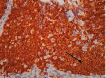
Figures 1, 2 and 3 show the expression of immunohistochemical markers.
There was no statistically significant diffe-rence between age, smoking, parity, age of first sexual intercourse and number of sexual partners and the different histological grades ordisease recurrence. When comparing the markers between subjects with and without recurrence, there were also no statistically significant differences.
DISCUSSION
Cervical carcinoma has a high potential for prevention. In our country, however, it is a public health problem.Studies have shown high incidence
and mortality in developing countries, andgood control in developed countries whose screening programs are effective18.
Works have found that the immunohistoche-mical markers p16ink4a and Ki67 are more accurate
in identifying precursor lesions in patients under 30 years with cervical cytopathological examination compatible with low grade intraepithelial lesion. The expression of these markers is associated with the severity of histological lesions. In the Possati-Resende et al. study18, patients underwent cervical biopsy,
Table 1. Histological grade and the expression of Immunohistochemistry markers
CIN I CIN I/II* and II CIN III CIN II and III P
p16ink4a 0.026
≤10 3 (23.1) 5 (38.5) 8 (16.7)
-11-50 - 1 (7.7) 3 (6.2) 2 (16.7)
>50 3 (23.1) 3 (23.1) 28 (58.3) 4 (33.3)
Absent 7 (53.8) 4 (30.8) 9 (18.8) 6 (50.0)
Ki67 0.032
≤10 9 (64.3) 5 (38.5) 11 (22.4) 5 (41.7)
11-50 3 (21.4) 1 (7.7) 9 (18.4)
->50 2 (14.3) 7 (53.8) 27 (55.1) 5 (41.7)
Absent - - 2 (4.1) 2 (16.7)
hTert 0.677
Positive 7 (50.0) 7 (53.8) 30 (62.5) 5 (45.5)
Negative 7 (50.0) 6 (46.2) 18 (37.5) 6 (54.5)
Data presented by n (%) and compared with the Chi-square test. Data in bold are data whose adjusted residue had absolute value greater than 1.96.
* High grade cervical intraepithelial lesion associated with the one of low grade.
Figure 1. p16ink4a; 200x magnification. Nuclear cells with marker
and the expression of p16ink4a and Ki67 was present
in 46.5% of CIN I, 82.8% of CIN II and 92.8% of CIN III. In our study, p16ink4a was overexpressed by
the immunohistochemical method in more than 50% of the cells of each sample in 43.3% of the cases, presenting a strong intensity in 48.9% of the exams. The Ki67 marker was expressed in 43.6% of the cases (positive in more than 50% of the sample). The hTert was positive in 56.5% of the cases studied.
Although the incidence of cervical neoplasms has decreased, there are still many diagnoses of cancer in cervical lesions with a high potential for regression and, consequently, of inadequate and excessive treatments. In this context, the use of immunohistochemical markers helps the pathologist in the histological evaluation of such lesions19. According to American guidelines, patients
with suspected high-grade cervical intraepithelial neoplasia and overexpressed p16ink4a benefit from
surgical treatment. The positivity for this marker is linked to the onset of oncogenic transformation for the development of cancer and its use would allow the detection of pre-neoplastic lesions and the reduction of unnecessary aggressive treatments19.
The importance of studying these markers is due to the wide variability of interpretation among observers and the poor reproducibility of cytomorphological criteria, mainly regarding the use of hematoxylin and eosin for the histological diagnosis of cervical intraepithelial neoplasias, as well
as its classification in the categoriesCIN I, II and III. We minimized this limitation in this study through the evaluation and concordance of diagnosis by two pathologists, one original interpreter of the case and one reviewer. The use of these markers complements the doubtful diagnoses, avoiding unnecessary treatments20,21. The presence of Ki67 in CIN I and CIN
II in uterine cervix specimens is a strong independent predictive factor for graduation and disease progression, in addition to displaying an interobserver reproducibility of 100%22. The literature shows that
the evaluated immunohistochemical markers, p16ink4a,
Ki67 and hTert, are clearly associated with histological grade. Some studies have found an association with the risk of progression to high-grade neoplasias and cancer, but none showed that their overexpression is associated with recurrence after treatment.
The CIN recurrence rate usually ranges from 5% to 10%4,12,13. However, in the study by Serati et
al.23, who followed patients for ten years, recurrence
occurred in 19.4% of the free-margin excisions and was twice as common with the LEEP technique. Although we included in our study only surgical pieces with free margins, the incidence of recurrence of 24.5% was high when compared with the rates described in the literature. On the other hand, Malapati et al.24
followed 717 eligible patients after LEEP and observed postoperative persistence/recurrence of 24.7%. The most common type of recurrence was CIN I (47.8%), followed by CIN III (34.8%), and most occurred within Figure 2. Ki67; 200x magnification. Nuclear cells with marker
expres-sion in more than 50%.
the first six months after treatment (56%).
There was no difference between the expression of the analyzed immunohistochemical markers and disease recurrence, even when evaluated individually at each histological grade. This fact may be due to the small number of cases and the subdivision of the markers expression positivity in below 10%, 11-50% and greater than 11-50%, unlike other studies in which the quantitative analysis was made in absolute numbers, a method that still presents limitations for implementation in the surgical pathology diagnostic routine. The study by Cardoso et al.4 was one of the few
that showed statistical significance between p16ink4a
overexpression and disease recurrence, as did Nam et al.25. Fonseca et al.26 demonstrated that p16ink4a was
overexpressed in 43% of the recurrences, with high intensity in 58% of them, though this finding was not statistically significant. In a group of 90 patients with cervical intraepithelial neoplasia with overexpressed p16ink4a and Ki67, there was progression in 15 cases
(17%), two cases in the group of 25 patients with CIN I (8%) and 13 cases in the group of 65 patients with CIN II (20%)22. In our study, Ki67 expression was
positive in 10% or less in CIN I and overexpressed in more than 50% of each sample in 53% of CIN II and 55% of CIN III (p=0.032).
Age, smoking, number of children, age of first intercourse, and number of sexual partners showed no significant difference in disease recurrence. Contrary to our findings, the literature shows that age greater than or equal to 35 years and smoking are risk factors for recurrence, as well as larger cytological alterations, high-risk preoperative HPV infection, positive endocervical margins, and HIV infection24,27,28.
The expression of p16ink4a related significantly
to the histological grade in this study. It was negative in 53% of CIN I, overexpressed in more than 50% of CIN III in 58% of cases and expressed in a lower percentage (equal or less than 10%) in 38.5% of the cases of CIN II (p=0.026). We observed a lower percentage of positivity for p16ink4a in CIN II than in
CIN III, although both overexpressed this marker with high intensity (53.8% of CIN II and 62.5% of CIN III). In the study of Genovés6, evaluating 92 patients,
p16ink4a was overexpressed in seven of the 54 patients
with CIN I and in 17 of the 23 patients with CIN II. The study emphasizes that the diagnosis of CIN II should not rely solely on the positivity of this marker, and its absence does not imply lesionregression.
Regarding the risk of progression, a study evaluating 52 patients with CIN II showed disease regression in 28 cases, progression to CIN III in 13 and persistence of the lesion in 11 cases. In those who progressed, 91% of the lesions had overexpressed p16ink4a, with strong intensity29. As for hTert, the
literature also describes its overexpression in high-grade intraepithelial lesions (p<0.001) in 88% to 90% of the samples, while low-grade intraepithelial lesions have low expression in 94% of cases30. It
can be inferred that the expression of this marker represents an early manifestation of the dysplastic process, therefore being overexpressed in 45% of CIN I, 70% of CIN II and 80% (p=0.024)31. In our
study, hTert did not show statistical significance in relation to the histological grade (p=0.677), although it was positive in 62.5% of the NCI III, 53.8% of CIN II and 50% of CIN I. Although the expression of immunohistochemical markers was positive in CIN II and CIN III, we observedvariable expression, showing a more intense and higher percentage of cells in high grade dysplasia. Such behavior of CIN II shows that this lesion probably has a behavior of lower aggressiveness when compared to CIN III. According to Wilkinson et al.32, the regression rate of CIN II is similar to that of
CIN I in a follow-up of two years (17%x12%). This poses a risk of overtreatment of such lesions, mainly in women under 25 years of age, since the risk of developing cervical cancer in untreated women with high-grade dysplasia is less than 1.5%.
REFERENCES
1. Wise J. Pilot study will assess whether HPV test should replace smears to screen for cervical cancer. BMJ. 2012;344:e3744.
2. Ministério da Saúde. Instituto Nacional de Câncer. Diretrizes brasileiras para o rastreamento do câncer do colo do útero. Rio de Janeiro: INCA; 2011. 3. zur Hausen H. Condylomata acuminata and human
genital cancer. Cancer Res. 1976;36(2 Pt 2):794. 4. Cardoso FA, Campaner AB, Silva MA. Prognostic
value of p16(INK4a) as a marker of clinical evolu-tion in patients with cervical intraepithelial neopla-sia grade 3 (CIN 3) treated by cervical conization. APMIS. 2014;122(3):192-9.
5. Yildiz IZ, Usubütün A, Firat P, Ayhan A, Küçükali T. Efficiency of immunohistochemical p16 expression and HPV typing in cervical squamous intraepithelial lesion grading and review of the p16 literature. Pa-thol Res Pract. 2007;203(6):445-9.
6. Genovés J, Alameda F, Mancebo G, Solé JM, Bello-sillo B, Lloveras B, et al. Human papillomavirus detec-tion and p16INK4a expression in cervical lesions: a comparative study. Hum Pathol. 2014;45(4):826-33. 7. Martin CM, O’Leary JJ. Histology of cervical intraepithelial
neoplasia and the role of biomarkers. Best Pract Res Clin Obstet Gynaecol. 2011;25(5):605-15.
8. Hsu CG, Wang PH, Ko JL, Chen GD, Chang H, Yang SF, et al. Concurrent high expression of human telo-merase reverse transcriptase and human nonmetas-tatic clone 23 in high-grade squamous intraepithe-lial neoplasia and squamous cell carcinoma of
ute-rine cervix. Int J Gynecol Cancer. 2007;17(4):851-7. 9. Saha B, Chaiwun B, Tsao-Wei DD, Groshen SL,
Naritoku WY, Atkinson RD, et al. Telomerase and markers of cellular proliferation are associated with the progression of cervical intraepithelial neoplasia lesions. Int J Gynecol Pathol. 2007;26(3):214-22. 10. Lindeque BG. Management of cervical
premalig-nant lesions. Best Pract Res Clin Obstet Gynaecol. 2005;19(4):545-61.
11. Massad LS. Preinvasive disease of the cervix. In: Di Saia PJ, Creasman WT, Mannel RS, McMeekin DS, Mutch DG, editors. Clinical gynecologic oncology.
8th ed. Philadelphia: Elsevier; 2012. p. 1-30.
12. Sarian LO, Derchain SF, Andrade LA, Tambascia J, Morais SS, Syrjänen KJ. HPV DNA test and Pap smear in detection of residual and recurrent disease following loop electrosurgical excision procedure of high-grade cervical intraepithelial neoplasia. Gynecol Oncol. 2004;94(1):181-6. 13. Prato B, Ghelardi A, Gadducci A, Marchetti I, Di
Cristofano C, Di Coscio G, et al. Correlation of re-currence rates and times with posttreatment hu-man papillomavirus status in patients treated with loop electrosurgical excision procedure conization for cervical squamous intraepithelial lesions. Int J Gynecol Cancer. 2008;18(1):90-4.
14. Kyrgiou M, Koliopoulos G, Martin-Hirsch P, Ar-byn M, Prendiville W, Paraskevaidis E. Obste-tric outcomes after conservative treatment for intraepithelial or early invasive cervical lesions: systematic review and meta-analysis. Lancet. 2006;367(9509):489-98.
Objetivo: estudar a associação entre a graduação histológica das neoplasias intraepiteliais cervicais (NIC I, NIC II e NIC III) e a expressão imuno-histoquímica para p16ink4a, hTert e Ki67, assim como, avaliar a relação destes marcadores com o risco de recorrência após
trata-mento cirúrgico. Métodos: estudo de coorte histórica de 94 mulheres portadoras de lesões intraepiteliais NIC I (baixo grau), NIC II e NIC III (altos graus), submetidas à conização ou à excisão eletrocirúrgica da zona de transformação. Todas as peças cirúrgicas foram avaliadas quanto à expressão imuno-histoquímica para p16ink4a, hTert e Ki67. Resultados: a média de idade das pacientes foi 38,2 anos. Nas
pacientes NIC I, a p16ink4a estava ausente na maioria dos casos; nas pacientes NIC II ou I/II (associação de lesões de baixo e alto graus),
observou-se frequência de p16ink4a≤10%. Nas pacientes NIC III, observou-se maior frequência de expressão de p16ink4a>50%. Na categoria NIC I, a maioria apresentava Ki67≤10% e baixa frequência de Ki67>50%. Na categoria NIC III houve menor número de pa-cientes com Ki67≤10%, sendo que a maior parte das pacientes tinha Ki67 ausente nos grupos NIC II e III. Não houve associação entre a expressão do marcador imuno-histoquímico hTert e a graduação histológica. Não houve diferenças estatisticamente significativas entre as expressões dos marcadores em pacientes com e sem recorrência. Conclusão: houve associação estatisticamente significativa apenas de p16ink4a e Ki67 com a graduação histológica. A expressão dos marcadores em relação à recorrência da doença não foi estatisticamente
significativa no período avaliado.
Descritores: Neoplasia Intraepitelial Cervical. Telomerase. Imuno-Histoquímica. Recidiva.
15. Darragh TM, Colgan TJ, Thomas Cox J, Heller DS, Henry MR, Luff RD, et al. The Lower Anogenital Squamous Terminology Standardization project for HPV-associated lesions: background and con-sensus recommendations from the College of American Pathologists and the American Society for Colposcopy and Cervical Pathology. Int J Gy-necol Pathol. 2013;32(1):76-115. Erratum in: Int J Gynecol Pathol. 2013;32(4):432. Int J Gynecol Pathol. 2013;32(2):241.
16. Fujii T, Saito M, Hasegawa T, Iwata T, Kura-moto H, Kubushiro K, et al. Performance of p16INK4a/Ki-67 immunocytochemistry for identifying CIN2+ in atypical squamous cells of undetermined significance and low-grade squa-mous intraepithelial lesion specimens: a Japa-nese Gynecologic Oncology Group study. Int J ClinOncol. 2015;20(1):134-42.
17. Solomon D, Nayar R. Sistema Bethesda para
ci-topatologia cervicovaginal. 2a ed. Rio de Janeiro:
Revinter; 2005.
18. Possati-Resende JC, Fregnani JH, Kerr LM, Mauad EC, Longatto-Filho A, Scapulatempo-Neto C. The Accuracy of p16/Ki-67 and HPV Test in the Detec-tion of CIN2/3 in Women Diagnosed with ASC-US or LSIL. PLoS One. 2015;10(7):e0134445.
19. Pacchiarotti A, Galeotti S, Bellardini P, Chi-ni F, Collina G, Dalla Palma P, et al. Impact of p16(INK4a) immunohistochemistry staining on interobserver agreement on the diagnosis of cer-vical intraepithelial neoplasia. Am J Clin Pathol. 2014;141(3):367-73.
20. Calil LN, Edelweiss MI, Meurer L, Igansi CN, Bozzetti MC. p16 INK4a and Ki67 expression in normal, dysplastic and neoplastic uterine cervical epithelium and human papillomavirus (HPV) infection. Pathol Res Pract. 2014;210(8):482-7. 21. Walts AE, Bose S. p16, Ki-67, and BD ProExC
immunostaining: a practical approach for diagno-sis of cervical intraepithelial neoplasia. Hum Pa-thol. 2009;40(7):957-64.
22. Kruse AJ, Baak JP, Janssen EA, Kjellevold KH, Fia-ne B, Lovslett K, et al. Ki67 predicts progression in early CIN: validation of a multivariate progression--risk model. Cell Oncol. 2004;26(1-2):13-20.
23. Serati M, Siesto G, Carollo S, Formenti G, Riva C, Cromi A, et al. Risk factors for cervical intraepi-thelial neoplasia recurrence after conization: a 10-year study. Eur J Obstet Gynecol Reprod Biol. 2012;165(1):86-90.
24. Malapati R, Chaparala S, Cejtin HE. Factors in-fluencing persistence or recurrence of cervical intraepithelial neoplasia after loop electrosur-gical excision procedure. J Low Genit Tract Dis. 2011;15(3):177-9.
25. Nam EJ, Kim JW, Hong JW, Jang HS, Lee SY, Jang SY, et al. Expression of the p16 and Ki-67 in rela-tion to the grade of cervical intraepithelial neopla-sia and high-risk human papillomavirus infection. J Gynecol Oncol. 2008;19(3):162-8.
26. Fonseca FV, Tomasich FD, Jung JE, Maestri CA, Carvalho NS. The role of P16ink4a and P53 immunostaining in predicting recurrence of HG--CIN after conization treatment. Rev Col Bras Cir. 2016;43(1):35-41.
27. Fu Y, Chen C, Feng S, Cheng X, Wang X, Xie X, et al. Residual disease and risk factors in patients with high-grade cervical intraepithelial neoplasia and positive margins after initial conization. Ther Clin Risk Manag. 2015;11:851-6.
28. de Mello Silva MV, Coutinho IC, de Andrade He-ráclio S, Fittipaldi HM Jr, Katz L. Factors associa-ted with the persistence/recurrence of CIN2/3 in women submitted to loop electrosurgical excision procedure in a teaching hospital in northeastern Brazil: a case-control study. J Low Genit Tract Dis. 2014;18(4):286-90.
29. Omori M, Hashi A, Nakazawa K, Yuminamochi T, Yamane T, Hirata S, et al. Estimation of prog-noses for cervical intraepithelial neoplasia 2 by p16INK4a immunoexpression and high-risk HPV in situ hybridization signal types. Am J Clin Pathol. 2007;128(2):208-17.
31. Bravaccini S, Sanchini MA, Amadori A, Medri L, Saragoni L, Calistri D, et al. Potential of te-lomerase expression and activity in cervical specimens as a diagnostic tool. J Clin Pathol. 2005;58(9):911-4.
32. Wilkinson TM, Sykes PH, Simcock B, Petrich S. Recurrence of high-grade cervical abnormalities following conservative management of cervical intraepithelial neoplasia grade 2. Am J Obstet Gy-necol. 2015;212(6):769.e1-7.
Received in: 11/08/2016
Accepted for publication: 26/09/2016 Conflict of interest: none.
Source of funding: none.
Mailing address:
Ana Paula Szezepaniak Goulart E-mail: [email protected]