Robot-Assisted Extended Pelvic Lymph Nodes Dissection for
Prostate Cancer: Personal Surgical Technique and Outcomes
_______________________________________________
Porpiglia Francesco
1, De Luca Stefano
1, Bertolo Riccardo
1, Passera Roberto
2, Mele Fabrizio
1,
Manfredi Matteo
1, Amparore Daniele
1, Morra Ivano
1, Fiori Cristian
11 Divisione di Urologia, Dipartimento di Oncologia, Università di Torino, San Luigi Gonzaga, Regione Gonzole
10, 10043 Orbassano (Torino), Italia; 2 Divisione di Medicina Nucleare, Dipartimento di Internal Medicina, Università di Torino, Ospedale San Giovanni Battista, Corso AM Dogliotti 14, 10126 Torino, Italia
ABSTRACT
ARTICLE
INFO
______________________________________________________________ ______________________
Objective: Extended pelvic lymph nodes dissection (EPLND) allows the removal of a higher number of lymph nodes than limited PLND. The aims of this study were to des-cribe our robot-assisted EPLND (RAEPLND) technique with related complications, and to report the number of lymph nodes removed and the rate of lymph nodal metastasis.
Materials and Methods: 153 patients underwent RAEPLND prior to robot-assisted radi-cal prostatectomy (RARP). Indications were defined according to Briganti nomogram, to predict risk of lymph-nodal metastasis. Lymphatic packages covering the distal tract of the common iliac artery, the medial portion of the external iliac artery, the external iliac vein and the internal iliac vessels, together with the obturator and the presacral lymphatic packages were removed on both sides.
Results: Median preoperative PSA was 7.5 ng/mL (IQR 5.5-11.5). Median operative time was 150 min (135-170). Median RAEPLND alone operative time was 38 min (32.75-41.25); for right and left side, 18 (15-29) and 20 min (15.75-30) (p=0.567). Median number of lymph nodes retrieved per patient was 25 (19.25-30); 13 (11-16) and 11 (8-15) for right and left side. In 19 patients (12.41%) metastasis was found at the level of pelvic lymph nodes. Median number of positive lymph nodes was 1 (1-4.6) per patient. Complications occurred in 11 patients (7.3%).
Conclusions: the number of lymph nodes removed was comparable to published data about open series, allowing the increase of detection rate of lymph nodal metastasis for minimally invasive approach without compromising complications’ rate if performing the procedure following reported technique.
Key words:
Complications [Subheading]; Laparoscopy; Prostatic Neoplasms; Lymph Nodes; Lymph Node Excision; Surgical Procedures, Operative
Int Braz J Urol. 2015; 41: 1209-19
_____________________
Submitted for publication: January 29, 2015
_____________________
Accepted after revision: July 06, 2015
INTRODUCTION
Pelvic lymph nodes dissection (PLND) is considered the surgical standard for staging of prostate cancer (PCa). The nomenclature and an-atomic boundaries of PLND vary. Limited PLND is defined by many surgeons as the removal of lymphatic packages along the external iliac artery and vein, obturator fossa, and obturator nerve (1,
The American Urological Association (AUA) guidelines consider that PLND should be reserved for patients with higher risk of nodal in-volvement with no clear cut-off (14).
For these reasons some controversies have risen about the appropriateness of limited PLND as a staging tool and more recently increasing evi-dences support EPLND if PSA level is >10ng/mL or the Gleason Score is ≥7 (3, 5, 7, 10).
It is known that patients affected by high-risk PCa have a high-risk of lymph nodal metastasis of about 38% (10) but available literature data are derived from open and pure laparoscopic experi-ences.
Since the introduction of robotic Da-Vinci® system in urologic surgery, robot-assisted radical prostatectomy (RARP) has been becoming an increasingly popular procedure throughout Eu-rope and the United States (10, 15-19).
In parallel with the beginning of RARP case-studies, experiences with robot-assisted PLND have started. The intraoperative magnifica-tion together with the higher degrees of freedom in movements allowed by robotic system have boosted performing of EPLND from the beginning in the majority of centers specialized in radical prostatectomy. To date, literature still lacks data about robot-assisted EPLND.
In our study, the primary aim was to de-scribe our surgical technique for RAEPLND and to report surgery-related complications with dis-cussion about how to prevent them; the second-ary aim was to report the number of lymph nodes removed by this technique and the detection rate and location of lymph nodal metastasis.
MATERIALS AND METHODS
From January 2011 to December 2013, 153 patients consecutively underwent RAEPLND for PCa. Indications for RAEPLND were given ac-cording to the updated nomogram for prediction of lymph nodes invasion (LNI) by Briganti et al. (13). Patient’s demographics were collected and reported in Table-1. All patients preoperatively underwent staging examinations by computed tomography (CT) scanning and/or magnetic reso-nance imaging (MRI) of the abdomen and pelvis.
In patients with a preoperative serum PSA le-vel above 20ng/mL, a total-body bone scanning was performed. Routine postoperative imaging assessment included ultrasonography at 1 mon-th and mon-three monmon-ths to evaluate surgery-related complications.
Technique step-by-step
All procedures were performed by trans-peritoneal approach. Patient was placed in a 30º Trendelenburg position. Using a four-arm Si HD Da Vinci robotic system (Intuitive Surgical, Sunnyvale, CA, USA), six trocars were placed. A 10-mm port for the camera was placed just cra-nially the umbilicus through a midline incision; two 8 mm ports for the robotic working instru-ments were placed on the right and left pararectal lines at their intersection with umbilical line. The third 8 mm port (for the fourth robotic arm) was placed 8 cm laterally to the left robotic port. Two additional trocars were placed for the assistant: the first one (5-mm) placed between the camera and the right robotic port; the second one (10 mm)
Table 1 - Patient characteristics.
No. of patients 153 Median age, yr (IQR) 64 (59-68) BMI, median (IQR) 26 (24-28.1) Preoperative PSA, median, ng/mL (IQR) 7.5 (5.5-11.5)
Clinical T stage, No (%)
T1 75 (49.0%) T2a 31 (20.3%) T2b 32 (20.9%) T2c 9 (5.9%)
T3 6 (3.9%)
Preoperative Gleason Score, No (%)
5 2 (1.3%)
6 22 (14.4%)
7a (3+4) 40 (26.2%) 7b (4+3) 39 (25.5%)
8 41 (26.7%)
9 9 (5.9%)
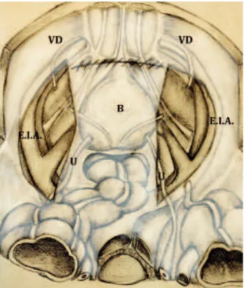
about 8 cm laterally to the right robotic trocar and about 4 cm cranially to the right anterior superior iliac spine (see Figure-1).
The anatomic landmarks of our technique of RAEPLND were the umbilical artery and the iliac vessels.
The anatomical limits were the bifurcation of the common iliac arteries, including the identi-fication of the ureter cranially, the Cloquet’s lymph node caudally, the external iliac artery laterally, and the bladder wall medially. The lymph nodes dissec-tion included the lymphatic packages located at the angle between external and internal iliac artery and along the obturator nerve. The dissection was per-formed by bipolar forceps and monopolar scissors. We here report the specific steps of the procedure:
1-Conventionally, the RAEPLND is begun on the right side. After mobilizing the sig-moid colon, PLND starts with the incision of peritoneum, laterally to the umbilical ligament overlying the common iliac ar-tery and parallel to the external iliac arar-tery until the ureter.
Incision is performed at the level of the pubic bone until the crossing of the ureter with common iliac vessels. External iliac vessels are identified and exposed. Vas deferens is identified and sectioned.
A blunt dissection is performed (preserving pre-vesical fascia) in order to enlarge the opera-tive field among lateral bladder wall, iliac vessels and lateral pelvic wall. The peritoneum covering hypogastric vessels and sacrum is medialized.
2-The ureter is identified at its crossing with common iliac artery, dissected, sus-pended (if necessary by using a vessel loop) and then displaced (we underline that ves-sel loop is generally used on the left side only because on the right side ureter ad-heres to peritoneum so that medialization of peritoneum itself is enough for ureter displacement). The operative field is now well prepared. We underline that presacral area is easy to get and dissect on the right side, while on the left side sacrum prom-ontorium is usually covered by common iliac vein that limit the intraoperative vi-sion and the dissection of presacral lymph nodes.
Once splitted the fibro-fatty tissue overly-ing the distal portion of the common and external iliac vessels, common iliac artery and its bifurca-tion are visible. The fibro-fatty tissue containing the lymphatics overlying the internal iliac artery, its medial vesical branches and the presacral lymph nodes are identified and dissected (Figure-2).
3-The external iliac lymph nodes are pro-gressively dissected. The dissection of the external iliac packages starts with the di-vision of the adventitia overlying the ex-ternal iliac vein distally. Then dissection is carried out from the crossing of the ure-ter over the common iliac arure-tery until the pubic bone at the level of circumflex vein
that usually is preserved and dissected (one Hem-o-lok clip is placed just cranially to the Cloquet’s lymph node, preserved in order to prevent lymphocele and lymphoe-dema) (Figure-3). Lateral limit of such a dissection is the medial portion of the ex-ternal iliac artery: the tissue covering the lateral part of the artery is spared in order to prevent lymphoedema.
4-Then the obturator fossa is reached and the lymph nodes are progressively dissect-ed until complete exposure of obturator nerve. Dissection is here performed with care in order to avoid any neural injury.
The dissection is started at the angle be-tween the external iliac vein and the pubic bone. The lymphatic package is dissected beneath the external iliac vein, proceed-ing until the pelvic side wall, which is the lateral limit of the dissection. The proximal attachments of the lymphatic packages are dissected by using a combination of either sharp or blunt dissection, paying attention in order to avoid any sharp, blunt, or ther-mal injury to the obturator nerve. The same technique is performed contra-laterally.
Figure 2 - Overview after suspension of the ureter (U) and removal of the fibrofatty tissue overlying the distal portion of the common (C.I.A) and external (E.I.A) iliac vessels (the bifurcation of the common iliac artery is now visible); the presacral (P.L.N) and hypogastric (H.L.N) lymph nodes are identified and dissected (left side). H.A hypogastric artery.
5-Once prostatectomy and its reconstruc-tive phase is completed, the anterior peri-toneum is sutured by using a running 3/0 “barbed” suture. At the end of the suture the peritoneal cavity and retropubic space are not in communication yet thanks to previously preserved pre-vesical fascia (Figure-4). Bilateral incisions of peritone-um done in order to perform RAEPLND are not sutured: at the end of the procedure, reconstruction is performed only at mid-line where parietal peritoneum covers the retropubic space.
Two drains are placed: one intraperitone-ally and one, extraperitoneintraperitone-ally, in the Retzius spa-ce: they are usually removed at the 1st and the 2nd
postoperative day, respectively.
Specifically for the purpose of the study, in order to assess the location of lymph-nodal me-tastasis, lymph nodes were sent in two separated packages removed by two laparoscopic endo-ca-tches from both sides (for convention, the right
sided ones are secured by hem-o-lok clips to label them): one containing the lymphatics overlying the distal portion of the common, external (only medial portion) and obturator fossa’s lymph nodes (they will be disposed on histopathological analy-sis Table from the caudalest to the cranialest in order to be recognized by pathologist); the other containing the internal iliac artery and the presa-cral lymphatic packages.
Histopathological analysis
A dedicated uro-pathologist performed all histopathological analysis. Tissue was submitted for permanent sectioning. Frozen section analysis was not routinely performed unless that in case of enlarged and/or clinically suspicious nodes.
Pathologic work-up to detect lymph nodes as well as lymph nodal metastases included di-rect visualization, palpation and standard hema-toxylin-eosin staining.
Outcome measurements
Skin-to-skin time, RAEPLND (right, left and overall) operative time, estimated blood los-ses, intraoperative complications (as classified by modified Satava system (20)), postoperative com-plications (as classified according to the modified Clavien system (21)), duration of hospitalization, catheterization time and transfusion rate were collected and analyzed.
The number and the locations of dissected lymph nodes on each side and the rate of lymph nodal metastases were recorded. The daily amount of drainage secretion (mL) and duration of draina-ge (days) were registered. In case of patient with a drained volume over 200mL/24 hours, urinary leakage was excluded by creatinine measurement.
Statistical analysis
The descriptive statistics of patients cha-racteristics are presented as median (IQR, inter quartile range) for continuous covariates, while as frequency (percentage) for categorical ones. No formal inferential test was performed, since the patients came from a single series. The data were analyzed by R 3.0.2 (R Foundation for Statistical Computing, Vienna-A, www.R-project.org).
RESULTS
Preoperative diagnostics were negative for metastasis in every case. All enrolled patients underwent RAEPLND+RARP. No patient received neo-adjuvant hormonal therapy. Median over-all operative time was 150 (IQR 135-170) min. Median RAEPLND alone operative time was 38 (IQR 32.75-41.25) min; for right and left side, 18 (IQR 15-29) and 20 (IQR 15.75-30), respectively, p=0.567. No case was converted to open surgery. Patients were discharged after a median hospital stay of 5 days (IQR 4-7).
Median number of lymph nodes retrieved per patient was 25 (IQR: 19.25-30), specifically 13 (IQR: 11-16) and 11 (IQR: 8-15), right and left side, respectively. In 19 patients (12.41%) metastasis
was found at the level of pelvic lymph nodes. Me-dian number of positive lymph nodes was 1 (IQR: 1-4.6) per patient.
In lymph nodal metastatic patients, medi-an PSA level was 8.2 (IQR 5.5-16.5) ng/mL versus 7.3 (IQR 5.3-11.4) ng/mL in negative lymph node-patients. Distribution of metastatic lymph nodes according to pathological stage and final Gleason Score is reported in Tables 2 and 3. Location and number of metastases per anatomic region are re-ported in Table-4.
In 11 patients (7.3%), RAEPLND-associated complications occurred: one patient (0.7%) had a temporary and reversible neuropraxia (involving ischiatic and obturator nerve). Ten (6.6%) patients were found to have lymphocele at ultrasonogra-phy performed at 1st month postoperatively with a
Table 3 - Metastatic lymph nodes according to pathologic grading.
Pathologic Gleason Score Nº of patients Nº of patients with LNI (%)
Overall 153 19 (12.41)
5 0 0
6 5 0
7a (3+4) 59 2 (3.38)
7b (4+3) 47 7 (14.89)
8 36 5 (13.88)
9 6 5 (83.33)
10 0 0
Table 2 - Metastatic lymph nodes according to pathologic staging.
Pathologic T stage N° of patients N° of patients with LNI (%)
Overall 153 19 (12.4)
pT2a 17 0
pT2b 5 0
pT2c 41 1 (2.4)
Total pT2 63 1 (1.6)
pT3a 56 6 (10.7)
pT3b 34 12 (35.3)
Total pT3 90 18 (20.0)
ranging size from 3.2 to 8.0cm. Among them only 5 (3.3%), with a lymphocele with ranging size from 4.5 to 8.0cm, were symptomatic complain-ing of lower abdominal pain and required percu-taneous drainage. Non symptomatic lymphoceles (5 patients) measured lower than 4.5cm maximum diameter and were located in obturator fossa. At 3rd month control these lymphoceles were stable.
The overall median blood loss for RARP including the RAEPLND was 200 (IQR 150-350) mL; 1 patient (0.7%) who had preoperative serum haemoglobin concentration of 9.2g/dL received intraoperative blood transfusions (1 unit) during prostatectomy phase.
DISCUSSION
In men diagnosed with localized PCa who have opted for surgical treatment by radical pros-tatectomy, one of the key decision points of uro-logist is whether to include or not a staging PLND. Whether or not such a procedure has a therapeutic role in prostate cancer management still remains under investigation and even Guidelines do not agree on a uniform approach about it (22).
Up to date, standard imaging technologies (e.g. CT and MRI) are still able to detect enlarged lymph nodes over 1cm in diameter (23).
The rationale for regional lymph nodes dissection in prostate cancer would be the detec-tion of occult micro-metastases for a proper sta-ging of patients and identification of those who might benefit from adjuvant treatments.
Current indications for PLND vary. Incre-asing evidences support EPLND in patients with PCa if PSA level is over 10ng/mL or Gleason
Sco-re ≥7 (2, 9-12). Accordingly, recent data suggest avoiding lymph nodes dissection in low-risk pa-tients (2, 8).
Briganti et al. reported that using a 5% no-mogram cut-off for risk of LNI, about 70% of pa-tients would be spared of EPLND, and LNI would be missed in only 1.5% (19). Indeed, we avoided EPLND in all patients with a nomogram-derived LNI risk <5%.
It is known that Briganti et al. predictive nomogram is based on easily available clinical pa-rameters, such as pretreatment PSA, clinical stage, primary and secondary biopsy Gleason score, and percentage of positive cores (13). On the other side, the incidence of lymph nodal metastases is not exclusively dependent by such parameters: qua-lity of surgical performance and extent of PLND have a crucial role. The lack of standardization in terminology and definitions of anatomic dissec-tion landmarks has caused difficult comparisons among published data about this topic.
For this reason, some authors stressed the importance of evaluating the number of removed lymph nodes as a measure of the quality of PLND. The study by Weingärtner et al. on cadavers con-sidered a total number of removed lymph nodes equal to twenty to be sufficient for accurate sta-ging (24). However, given the fact that during the procedure nodes count is not available for sur-geon, definition of extended rather than limited PLND is not based on the number of nodes remo-ved but on the anatomical template.
Nowadays EPLND has been widely accep-ted as the standard of care when a regional surgi-cal staging is required during surgery for PCa (25). It has been described as involving the lymphatic
Table 4 - The location and number of metastases per anatomic region.
Anatomic region Total lymph nodes, No Metastatic lymph nodes, No (%) Number of exclusively metastatic lymph nodes in this region Iliac-obturator left 1611 31 (1.92) 13
Hypogastric-presacral left 236 8 (3.38) 2* and 1**
Iliac-obturator right 1537 22 (1.43) 7
Hypogastric-presacral right 524 7 (1.33) 2* and 1**
Total 3908 68 (1.74)
packages along the external iliac artery and vein, the obturator fossa, along the obturator nerve, along the internal iliac artery and presacral lym-ph nodes. Its metastases-detection performance is two- to three-fold the limited PLND one, increa-sing the diagnostic value of lymph nodes dissec-tion (11, 15, 19).
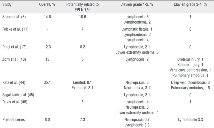
Extending PLND template may increase the risk of complications: this have to be coun-terbalanced by potential benefits. Overall and EPLND-related complications during RARP se-ries are reported in Table-5. As expected the most frequently reported complication in literature was lymphocele formation, proportional with the number of lymph nodes removed (10.3% with >10 nodes versus 4.6% with <10 nodes) (8, 12, 14). Previous reports showed complications’ rates for limited and standard PLND ranging from 2 to 9.8%, while EPLND complication’s rates vary from 19.8% to 75% (15, 20). Seventy-five percent of complications in EPLND may be due to extensive dissection of lymphatic tissue along the external
iliac artery that primarily supplies the lower extre-mities. Moreover, this area has never been shown to be affected by metastases in previous anatomi-cal studies (7, 8).
To reduce or to prevent PLND-associated morbidities, advices regarding meticulous sur-gical technique have been provided by several authors (8, 9): first, all the lymphatic vessels co-ming from the lower extremities should be tied by using ligatures instead of clips. In our experience a Hem-o-lok clip only is placed just cranially to the Cloquet’s lymph node, preserved in order to prevent lymphocele. Second, all lymphatics lateral to the external iliac artery should be spared in or-der to prevent lymphoedema. Third, two drainages should be placed, one per side of the pelvis and should not be removed until the total amount of fluid drained is below 50mL per 24 hours.
In the present prospective study we repor-ted our experience with RAEPLND. Transperitone-al approach was chosen in Transperitone-all cases Transperitone-allowing an excellent working space. Our median number of
Table 5 - Overall and EPLND-related complications in RARP series.
Study Overall, % Potentially related to EPLND,%
Clavien grade 1-2, % Clavien grade 3-4, %
Stone et al. (8) 14.6 10.6 Lymphocele, 6 Lymphoedema, 2
1
Feicke et al. (11) - 7 Lymphatic fistula, 1 Lymphoedema, 2
Lymphocele, 4
0
Patel et al. (17) 12.3 8.2 Lymphocele, 2.1 Lower extremity oedema, 3
0
Zorn et al. (18) 13 5 Lymphocele, 2 Ureteral injury, 1 Bladder injury, 1 Vena cava compression, 1
Pulmonary embolus, 1 Katz et al. (44) 35.1 Limited: 8.1
Extended: 3.1
Neuropraxia, 3 Neuropraxia, 3.1
Deep vein thrombosis, 3 Pulmonary embolus, 1.6 Sagalovich et al. (45) - - Lymphocele, 2.1 0
Davis et al. (46) - 5 Lymphocele, 4 Neuropraxia, 3 Lower extremity oedema, 4
1
Present series 8.0 7.3 Neuropraxia 0.7 Lymphocele 3.3
lymph nodes removed was 25, in line with publi-shed data on open series (1, 3, 17, 26-30) and com-parable to the similar study published by Feicke et al. on robot-assisted approach who reported a me-dian number of lymph nodes removed of 19 (11).
Among the 68 metastatic lymph nodes, 15 (22.05%) of them were localized either in the hy-pogastric or presacral regions (both not included in the limited PLND). In the vast majority, they were associated to the presence of lymph node metastases in the external/obturator iliac area. In our series, we observed just one patient (5.26%) with positive lymph nodes in bilateral presacral region only and two patients (10.52%) with hypo-gastric artery positive nodes only.
A dedicated comment about this finding is needed. It could seem this paper is actually an ar-gument for not doing EPLND due to such a large number needed to treat (51:1) in order to record a benefit in undergoing EPLND (just 3 patients out of 153, 1.96%, exclusively found metastasis in hypo-gastric/presacral region). Moreover we underline that, in 5 patients having metastatic iliac-obtura-tor lymph-nodes, 11 positive hypogastric/presacral region lymph-nodes were retrieved (3.26%). If we sponsor the therapeutic role of lymph-nodes dis-section, this is an important finding. On the other side, we daily perform PLND in all patients, with a probability of lymph-nodal metastasis according to Briganti updated nomogram over 5%, which is not so different from this case-study percentages. If we add that our described technique was safe and that EPLND consumed just half an hour in the overall operative time, we believe that this is the right direction. Moreover, by performing hy-pogastric/presacral lymph-nodes removal, we are convinced about the fact that, in case of bioche-mical recurrence, further exams such as total body choline-PET will be more reliable.
After more than 100 procedures we suggest a possible solution to the most important surgical matter: the complete reconstruction of peritoneum at the end of RARP, in order to avoid communica-tion between the peritoneal cavity and retropubic space is paramount. We underline that the bila-teral peritoneal incisions above the iliac vessels are not deliberately sutured in order to facilitate lymphatic reabsorption by the peritoneum.
We believe that EPLND-related compli-cations occur due to the fact that peritoneal end extraperitoneal space remains in communication at the end of the procedure, and this is particu-larly true for transperitoneal laparoscopic (pure or robot-assisted) approach.
The strength of our technique is the ana-tomical reconstruction of the two operative fields, that will be separated again thanks to the perito-neum reconstruction and the previous sparing of prevesical fascia.
The potential advantages are: first, the avoided risk of lymphatic leakage into the retropu-bic extraperitoneal space; second, the displacement of bowel loops into the retropubic space is avoi-ded; third, eventual future surgeries are facilitated thanks to preserved and separated anatomical spa-ces. How were we able to remove a high number of lymph nodes in a relatively short operative time? Again thanks to our experience, we here underli-ne some crucial technical steps: first, on the right side, ureter should be identified and then it should always remain inside intraoperative view; in case of any doubts, it should be suspended; second, on the left side, surgeon should know that ureter will not be sufficiently mobilized by medicalization of peritoneum only: indeed it is paramount to sus-pend it on this side; third, left iliac vessels anatomy is different: hypogastric vein partially covers sa-crum: for this reason presacral lymp-nodes dissec-tion is more challenging on this side.
Even if we were not able to perform long--term functional evaluation, our experience taught us that careful presacral lymph nodes dissection (thanks to robotic-system optical magnification) allows better functional outcomes.
for unreporting functional outcomes: they will be object of future researches.
CONFLICT OF INTEREST
None declared.
REFERENCES
1. Heidenreich A, Ohlmann CH, Polyakov S. Anatomical extent of pelvic lymphadenectomy in patients undergoing radical prostatectomy. Eur Urol. 2007; 52:29-37.
2. Clark T, Parekh DJ, Cookson MS, Chang SS, Smith ER Jr, Wells N, et al. Randomized prospective evaluation of extended versus limited lymph node dissection in patients with clinically localized prostate cancer. J Urol. 2003; 169:145-7.
3. Schumacher MC, Burkhard FC, Thalmann GN, Fleischmann A, Studer UE. Is pelviclymph node dissection necessary in patients with a serum PSA<10ng/mL undergoing radical prostatectomy for prostate cancer? Eur Urol. 2006; 50:272-9.
4. Golimbu M, Morales P, Al-Askari S, Brown J. Extended pelvic lymphadenectomy for prostatic cancer. J Urol. 1979; 121:617-20.
5. Joslyn SA, Konety BR. Impact of extent of lymphadenectomy on survival after radical prostatectomy for prostate cancer. Urology. 2006; 68:121-5.
6. Jeschke S, Burkhard FC, Thurairaja R, Dhar N, Studer UE. Extended lymph node dissection for prostate cancer. Curr Urol Rep. 2008; 9:237-42.
7. Allaf ME, Palapattu GS, Trock BJ, Carter HB, Walsh PC. Anatomical extent of lymph node dissection: impact on men with clinically localized prostate cancer. J Urol. 2004; 172:1840-4.
8. Stone NN, Stock RG, Unger P. Laparoscopic pelvic lymph node dissection for prostate cancer: comparison of the extended and modified techniques. J Urol. 1997; 158:1891-4.
9. Sivalingam S, Oxley J, Probert JL, Stolzenburg JU, Schwaibold H. Role of pelvic lymphadenectomy in prostate cancer management. Urology. 2007; 69:203-9.
10. Burkhard FC, Studer UE. The role of lymphadenectomy in high risk prostate cancer. World J Urol. 2008; 26:231-6. 11. Feicke A, Baumgartner M, Talimi S, Schmid DM, Seifert HH,
Müntener M, et al. Robotic-assisted laparoscopic extended pelvic lymph node dissection for prostate cancer: surgical technique and experience with the first 99 cases. Eur Urol. 2009; 55:876-83.
12. Kawakami J, Meng MV, Sadetsky N, Latini DM, Duchane J, Carroll PR; et al. Changing patterns of pelvic lymphadenectomy for prostate cancer: results from CaPSURE. J Urol. 2006; 176:1382-6.
13. Briganti A, Larcher A, Abdollah F, Capitanio U, Gallina A, Suardi N, et al. Updated nomogram predicting lymph node invasion in patients with prostate câncer undergoing extended pelvic lymph node dissection: the essential importance of percentage of positive cores. Eur Urol. 2012; 61:480-7.
14. Thompson I, Thrasher JB, Aus G, Burnett AL, Canby-Hagino ED, Cookson MS, et al. AUA Prostate Cancer Clinical Guideline Update Panel. Guideline for the management of clinically localized prostate cancer: 2007 update. J Urol. 2007; 177:2106-31.
15. Klevecka V, Musch M, Roggenbuck U, Stoerkel S, Kroepfl D. The incidence of lymph node metastases in prostate carcinoma depends not only on tumor characteristics but also on surgical performance and extent of pelvic lymphadenectomy. Medicina (Kaunas). 2008; 44:601-8. 16. Menon M, Shrivastava A, Kaul S, Badani KK, Fumo M,
Bhandari M, et al. Vattikuti Institute prostatectomy: contemporary technique and analysis of results. Eur Urol. 2007; 51:648-57.
17. Patel VR, Thaly R, Shah K. Robotic radical prostatectomy: outcomes of 500 cases. BJU Int. 2007; 99:1109-12. 18. Zorn KC, Gofrit ON, Orvieto MA, Mikhail AA, Zagaja GP,
Shalhav AL. Robotic-assisted laparoscopic prostatectomy: functional and pathologic outcomes with interfascial nerve preservation. Eur Urol. 2007; 51:755-62.
19. Bogdanovic J, Sekulic V. Re: Alberto Briganti, Umberto Capitanio, Felix K.-H. Chun, et al. Impact of surgical volume on the rate of lymph node metastases inpatients undergoing radical prostatectomy and extended pelvic lymph node dissection for clinically localized prostate cancer. Eur Urol 2008; 54:794-804.
20. Kazaryan AM, Røsok BI, Edwin B. Morbidity assessment in surgery: refinement proposal based on a concept of perioperative adverse events. ISRN Surg. 2013; 2013:625093.
21. Dindo D, Demartines N, Clavien PA. Classification of surgical complications: a new proposal with evaluation in a cohort of 6336 patients and results of a survey. Ann Surg. 2004; 240:205-13.
22. Briganti A, Blute ML, Eastham JH, Graefen M, Heidenreich A, Karnes JR, et al. Pelvic lymph node dissection in prostate cancer. Eur Urol. 2009;55:1251-65.
24. Weingärtner K, Ramaswamy A, Bittinger A, Gerharz EW, Vöge D, Riedmiller H. Anatomical basis for pelvic lymphadenectomy in prostate cancer: results of na autopsy study and implications for the clinic. J Urol. 1996; 156:1969-71.
25. Ploussard G, Briganti A, de la Taille A, Haese A, Heidenreich A, Menon M, et al. Pelvic lymph node dissection during robot-assisted radical prostatectomy: efficacy, limitations, and complications-a systematic review of the literature. Eur Urol. 2014; 65:7-16.
26. Heidenreich A, Varga Z, Von Knobloch R. Extended pelvic lymphadenectomy in patients undergoing radical prostatectomy: high incidence of lymph node metastasis. J Urol. 2002; 167:1681-6.
27. Wyler SF, Sulser T, Seifert HH, Ruszat R, Forster TH, Gasser TC, et al. Laparoscopic extended pelvic lymph node dissection for high-risk prostate cancer. Urology. 2006; 68:883-7.
28. Touijer K, Rabbani F, Otero JR, Secin FP, Eastham JA, Scardino PT, et al. Standard versus limited pelvic lymph node dissection for prostate cancer in patients with a predicted probability of nodal metastasis greater than 1%. J Urol. 2007; 178:120-4.
29. Sagalovich D, Calaway A, Srivastava A, Sooriakumaran P, Tewari AK. Assessment of required nodal yield in a high risk cohort undergoing extended pelvic lymphadenectomy in robotic-assisted radical prostatectomy and its impact on functional outcomes. BJU Int. 2013; 111:85-94.
30. Davis JW, Shah JB, Achim M. Robot-assisted extended pelvic lymph node dissection (PLND) at the time of radical prostatectomy (RP): a video-based illustration of technique, results, and unmet patient selection needs. BJU Int. 2011; 108:993-8.