CASE REPORT
Intrasellar Rupture of a Paraclinoid Aneurysm
with Subarachnoid Hemorrhage: Usefulness of
MR Imaging in Diagnosis
M. Ribeiro P. Howard R. Willinsky K. ter Brugge R. Agid L. Thines L. da Costa
SUMMARY:Characterization of paraclinoid aneurysms may be difficult because of the complexity of anatomic structures involved, and differentiation between intradural and extradural lesions is crucial. We report a case of a patient with a unique presentation of a paraclinoid aneurysm with intrasellar hemorrhage in which the presence of intrasellar blood and the relationship of the paraclinoid aneurys-mal neck and sac to the dural rings were elegantly demonstrated on MR imaging and were critical in choosing the target lesion for treatment.
T
he anatomy of the paraclinoid region is complex, and sometimes it is hard to properly characterize aneurysms arising in that region. Differentiation between an intradural and extradural location is of the utmost importance because it may determine patient prognosis and management strategy. Intracavernous internal carotid artery (ICA) aneurysms usu-ally have a benign natural history, and treatment is reserved for patients with unbearable pain or progressive neurologic deficits. An intradural aneurysm carries a risk for subarach-noid hemorrhage and may require treatment. We report an unusual case of multiple intracranial aneurysms including a ruptured paraclinoid carotid aneurysm presenting with intra-sellar bleeding followed by subarachnoid extension.Case Report
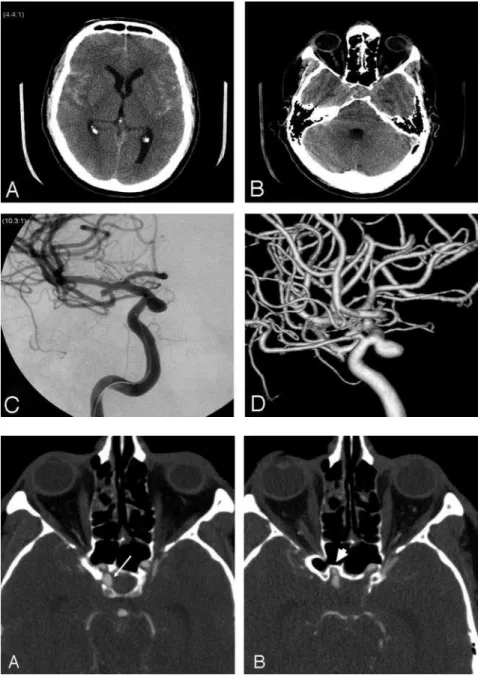
A 45-year-old man presented with good clinical grade subarachnoid hemorrhage. CT revealed blood in the basal cisterns and Sylvian fis-sures with symmetric distribution (Fig 1A,B). A digital subtraction angiography (Fig 1C, D) and a CT angiogram (CTA) demonstrated 2 aneurysms, a larger one arising from the right paraclinoid carotid and a smaller one at the right middle cerebral artery (MCA) bifurcation. The neck of the paraclinoid aneurysm could be seen at the level of the optic strut, with the sac pointing posteriorly and inferomedially, in close relationship with the sella turcica. (Fig 2). MR imaging was performed with the hope of being able to clarify if the paraclinoid aneurysm was intradural or extradural; results showed the aneurys-mal neck to be intradural and its dome below the dural ring, extra-dural. In addition, an enlarged sella turcica was noted, with inferior displacement of the pituitary gland and a large amount of blood filling the sella and extending into the subarachnoid space (Fig 3). We con-cluded that the paraclinoid aneurysm had ruptured into the sella with subarachnoid extension. The paraclinoid aneurysm was successfully coiled.
Discussion
The establishment of either an intradural or extradural loca-tion of paraclinoid aneurysms has critical implicaloca-tions for treatment and prognosis. Extradural aneurysms have a good prognosis and very low risks for a subarachnoid hemorrhage and major neurologic complications.1Treatment is reserved
for symptomatic lesions or for those lesions with extension into the subarachnoid space.2Intradural aneurysms carry a
risk for subarachnoid hemorrhage and may require treatment. Most paraclinoid aneurysms can be localized by their direc-tion of projecdirec-tion on angiograms.3 However, the complex
anatomy of the juxtadural ring area and individual variability in this region can sometimes make it difficult to define the relationship of the aneurysm to the dural ring, which differen-tiates extradural (below the dural ring) and intradural (above the dural ring) lesions.
Different techniques with several anatomic references have been used to differentiate intradural from extradural aneu-rysms. The simplest method uses the ophthalmic artery as the landmark for the transition of the extradural and intradural location. The major drawback of this method is that the origin of the ophthalmic artery is extradural in 10% of cases,4leading
to the erroneous assumption that an aneurysm is intradural when it is intracavernous. In our patient, the aneurysm arises distal to the ophthalmic artery, but its inferior direction places it below the plane of this artery (Fig 1D).
A reliable landmark for identification of the proximal dural ring, which defines the roof of cavernous sinus, is the optic strut.4-6Lesions above this level are considered intradural, and
below, intracavernous, extradural. In our case, the neck of the aneurysm is located at the exact level of the optic strut, and its fundus projects downward and medially, into the sella turcica. The aneurysm could be classified as a carotid cave aneurysm, arising between the 2 dural rings and growing into the cavern-ous sinus. The carotid cave is a pouch located in the postero-medial side of the distal dural ring,7above the proximal ring,
which may or may not have a communication with the sub-arachnoid space.
The diffuse subarachnoid hemorrhage demonstrated on CT could have been caused by either lesion, so it was impor-tant to determine if the proximal aneurysm was intradural or extradural. If the paraclinoid aneurysm was extradural, our treatment choice for the MCA aneurysm (Fig 1C) would have Received November 20, 2007; accepted December 20.
From the Department of Neuroradiology (M.R.), Hospital Sa˜o Marcos, Braga, Portugal; and Department of Medical Imaging (P.H., R.W., K.t.B., R.A., L.d.C.) and Division of Neurosur-gery (L.T.), Department of SurNeurosur-gery, Toronto Western Hospital, University of Toronto, Toronto, Ontario, Canada.
Please address correspondence to Manuel Ribeiro, MD, Hospital Sa˜o Marcos, Largo Engenheiro Carlos Amarante, Apartado 2242, Braga, Portugal, 4701–965; e-mail: [email protected]
DOI 10.3174/ajnr.A1022
been clipping (small size, relatively wide neck), but if it was intradural, our first choice of treatment would be coiling. CT and CTA alone were insufficient to define the location of the paraclinoid aneurysm and did not reveal the large intrasellar hemorrhage. In retrospect, after MR imaging revealed the in-trasellar hemorrhage, inin-trasellar blood could be suspected on the plain CT head (Fig 1B).
MR imaging at 3T was used to visualize the dural ring8and showed that the neck of the aneurysm was intradural and its dome, clearly extradural. More importantly, it revealed a large amount of blood inside an enlarged sella turcica, confirming that the paraclinoid aneurysm had bled. The large amount of intrasellar blood, the relatively small volume of blood in the subarachnoid space, and the empty sella syndrome lead to the hypothesis that the aneurysm had bled primarily into the sella and secondarily through an incompetent diaphragma sellae into the subarachnoid space. Absence of a bony septum in the posteromedial segment of the carotid cave9allows direct
con-tact of the pulsatile aneurysmal dome with the dura, which
eventually could cause its progressive erosion and the intrasel-lar hemorrhage. Advanced MR imaging techniques have im-proved our ability to identify the ruptured lesion in cases of multiple intracranial aneurysms, where MR imaging may identify a parenchymal clot adjacent to an aneurysm and re-cently has been used to determine an intradural or extradural location in unruptured paraclinoid aneurysms.
Conclusion
We report the case of a patient with a unique presentation of a paraclinoid aneurysm with intrasellar hemorrhage. Lesions located in the paraclinoid carotid can be difficult to clearly define as intradural or extradural. In this case, localization of the paraclinoid lesion (intradural vs extradural) and identifi-cation of the aneurysm responsible for the subarachnoid hem-orrhage were critical in choosing a treatment strategy. The presence of intrasellar blood and the relationship of the para-clinoid aneurysm to the dura elegantly demonstrated on MR
Fig 1.A, Axial unenhanced CT shows diffuse symmetric
subarachnoid hemorrhage.B, Questionable hyperattenuation at the level of the sella turcica is difficult to interpret given adjacent streak artifact.C, Frontal view of right internal carotid angiogram shows MCA bifurcation and paraclinoid aneurysms.D, 3D right ICA angiogram in lateral view reveals the paraclinoid aneurysm arising distal to the ophthalmic artery but pointing inferiorly below the plane of the ophthal-mic artery.
Fig 2.A, Axial CT angiography shows the sac of the
para-clinoid aneurysm (arrow) adjacent to the sella, without inter-vening septum.B, At the level of the superior margin of the optic strut (arrowhead), the aneurysmal neck and body extend posteriorly from the ICA.
INTERVENTIONAL
CASE
REPORT
imaging were critical in choosing the target lesion for treatment.
References
1. Kupersmith MJ, Stiebel-Kalish H, Huna-Baron R, et al.Cavernous carotid an-eurysms rarely cause subarachnoid hemorrhage or major neurologic morbid-ity.J Stroke Cerebrovasc Dis2002;11:9 –14
2. Linskey ME, Sekhar LN, Hirsch WL Jr, et al.Aneurysms of the intracavernous carotid artery: natural history and indications for treatment.Neurosurgery
1990;26:933–37
3. Nutik SL.Subclinoid aneurysms.J Neurosurg2003;98:731–36
4. Beretta F, Sepahi AN, Zuccarello M, et al.Radiographic imaging of the distal
dural ring for determining the intradural or extradural location of aneu-rysms.Skull Base2005;15:253– 61
5. Gonzalez LF, Walker MT, Zabramski JM, et al.Distinction between paracli-noid and cavernous sinus aneurysms with computed tomographic angiogra-phy.Neurosurgery2003;52:1131–39
6. Hashimoto K, Nozaki K, Hashimoto N.Optic strut as a radiographic landmark in evaluating neck location of a paraclinoid aneurysm. Neurosurgery
2006;59:880 –95
7. Oikawa S, Kyoshima K, Kobayashi S.Surgical anatomy of the juxta-dural ring area.J Neurosurg1998;89:250 –54
8. Thines L, Gauvrit JY, Leclerc X, et al.Usefulness of MR imaging for the assess-ment of nonophthalmic paraclinoid aneurysms.AJNR Am J Neuroradiol
2008;29:125–29
9. Hitotsumatsu T, Natori Y, Matsushima T, et al.Micro-anatomical study of the carotid cave.Acta Neurochir (Wien)1997;139:869 –74
Fig 3.Coronal T1- and T2-weighted 3T MR imaging through
anterior (A,C) and posterior (B,D) sella.C, Junction of aneu-rysmal neck and body (*), which projects partly superior to dural ring (interface with CSF) and partly inferior to the dural ring (interface with blood within sella). Note the large sella turcica filled with blood (arrows) showing homogeneous hypointensity on T2-weighted images and isointensity on T1-weighted images, consistent with intracellular deoxyhe-moglobin. Note that the blood in the suprasellar cisterns has different signal intensity.D, The infundibulum can be traced to the compressed pituitary.