www.jped.com.br
ORIGINAL ARTICLE
Timing in resolution of left heart dilation according to the degree
of mitral regurgitation in children with ventricular septal defect
after surgical closure
夽
Hwa Jin Cho
a, Jae Sook Ma
a, Young Kuk Cho
a, Byoung Hee Ahn
b, Kook Joo Na
b,
In Seok Jeong
b,∗aDepartment of Pediatrics, Chonnam National University Hospital, Chonnam National University Medical School, Gwangju, South
Korea
bDepartment of Thoracic and Cardiovascular Surgery, Chonnam National University Hospital, Chonnam National University
Medical School, Gwangju, South Korea
Received 19 March 2013; accepted 5 June 2013 Available online 16 October 2013
KEYWORDS
Ventricular septal defect;
Mitral regurgitation; Left heart dilation; Echocardiography; Children
Abstract
Objective: children with ventricular septal defects (VSD) can have chronic volume overload, which can result in changes of left heart echocardiographic parameters. To evaluate the changes before and after surgical closure, the children were divided into three groups according to the degree of mitral regurgitation (MR), and their echocardiographic characteristics were reviewed at serial follow-up after surgical closure.
Methods: the preoperative, and one-, three-, and 12-month postoperative echocardiographic data of 40 children who underwent surgical closure of VSD were retrospectively reviewed. Left ventricular end-diastolic volume (LVEDV), left ventricular end-diastolic dimension (LVEDD), left ventricular end-systolic dimension (LVESD), mitral valvular characteristics, including degree of MR and mitral valve annulus, and left atrial (LA) characteristics, including volume and dimensions, were observed.
Results: preoperative LVEDV, LVEDD, LVESD, mitral valvular annulus, LA volume, and LA dimen-sions were significantly larger in children with MR. Additionally, there were significant decreases in LVEDV, LVEDD, LA volume, and LA dimensions at one, three, and 12 months postoperatively. The degree of MR also improved to a lower grade after surgical closure of the VSD without additional mitral valve repair.
Conclusion: the echocardiographic parameters of left heart dilation and MR in children with VSD improved within the first year after surgical closure without additional mitral valve repair. Furthermore, in all of the patients with VSD, regardless of MR, LA dilation was reduced within
夽 Please cite this article as: Cho HJ, Ma JS, Cho YK, Ahn BH, Na KJ, Jeong IS. Timing in resolution of left heart dilation according to the
degree of mitral regurgitation in children with ventricular septal defect after surgical closure. J Pediatr (Rio J). 2014;90:71---7.
∗Corresponding author.
E-mail:[email protected] (I.S. Jeong).
three months after surgical closure of the VSD; however, LV and mitral valve annular dilatation decreased within 12 months.
© 2013 Sociedade Brasileira de Pediatria. Published by Elsevier Editora Ltda. All rights reserved.
PALAVRAS-CHAVE
Defeito do septo ventricular;
Regurgitac¸ão mitral; Dilatac¸ão do corac¸ão esquerdo;
Ecocardiografia; Crianc¸as
Momento da resoluc¸ão da dilatac¸ão do corac¸ão esquerdo segundo o grau de regurgitac¸ão mitral em crianc¸as submetidas a fechamento cirúrgico de defeito do septo ventricular
Resumo
Objetivo: crianc¸as com defeito do septo ventricular (DSV) podem apresentar sobrecarga de volume crônica, que pode resultar em mudanc¸as nos parâmetros ecocardiográficos do corac¸ão esquerdo. Para avaliar as mudanc¸as antes e depois do fechamento cirúrgico, as crianc¸as foram divididas em 3 grupos segundo o grau de regurgitac¸ão mitral (RM) e suas características eco-cardiográficas foram analisadas com acompanhamento em série após o fechamento cirúrgico. Método: revisamos retrospectivamente os dados ecocardiográficos de 40 crianc¸as submetidas a fechamento cirúrgico de DSV antes da cirurgia e nos meses 1, 3 e 12 após a cirurgia. Observamos o volume diastólico final do ventrículo esquerdo (VDFVE), dimensão diastólica final do ventrículo esquerdo (DDFVE) e dimensão sistólica final do ventrículo esquerdo (DSFVE), características da válvula mitral, incluindo grau de RM e o anel da válvula mitral, e características do átrio esquerdo (AE), incluindo volume e dimensões.
Resultados: os resultados para VDFVE, DDFVE, DSFVE, anel da válvula mitral, volume do AE e dimensões do AE foram significativamente maiores em crianc¸as com RM. Além disso, não houve reduc¸ão significativa no VDFVE, DDFVE, volume do AE e nas dimensões do AE nos meses 1, 3 e 12 após a cirurgia. O grau de RM também apresentou melhoria para um grau menor após o fechamento cirúrgico do DSV sem reparo adicional da válvula mitral.
Conclusão: os parâmetros ecocardiográficos de dilatac¸ão do corac¸ão esquerdo e a RM em crianc¸as com DSV haviam apresentado melhora no primeiro ano após o fechamento cirúrgico sem reparo adicional da válvula mitral. Além disso, em todos os pacientes com DSV, indepen-dentemente de RM, a dilatac¸ão do AE reduziu em três meses após o fechamento cirúrgico do DSV; contudo, a dilatac¸ão do VE e do anel da válvula mitral reduziu em 12 meses.
© 2013 Sociedade Brasileira de Pediatria. Publicado por Elsevier Editora Ltda. Todos os direitos reservados.
Introduction
It is known that left-to-right shunting in ventricular septal defects (VSD) generally increases pulmonary arterial blood flow and pulmonary venous return to the left heart. This pathophysiologic sequela may result in volume overload of the left atrium (LA) and left ventricle (LV), and subsequent LV enlargement, mitral annular dilation, mitral regurgita-tion (MR), and consequent LA enlargement to allow for the homeostatic balance of LA pressure.1---3 In the natural course of these changes after surgical closure, it has been demonstrated that the left ventricular end-diastolic volume (LVEDV) returns to normal within the first 2 years of life. However, the left atrial volume (LAV) remains elevated.4 The natural course of MR in children with VSD has also been studied, and it is believed that MR in children with a normal mitral valve (MV) apparatus and hemodynamically large VSD resolves spontaneously after the surgical closure of VSD.5 However, limited information is available on the relationship between MR and left heart volume overload.
The hypothesis of the present study was that a higher degree of MR is associated with more severe left heart dila-tion, and that the reversibility of the myocardium damage might take a longer period with a more severe degree of left heart dilation. Thus, this study aimed to investigate the
timing in resolution of left heart dilation according to the degree of MR in children who underwent surgical closure of VSD.
Materials and methods
Subject characteristics
three groups at serial follow-up after surgical closure of VSD were retrospectively reviewed.
Assessment of MR and left heart echocardiographic changes
MR was classified as none, trivial, mild, moderate, or severe based on qualitative color flow mapping. The changes in the degree of MR preoperatively and at one, three, and 12 months postoperatively in those who had MR before the surgical closure of VSD were also reviewed.
The following parameters through echocardiography at approximately one month preoperatively were reviewed: type of VSD, sizes and peak Doppler gradients of the VSD, and degree of MR. Body weight, height, body surface area (BSA), and postoperative survival were also examined. At all times, including approximately one month preoperatively and at one, three, and 12 months postoperatively, the following parameters were evaluated in echocardiography: LV charac-teristics, including the LV end-diastolic dimension (LVEDD), LV end-systolic dimension (LVESD), and LV end-diastolic vol-ume (LVEDV), MV annulus, LA end-systolic dimensions, and LA end-systolic volume (LA volume). The changes in echocar-diographic parameters over serial follow-up times were then evaluated and compared among the three groups. LVEDD and LVESD were obtained by M-mode echocardiography, and they were converted to Z-scores. The LVEDD Z-score (LVEDD-Z) and LVESD Z-score (LVESD-Z) were calculated as the number of standard deviations from the mean value of the normal population relative to the BSA. The LVEDV was indexed to the BSA and defined as the LVEDV index. The mitral valve annulus was measured from the apical four-chamber view at mid-diastole, and the data were converted to Z-scores for inter-individual comparisons. The dimensions of three planes of linear LA were also measured: the parasternal long axis (PLAX), lateral (LAT), and superoinferior (SI).6 The LA volume was measured following recommendations for chamber quantification7 by the following equation: LA volume = 4/3×(PLAX/2)×(LAT/2)×(SI/2). All linear LA dimensions and LA volume to the BSA were indexed and defined as the PLAX index, LAT index, SI index, and LAV index.
Statistical analyses
The chi-squared test and one-way analysis of variance (ANOVA) with Tukey’s post-hoc analysis were used to test for differences in subject characteristics among the three groups of MR. Two-way repeated measures ANOVA with Tukey’s post-hoc analysis was used to compare the left heart echocardiographic parameters among three groups of MR according to the time. The Statistical Package for Social Sci-ences (SPSS) version 20.0 (SPSS Inc. - Chicago, IL, USA) was used for all data analyses.
Results
Subject characteristics
A total of 40 patients with VSD met the inclusion criteria: 16 patients had VSD with no preoperative MR, 15 patients had
4
3
Degree of mitral regurgitation
2
1
0
Preoperatively One month postoperatively
Three months postoperatively
12 months postoperatively
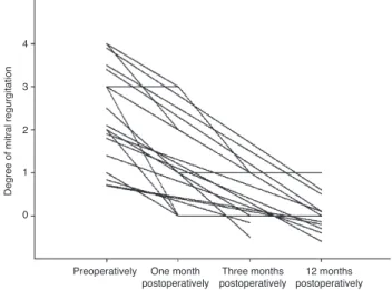
Figure 1 The regression of mitral regurgitation (MR) to various extents preoperatively through one, three, and 12 months postoperatively. 0 = no MR, 1 = trivial MR, 2 = mild MR, 3 = moderate MR, 4 = severe MR.
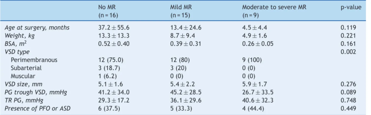
trivial to mild MR, and nine patients had moderate to severe MR. In the group with no MR, 12 patients (75.0%) had per-imembranous VSD, three (18.7%) had subarterial VSD, and one (6.2%) had outlet muscular VSD. In the group with triv-ial to mild MR, 12 patients (80.0%) had perimembranous VSD and three (20.0%) had subarterial VSD. In the group with moderate to severe MR, all nine patients (100.0%) had per-imembranous VSD (p = 0.002). There were no statistically significant differences in age, body weight, BSA, pressure gradient through VSD, pressure gradient of tricuspid regur-gitation, or the presence of ASD or PFO among the three groups (Table 1). There was no postoperative mortality.
Regression in degree of MR
Patients who did not have MR preoperatively did not progress to new-onset MR after surgical closure of the VSD. All patients improved regarding the degree of MR. In the mild MR group (n = 15), 11 patients had trivial MR and four had mild MR preoperatively; at one month postopera-tively (n = 15), 12 patients were resolved and three patients improved to a trivial degree of MR; at three months postop-eratively (n = 12), 11 patients were resolved and one patient remained with trivial MR until 12 months after surgery. In the moderate to severe MR group (n = 9), six patients had moderate MR and three had severe MR preoperatively; at one month postoperatively (n = 9), MR had resolved in three patients, improved to trivial in three patients, decreased to moderate in two patients, and one patient remained with moderate MR. At three months (n = 7), four patients improved to trivial and three patients remained without MR. At 12 months, follow-up data were available for six patients; among them, MR remained trivial in two patients (Fig. 1).
Left heart echocardiographic characteristics
Table 1 Subject characteristics according to the grade of mitral regurgitation.
No MR (n = 16)
Mild MR (n = 15)
Moderate to severe MR (n = 9)
p-value
Age at surgery, months 37.2±55.6 13.4±24.6 4.5±4.4 0.119
Weight, kg 13.3±13.3 8.7±9.4 4.9±1.6 0.221
BSA, m2 0.52±0.40 0.39±0.31 0.26±0.05 0.161
VSD type 0.002
Perimembranous 12 (75.0) 12 (80) 9 (100)
Subarterial 3 (18.7) 3 (20) 0 (0)
Muscular 1 (6.2) 0 (0) 0 (0)
VSD size, mm 5.1±1.6 5.4±2.2 5.9±1.7 0.276
PG trough VSD, mmHg 41.2±34.0 45.2±28.5 26.7±33.5 0.089
TR PG, mmHg 29.3±17.2 36.1±29.6 40.6±32.3 0.748
Presence of PFO or ASD 6 (37.5) 5 (33.3) 4 (44.4) 0.449
ASD, atrial septal defects; BSA, body surface area; MR, mitral regurgitation; PG, pressure gradient; PFO, patent foramen ovale; TR, tricuspid regurgitation; VSD, ventricular septal defect.
All data are presented as mean±standard deviation or n (%).
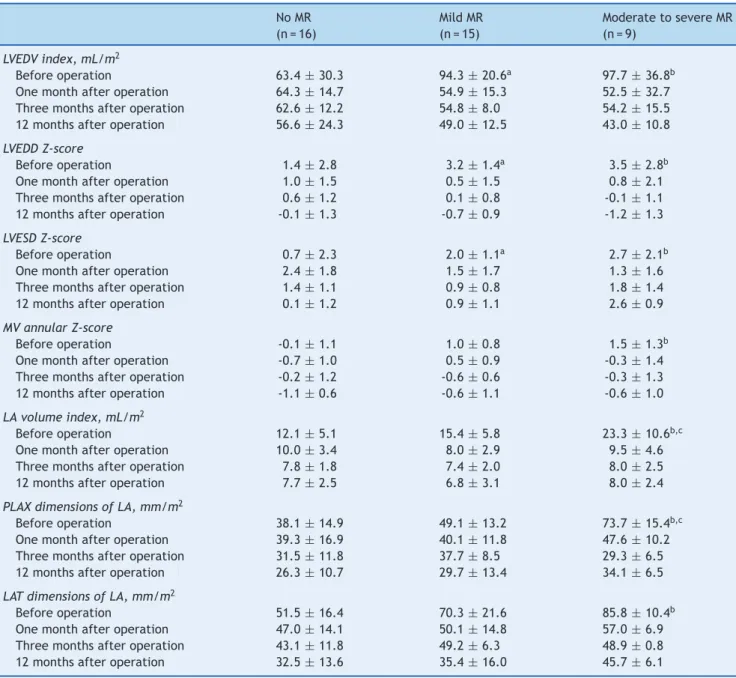
in the mild MR group (p = 0.034, p = 0.034, and p = 0.039, respectively) and moderate to severe MR group (p = 0.036, p = 0.035, and p = 0.020, respectively) than in the no MR group.
The preoperative MV annular Z-score in the moderate to severe MR group was significantly larger than that in the no MR group (p = 0.038).
There was a significant difference in the LA volume index and PLAX index of the moderate to severe MR group com-pared with that of the no MR group (p = 0.008, p = 0.001, respectively) and mild MR group (p = 0.047, p = 0.006, respectively) preoperatively.
The preoperative LAT index in the moderate to severe MR group was significantly higher than that in the no MR group (p = 0.001). There was no significant differ-ence in the echocardiographic values of LV, MV annulus, and LA parameters among the groups at any subsequent time.
Serial changes in postoperative left heart echocardiographic parameters
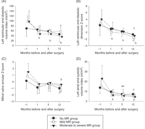
No groups showed any significant decrease in the LVEDV index, LVEDD-Z, LVESD-Z, and MV annulus Z-score at any time following closure of the VSD. The mild MR group demonstrated a significant reduction in the LVEDV index and LVEDD-Z at one month (p < 0.001, p < 0.001, respectively), three months (p = 0.004, p < 0.001, respectively), and 12 months postoperatively (p = 0.002, p < 0.001, respectively) when compared with one month preoperatively. The moder-ate to severe MR group demonstrmoder-ated a significant reduction in the LVEDV index and LVESD-Z only at 12 months postop-eratively (p = 0.035, p = 0.003, respectively) compared with one month preoperatively. There were significant reductions in LVEDD-Z at three months and 12 months postoperatively (p = 0.043, p = 0.027, respectively). There were no significant intergroup differences (Fig. 2A, B).
The mild MR group demonstrated a significant reduc-tion in the MV annular Z-score 12 months postop-eratively (p = 0.028) when compared with one month
preoperatively. There were no significant intergroup differ-ences (Fig. 2C).
The no MR group showed a significant decrease in the LA volume index only at three months (p = 0.023) follow-ing closure of the VSD. The mild MR group demonstrated a significant reduction in the LA volume index at one month (p = 0.011), three months (p = 0.020), and 12 months (p = 0.006) postoperatively when compared with one month preoperatively. The moderate to severe MR group demon-strated a significant decrease at three months (p = 0.021) and 12 months (p = 0.015). There was also a significant inter-group difference between the no MR inter-group and the moderate to severe MR group (p = 0.011), and between the mild MR group and the moderate to severe MR group (p = 0.027) (Fig. 2D).
The no MR group showed a significant decrease in the PLAX index at one month (p = 0.023) and three months (p = 0.014) postoperatively. The mild MR group demonstrated a significant reduction in the PLAX index at three months (p = 0.030), six months (p < 0.001), and 12 months (p < 0.001) postoperatively when compared with one month preoper-atively. The moderate to severe MR group also showed a significant decrease at one month (p = 0.034), three months (p = 0.006), and 12 months (p = 0.002). There was also a significant intergroup difference between the no MR and moderate to severe MR groups (p = 0.019)
The no MR group showed a significant decrease in the LAT index at three months (p = 0.041) and 12 months (p = 0.005) following closure of the VSD. The mild MR and the moderate to severe MR groups demonstrated a significant reduction in the LAT index at one month (p < 0.001, p = 0.002, respec-tively), three months (p = 0.003, p < 0.001, respecrespec-tively), and 12 months (p < 0.001, p < 0.001, respectively) postop-eratively when compared with one month preoppostop-eratively. There was also a significant intergroup difference between the groups with no MR and with moderate to severe MR (p = 0.028).
Table 2 Left heart echocardiographic parameters before and at one, three, and 12 months after surgical closure of ventricular septal defect, according to the degree of mitral regurgitation.
No MR (n = 16)
Mild MR (n = 15)
Moderate to severe MR (n = 9)
LVEDV index, mL/m2
Before operation 63.4±30.3 94.3±20.6a 97.7±36.8b
One month after operation 64.3±14.7 54.9±15.3 52.5±32.7 Three months after operation 62.6±12.2 54.8±8.0 54.2±15.5 12 months after operation 56.6±24.3 49.0±12.5 43.0±10.8
LVEDD Z-score
Before operation 1.4±2.8 3.2±1.4a 3.5±2.8b
One month after operation 1.0±1.5 0.5±1.5 0.8±2.1 Three months after operation 0.6±1.2 0.1±0.8 -0.1±1.1 12 months after operation -0.1±1.3 -0.7±0.9 -1.2±1.3
LVESD Z-score
Before operation 0.7±2.3 2.0±1.1a 2.7±2.1b
One month after operation 2.4±1.8 1.5±1.7 1.3±1.6 Three months after operation 1.4±1.1 0.9±0.8 1.8±1.4 12 months after operation 0.1±1.2 0.9±1.1 2.6±0.9
MV annular Z-score
Before operation -0.1±1.1 1.0±0.8 1.5±1.3b
One month after operation -0.7±1.0 0.5±0.9 -0.3±1.4 Three months after operation -0.2±1.2 -0.6±0.6 -0.3±1.3 12 months after operation -1.1±0.6 -0.6±1.1 -0.6±1.0
LA volume index, mL/m2
Before operation 12.1±5.1 15.4±5.8 23.3±10.6b,c
One month after operation 10.0±3.4 8.0±2.9 9.5±4.6 Three months after operation 7.8±1.8 7.4±2.0 8.0±2.5 12 months after operation 7.7±2.5 6.8±3.1 8.0±2.4
PLAX dimensions of LA, mm/m2
Before operation 38.1±14.9 49.1±13.2 73.7±15.4b,c
One month after operation 39.3±16.9 40.1±11.8 47.6±10.2 Three months after operation 31.5±11.8 37.7±8.5 29.3±6.5 12 months after operation 26.3±10.7 29.7±13.4 34.1±6.5
LAT dimensions of LA, mm/m2
Before operation 51.5±16.4 70.3±21.6 85.8±10.4b
One month after operation 47.0±14.1 50.1±14.8 57.0±6.9 Three months after operation 43.1±11.8 49.2±6.3 48.9±0.8 12 months after operation 32.5±13.6 35.4±16.0 45.7±6.1
LA, left atrium; LAT, lateral; LVEDD, left ventricular end-diastolic dimension; LVEDV, left ventricular end-diastolic volume; LVESD, left ventricular end-systolic dimension; MR, mitral regurgitation; MV, mitral valve; PLAX, parasternal long axis; SI, superior-inferior. All data are presented as mean±standard deviation.
a p < 0.05, VSD with mild MRvs. no MR.
b p < 0.05, VSD with moderate to severe MRvs. no MR. c p < 0.05, VSD with moderate to severe MRvs. mild MR.
when compared with one month preoperatively. There were no significant intergroup differences.
Discussion
In the present study, the LV, MV annulus, and LA dilation were evaluated by measuring the LVEDV, LVEDD, MV annulus, LA dimensions, and LA volume. It was observed that all of the echocardiographic parameters associated with left heart dilation decreased regardless of MR within one year.
LV dilation
160
(A) (B)
(C) (D)
140
120
100
80
60
40
20
8
6
4
2
0
–2
–4 1
b b
b
c
b b
b c
3 12 0
–1 –1 1 3 12
Left v
e
ntricular end-diastolic
volume index (ml/m
2)
Left v
e
ntricular end-diastolic
dimension Z-score
Months before and after surgery Months before and after surgery
20
10
0 30 40
ac b
b b b
c 1
1 3
3 2
12 0
–1
–1 –1 1 3 12
–2
–3
Mitral valve annular Z-score
Left atrial end-systolic volume index (ml/m 2)
Months before and after surgery Months before and after surgery
No MR group Mild MR group
Moderate to severe MR group
Figure 2 Changes in echocardiographic data in the three groups according to time: before the operation, and at one, three, and 12 months after ventricular septal defect operation (expressed as -1, 1, 3, and 12, respectively).A, left ventricular end-diastolic volume index (mL/m2).B, left ventricular end-diastolic dimension Z-score.C, mitral valve annular Z-score.D, left atrial volume
index (mL/m2). ap < 0.05vs. one month preoperatively in the group with no mitral regurgitation (MR). bp < 0.05vs. one month
preoperatively in the group with trivial to mild MR.cp < 0.05vs. one month preoperatively in the group with moderate to severe
MR.
study concerning the reversibility of left ventricular dilation demonstrated that children with moderately large VSD and LV volume overloads without pulmonary hypertension or con-gestive heart failure experienced a spontaneous decrease of LV dilation.8 In the present study, with a chronic left to right shunt, LVEDV, LVEDD, and LVESD were significantly greater in patients with MR compared to those without MR. After removal of the shunt burden, there were no statisti-cal differences among the three groups at any of the serial follow-up times. In addition, the LVEDV and LVEDD decreased significantly after surgical closure at all of the follow-up times in children with VSD and trivial to mild MR.
MV annular dilation and MR
MV annular dilation is considered to be a physiologic sequela to volume overload to the left heart, and MR develops sec-ondary to annular dilation.9Hisatomi et al.10reported cases of children with VSD who underwent MV repair, and con-cluded that if MR develops secondary to annular dilation, there is no need for MV repair. In contrast, Honjo et al.11 have also reported on 17 children who underwent MV repair, among whom were five patients with VSD, two with ASD, two with left ventricular diverticulum, one with partial anoma-lous pulmonary venous return, and one with coarctation of the aorta and VSD. They concluded that MV repair is feasi-ble and presents a low reoperation rate. However, Mahadin
et al.5stated that a more aggressive approach to MV repair would do harm to growing children if the natural course of MR was not fully recognized. They suggested that MR with a normal mitral valve apparatus improves after surgi-cal closure of VSD, and that MV repair should be taken under careful consideration. In this study, the MV annular Z-scores were measured and compared according to the degree of MR. The MV annulus was significantly larger in children with moderate to severe MR compared to those without MR. In addition, the MV annulus was found to decrease significantly after surgical closure in those with trivial to mild MR within one year. The present findings support the fact that children with VSD experience restoration to their normal valvular competency and the coaptation zone after surgical closure of VSD without MV repair.5It was also observed that MR was reversible after surgical closure of VSD, and this happened mostly within the first year after surgery. In particular, the degree of MR decreased within one month of surgical clo-sure. Based on these findings, an aggressive approach to MV repair in children with VSD is not necessary, considering the natural course of MR.
LA dilation
recently developed an LA volume-tracking method, and highlighted the importance of measuring LA volume in patients with chronic LV volume overload. Cordell et al.4 have measured LVEDV and maximal LA volume by catheter-ization, and demonstrated that LVEDV returned to normal after surgical closure of VSD; however, the maximal LA vol-ume remained elevated. They indicated this as a permanent change in the elasticity of LA. In contrast to that study, in the present study echocardiography was used. The dimen-sions of LA at the parasternal long axis view and at the apical four-chamber view were measured, the LA volume was cal-culated using the recommended formula and then indexed to the BSA. The preoperative LA volume indexed to the BSA was significantly larger in children with VSD and moderate to severe MR than in those with a lesser degree of MR. In contrast to other parameters of LV and MV annular dilata-tion, which did not show a significant decrease in children without MR at any subsequent time postoperatively, the LA volume and dimensions decreased significantly within three months after surgical closure in all degrees of MR, including VSD with no MR. There was no difference in the chamber size at any time after surgery relative to the degree of MR. The present study had a relatively small number of reviewed patients, especially among those with moderate to severe MR. This is due to the practice adopted in this institu-tion to not wait for surgery if the degree of MR is higher than mild to moderate. In addition, follow-up data were missing for nine of the 40 subjects at three months after surgery, and for seven of 40 subjects at 12 months after surgery. Quali-tative methods were used to determine the degree of MR, and misrepresentation of the severity is thus possible.
This study is the first to assess the serial changes in left heart echocardiographic parameters before surgery and at one, three, and 12 months after surgical closure in pre-dicting the natural course in children with surgical closure of VSD. The echocardiographic parameters, including the LVEDD, LVEDV, MV annulus, LA dimensions, and LA volume showed significant differences according to the degree of MR. Also, in all patients with VSD, regardless of MR, LA dila-tion was reduced within three months after surgical closure of the VSD; however, LV and MV annular dilatation decreased within 12 months, which tended to take more time only in those with MR. Further studies are needed to determine the reason for different times required for the resolution of LV, MV, and LA dilation.
Funding
This study was supported by a grant (CRI 13024-1) from the Chonnam National University Hospital Research Institute of Clinical Medicine.
Conflicts of interest
The authors declare no conflicts of interest.
References
1. Kizer JR, Bella JN, Palmieri V, Liu JE, Best LG, Lee ET, et al. Left atrial diameter as an independent predictor of first clinical cardiovascular events in middle-aged and elderly adults: the Strong HeartStudy (SHS). Am Heart J. 2006;151: 412---8.
2. Senzaki H, Kumakura R, Ishido H, Masutani S, Seki M, Yoshiba S. Left atrial systolic force in children: reference values for nor-mal children and changes in cardiovascular disease with left ventricular volume overload or pressure overload. J Am Soc Echocardiogr. 2009;22:939---46.
3. Ueda Y, Fukushige J, Ueda K. Congestive heart failure during early infancy in patients with ventricular septal defect relative to early closure. Pediatr Cardiol. 1996;17:382---6.
4. Cordell D, Graham Jr TP, Atwood GF, Boerth RC, Boucek RJ, Bender HW. Left heart volume characteristics following ven-tricular septal defect closure in infancy. Circulation. 1976;54: 294---8.
5. Mahadin DR, Srivastava S, Parness IA, Nguyen K, Love BA, Walsh R, et al. Outcomes of mitral regurgitation associated with large ventricular septal defect and a normal mitral valve appara-tus: does intact atrial septum have an impact? Pediatr Cardiol. 2011;32:1128---31.
6. Aurigemma GP, Gottdiener JS, Arnold AM, Chinali M, Hill JC, Kitzman D. Left atrial volume and geometry in healthy aging: the Cardiovascular Health Study. Circ Cardiovasc Imag-ing. 2009;2:282---9.
7. Lang RM, Bierig M, Devereux RB, Flachskampf FA, Foster E, Pellikka PA, et al. Recommendations for chamber quantifica-tion: a report from the American Society of Echocardiography’s Guidelines and Standards Committee and the Chamber Quan-tification Writing Group, developed in conjunction with the European Association of Echocardiography, a branch of the Euro-pean Society of Cardiology. J Am Soc Echocardiogr. 2005;18: 1440---63.
8. Kleinman CS, Tabibian M, Starc TJ, Hsu DT, Gersony WM. Spontaneous regression of left ventricular dilation in chil-dren with restrictive ventricular septal defects. J Pediatr. 2007;150:583---6.
9. Papadimitriou JM, Hopkins BE, Taylor RR. Regression of left ventricular dilation and hypertrophy after removal of volume overload. Morphological and ultrastructural study. Circ Res. 1974;35:127---35.
10. Hisatomi K, Isomura T, Sato T, Kosuga K, Ohishi K, Katoh H. Mitral valve repair for mitral regurgitation with ventri-cular septal defect in children. Ann Thorac Surg. 1996;62: 1773---7.
11. Honjo O, Ishino K, Kawada M, Akagi T, Sano S. Midterm outcome of mitral valve repair for congenital mitral regurgi-tation in infants and children. Interact Cardiovasc Thorac Surg. 2006;5:589---93.
12. Ogawa K, Hozumi T, Sugioka K, Iwata S, Otsuka R, Takagi Y, et al. Automated assessment of left atrial function from time-left atrial volume curves using a novel speckle tracking imaging method. J Am Soc Echocardiogr. 2009;22:63---9.