Clinicoradiological Session
Case 2/2011 - Forty-Three Months Old Child, Male, with Multiple
Ventricular Septal Defect, Unfavorable Clinical Course, Long Term
After Anatomical Correction
Edmar Atik
Hospital Sírio-Libanês, São Paulo, SP - Brazil
Mailing address: Edmar Atik •
Rua Dona Adma Jafet, 74 conj.73 - Bela Vista - 01308-050 - São Paulo, SP - Brazil
E-mail: eatik@cardiol.br, conatik@incor.usp.br
Manuscript received July 29, 2010; revised mansucript received January 13, 2011; accepted January 13, 2011.
Keywords
Congenital heart diseases, ventricular septal defect, heart failure, heart murmurs.
Diagnosis
Multiple ventricular septal defects in severe heart failure despite effective pulmonary banding.
Conduct
Upon the surgery, under prolonged cardiopulmonary bypass of 105 minutes, three large ventricular septal defects were closed, one in the ventricular inlet, with 10 mm, another in the muscular trabecular part, of 7 mm, both through the right atrium, and an apical one with 12 mm, by incision at the apical part of the left ventricle. The pulmonary artery debanding was performed with end-to-end anastomosis.
The postoperative course was complicated by low cardiac output that required mechanical ventilation for 4 days, vasoactive drugs for 06 days and peritoneal dialysis for 07 days. Atrioventricular block also complicated the picture with spontaneous reversal on day 14, and patient was discharged on the 16th day after the surgery.
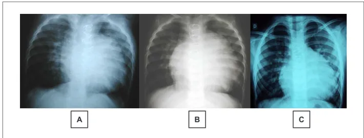
In the long term, evaluated after 14 and 26 months after the surgery, the presence of heart murmur from residual ventricular septal defect was clear, as confirmed by echocardiography with 4 mm and 2 mm each, besides the persistence of heart failure with fatigue, cardiomegaly (despite decreased cardiac area compared to the pre-operative period - Figure 1) and hepatomegaly 4cm from the right costal edge. Despite these findings, the child performs well tolerated physical activity and under use of furosemide, spironolactone, captopril, and carvedilol. Echocardiogram reveals increased left cavities and left ventricular dysfunction (fractional shortening of myocardial fiber of 27.0% and ejection fraction of 52.0%).
Notes
Even corrected, the multiple ventricular septal defects may have multiple unfavorable outcomes, as in the case presented. This outcome is due to several factors, such as severe systolic and diastolic overload on the heart before surgery for a long time, though relieved by pulmonary artery banding at 6 months, prolonged extracorporeal circulation, placement of multiple patches for closure of defects and surgical incision at the apical portion of the left ventricle. Evolution in a longer term is unknown, but it is possible to infer until improvement is achieved, as long as the continuity of medication can reverse the dysfunction found. This anomaly continues to be challenging, despite the latter inference is presumptively positive.
Clinical aspects
Since birth, persistent dyspnea was observed, even after pulmonary artery banding performed with 06 months of age. Moreover, low weight gain also motivated the correction of the defect with 16 months of age. Upon physical examination at that time, he was dyspneic, acyanotic and pulses were normal. His weight was 7,580 g, with a height of 74 cm. Precordium presented pronounced bulging, with mild systolic impulses and pectus carinatum (pigeon chest). Apical impulse was diffuse in the fourth and fifth intercostal spaces. There was systolic murmur and thrill in the third and fourth intercostal spaces at the left sternal edge irradiating to the upper sternal edge. The second sound was accentuated and split. The liver was palpable two centimeters in the right costal edge.
Complementary tests
Electrocardiogram revealed signs of severe biventricular overload and left anterior hemiblock.
Chest radiography revealed marked increase in heart size and pulmonary vasculature. The middle arch was also prominent (Figure 1).
Echocardiogram showed multiple ventricular septal defects in the ventricular inlet with 6 mm, 7 mm in the trabecular part and 10 mm in the apical section. The shunt was bidirectional and the pulmonary artery banding had pressure gradient of 77 mmHg. The left cavities were very dilated (Figure 2).
Clinico Radiological Session
Edmar Atik
Arq Bras Cardiol 2011; 96(2): e18-e19 Figure 1 -Chest radiography in the preoperative period (A), 14 months (B) and 26 months (C) after correction showed variable cardiomegaly with tendency to decrease, observed in the last radiography.
A
B
C
Figure 2 -Preoperative echocardiogram emphasizes the presence of large ventricular septal defects in the inlet, in the trabecular part, and in the apical portion (arrows). Abbreviations: RA - right atrium; LA - left atrium; RV - right ventricle; and LV - left ventricle.
