• Nenhum resultado encontrado

Arq. Bras. Cardiol. vol.98 número4 en v98n4a13

N/A
N/A
Protected

Academic year: 2018

Share "Arq. Bras. Cardiol. vol.98 número4 en v98n4a13"

Copied!
2
0
0

Texto

(1)

Clinicoradiological Session

Case 3/2012 - 16-Year Old Patient with Transposition of Great

Arteries and Pulmonary Stenosis in Natural Evolution

Edmar Atik

Instituto do Coração do Hospital das Clínicas da Faculdade de Medicina da Universidade de São Paulo, São Paulo, SP, Brazil

Clinical data: heart murmur, cyanosis and fatigue are observed from birth, with clear worsening in recent years. Without proper medical treatment, 83% hematocrit and hemoglobin concentration of 28 g were recently found. Repeated hemapheresis decreased hematocrit to 73%. No specific medication had been administered since birth.

Physical examination: Regular general condition, eupneic, marked cyanosis, normal pulses. Weight: 42.6 kg; height: 160 cm; BP: 105/65 mmHg; HR: 82 bpm, RR: 20 rpm. O2 saturation: 75%. The aorta was discreetly palpated at the suprasternal notch.

Precordial systolic impulsions were discrete, sharp systolic thrill across the left sternal border. Apical impulse was not palpable. Heart sounds were very loud and systolic murmur was sharp throughout the left sternal border with radiation to the aortic area. The liver was not palpable and the lungs were clear.

Complementary tests

Electrocardiogram showed sinus rhythm and signs of sharp right ventricular overload with rsR’s’ morphology and negative T wave in V1; abrupt transition of the QRS complex from V1 to V2; along with right atrial overload with P wave peaked with 3 mm amplitude in D2. AP: +60o, AQRS: +170o, AT:

+20o (Fig. 1).

Chest radiography showed cardiac area within normal limits, with rounded morphology. The middle arch is excavated and the pulmonary vasculature is slightly increased (Fig. 1).

Echocardiography (fig. 2) showed very hypertrophied cardiac chambers, especially the right ventricle. The atrioventricular valves were normal and inflow ventricular septal defect extending to the outflow measured 17 mm with bidirectional shunt. The biventricular function was preserved. The posterior pulmonary valve was thickened and had hypoplasia of the pulmonary ring. Straddling the anterior mitral valve at the top of the ventricular septum was clear. The measures were RV=22, LV=42, TV=29, MV=25, RPA=16, LPA=19 mm.

Magnetic resonance imaging showed the same aspects with ventricular septal defect of 19 mm; increased atria; Vd2RV = 90 ml/m2 and Vd2LV=112 ml/m2; RVEF = 62%; LVEF = 61%.

The ventricular septum measured 12 mm and the posterior wall, 7 mm. Pulmonary stenosis was severe.

Cardiac catheterization was not performed given the appropriate diagnostic conclusion by other tests.

Clinical diagnosis: transposition of great arteries with ventricular septal defect and pulmonary stenosis, and straddling of mitral valve at the top of the ventricular septum in severe hypoxia.

Clinical reasoning: The clinical elements led to cyanotic congenital heart disease of transposition of great arteries due to accentuated second sound in the presence of ventricular septal defect murmur and pulmonary stenosis radiating to the aortic area. Radiographic elements such as increased pulmonary blood flow and excavated middle arch led to the diagnosis of transposition of great arteries associated with the other defects mentioned. The elements of other images were decisive for the diagnosis of abnormalities.

Differential diagnosis: Other heart diseases that are associated with pulmonary stenosis should also be considered in the clinical differentiation, such as right ventricular outflow, double inlet ventricle and other similar functional anomalies.

Conduct: There were doubts as to the corrective technique to be employed considering the severe right ventricular hypertrophy, a limiting element in directing the left ventricle into the aorta through ventricular septal defect along with placing a tube in the right ventricle to the pulmonary trunk. However, as the MRI had revealed appropriately sized right ventricular cavity, the Rastelli technique was performed with good initial progress. Valve straddling was not an impediment to directing the flow from the left ventricle into the aorta and aortic homograft of 20 mm from the right ventricle to the pulmonary arteries. Cardiopulmonary bypass and anoxic period were prolonged, 4h and 3:20 h, respectively.

Comments: Among the anomalies associated with transposition of great arteries, ventricular septal defect and pulmonary stenosis are those which, when combined, allow evolution for a longer period. The evolution of this patient until 16 years of age without complications with no use of specific drugs attests this statement. Chronic hypoxia, however, was responsible for an exaggerated elevation of hematocrit at high risk of thrombosis and pulmonary stenosis to myocardial hypertrophy. These adverse evolutionary aspects entail adverse consequences even after corrective surgery, interfering with subsequent developments.

Mailing Address: Edmar Atik •

InCor - Av. Dr. Enéas Carvalho de Aguiar, 44 - 05403-000 - São Paulo, SP - Brazil

E-mail: conatik@incor.usp.br

Keywords

Transposition of great vessels; pulmonary valve stenosis; heart murmurs; anoxia.

(2)

Clinicoradiological Session

Atik Clinicoradiological Session

Arq Bras Cardiol 2012;98(4):e64-e65 Figure 2 - Echocardiogram shows marked right ventricular hypertrophy with reduced cavity in four-chamber section in A, subvalvar and annular pulmonary stenosis in

connection with the left ventricle in longitudinal section in B, anterior aorta larger than pulmonary trunk in suprasternal section in C, large inlow ventricular septal defect in four-chamber section with predominant low from left to right on color Doppler in D.

Figure 1 - Chest X-ray reveals normal cardiac area with rounded morphology, excavated middle arch and pulmonary vasculature increased in the pulmonary hila. Electrocardiogram reveals the signs of sharp right ventricular overload.

Imagem

Figure 1 -  Chest X-ray reveals normal cardiac area with rounded morphology, excavated middle arch and pulmonary vasculature increased in the pulmonary hila

Referências

Documentos relacionados

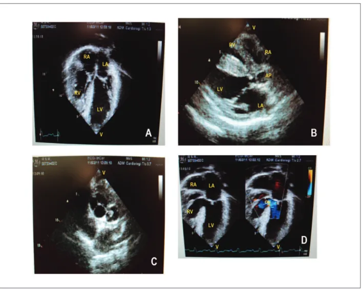
An 11-year-old female, 24.9 kg, with a diagnosis of transposition of great arteries with hypoplastic left ventricle and pulmonary stenosis, who underwent atrial septostomy

ches in the preoperative period, association with atrioventri- cular septal defect or agenesis of the pulmonary valve, significant residual ventricular septal defect, residual

In this study, we assessed the clinical evolvement, the echo- cardiographic, cineangiographic, and surgical characteristics of children with perimembranous ventricular septal defect

Based on the analysis of the cineangiocardiographic studies of patients with pulmonary atresia, ventricular septal defect, and aorto- pulmonary collateral arteries (group B),

Our management for patients with transposition of the great arteries with intact ventricular septum was to indicate systematic primary anatomic correction in all patients in

Clinical fi ndings strongly suggest the diagnosis of pulmonary atresia with ventricular septal defect because of the presence of an early systolic click in the aortic area and

The connection between the pulmonary arteries and the right ventricular outflow tract (RVOT) in pulmonary atresia (PA) with ventricular septal defect (VSD) may be corrected

Clinical findings are consistent with the diagnosis of atrial septal defect, considering the split second heart sound, the soft murmur in the pulmonary area, the right