Chronic pulmonary histoplasmosis mimicking
tuberculosis*
GISELA UNIS1, LUIZ CARLOS SEVERO2
Keywords: Histoplasmosis/diagnosis; Histoplasmosis/drug therapy; Histoplasma/isolation & purification; Tuberculosis, pulmonary/diagnosis; Itraconazole/drug therapy; Sputum/microbiology; Diagnosis, Differential
* Study conducted in the Mycology Laboratory of the Santa Casa Hospital Complex, Porto Alegre (RS) Brazil.
1. Doctoral student in the Postgraduate Pulmonary Medicine Program of the Universidade Federal do Rio Grande do Sul (UFRS, Rio Grande do Sul Federal University), Porto Alegre (RS) Brasil.
2. Researcher for CNPq.
Correspondence to: Dr. Luiz Carlos Severo. Laboratório de Micologia, Hospital Santa Rita, Santa Casa-Complexo Hospitalar. Annes Dias 285. CEP 90020-090. Fax: 55 51 3214-8435. E-mail: [email protected] Submitted: 30 April 2004. Accepted, after review: 16 September 2004.
ABSTRACT
Chronic pulmonary histoplasmosis mimicking tuberculosis 319
INTRODUCTION
The thermal dimorphic fungus Histoplasma capsulatum var. capsulatum causes various clinical manifestations depending on the anatomical and immunologic state of the host and on the quantity of fungal inoculum.(1-3) Chronic pulmonary
h i s t o p l a s m o s i s ( C P H ) i s a n o p p o r t u n i s t i c manifestation of the mycosis in pre-existing emphysematous air spaces of the lung parenchyma and caused by prolonged exposure to fungi. There are small amounts of fungi and limited tissue invasion. Parenchyma inflammation and the resulting necrosis and fibrosis represent an immune response of the host against the fungal antigens.(4)
In the present study, we report four new cases of CPH in the state of Rio Grande do Sul, and we review diagnostic aspects of other CPH cases published in the Brazilian literature.(5)
METHODS
Patient charts from the archives of the Laboratório de Micologia do Complexo Hospitalar Santa Casa de Porto Alegre (Mycology Laboratory of the Santa Casa Hospital Complex in Porto Alegre) were retrospectively reviewed regarding age, gender, race, epidemiological history, origin of referral, signs and symptoms, concomitant or predisposing conditions, treatment, evolution and complications. The criteria for the diagnosis of chronic pulmonary histoplasmosis and for the inclusion of cases in the study were: compatible clinical disease in individuals residing in the state of Rio Grande do Sul; laboratory evidence of histoplasmosis, that is, positive culture for H. capsulatum, histopathological findings showing fungal elements consistent with H. capsulatum or immunodiffusion test results revealing H or M bands; culture or histopathological evidence of H. capsulatum located exclusively in the lungs of patients presenting structural defects in lung anatomy.
Materials used for diagnosis were sputum, bronchoalveolar lavage fluid or lung biopsy sample. The smears on the slides were stained with silver methenamine according to the Grocott Gomori technique. Cultures were grown on Sabouraud agar (DIFCO, Detroit, MI, USA), 1% chloramphenicol (União Química Farmacêutica Nacional S.A., São Paulo, Brazil), and Mycosel® (BBL) media, processed in a model FVL, series 636 Class II biosafety laminar
flow hood (Trox do Brasil Ltda., São Paulo, Brazil), and incubated at 25ºC. Cultures testing positive for H. capsulatum var. capsulatum were confirmed by the micromorphological aspect (tuberculate macroconidia), and thermal dimorphism was characterized through conversion to the yeast phase on brain heart infusion (BHI) agar at 37ºC.
RESULTS
In the present study, the clinical charts of 212 patients diagnosed with histoplasmosis over a 25-year period (1997-2002) were obtained from the archives of the Mycology Laboratory of the Santa Casa Hospital Complex. Of these, ten files were eligible for evaluation. However, four of these had to be excluded because, although there were compatible clinical and radiological criteria, laboratory confirmation revealed positive results only for the immunodiffusion test. Of the six cases found, two have previously been reported in the literature,(2)
and four are reported in the present study: CASE 1- A 69-year-old male Caucasian, resident of the city of São Leopoldo, in the state of Rio Grande do Sul (RS) and a shoemaker, was admitted to the hospital with dyspnea, productive cough, fever and weight loss. The patient had been smoking 20 cigarettes a day for 47 years and had been diagnosed with chronic obstructive pulmonary disease a long time prior. He had also recently been diagnosed with diabetes mellitus due to the chronic use of corticoids. The patient had been submitted to empirical treatment for tuberculosis for six months, even including repeated negative sputum smear microscopy for acid-fast bacilli and tuberculin tests using the Mantoux technique. The progression of symptoms led to his hospitalization.
Epidemiological history: There was a chicken coop on his property.
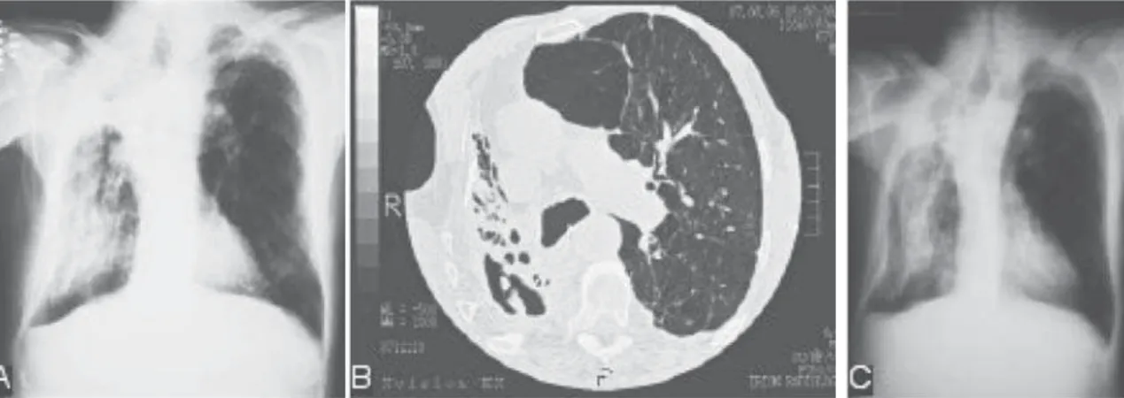
Imaging: Radiographic and tomographic findings indicating a mass with dense soft parts within the cavity in the upper right lobe and the post-treatment control thereof (Figure 1).
small, budding yeast-like organisms suggestive of H. capsulatum; culture was negative.
Treatment and evolution: Itraconazole (100 mg/ day) was given for four months. Five years later, there was exacerbation of the disease, and the patient received the same medication for an additional three years.
CASE 2 - A 63-year-old male Caucasian, resident of Novo Hamburgo, RS, a mason and a pack-a-day smoker since the age of 11 was hospitalized with progressive weight loss for nine months (14/62 kg), productive cough with hyaline sputum, progressive dyspnea, anorexia, asthenia and fever. The patient reported no hemoptysis, night sweats or chest pain. Symptoms began nine months prior to hospitalization. Physical examination revealed the patient was emaciated, dehydrated and pale. Three months later, the patient was discharged without a conclusive diagnosis and was treated for symptoms of chronic obstructive pulmonary disease. Due to worsening of clinical symptoms, the patient was rehospitalized eight months later for a second diagnostic investigation. Several sputum smear tests were carried out, and all were negative for acid-fast bacilli.
Epidemiological history: Some years prior, the patient had participated in the demolition of an old chicken coop, where bats resided.
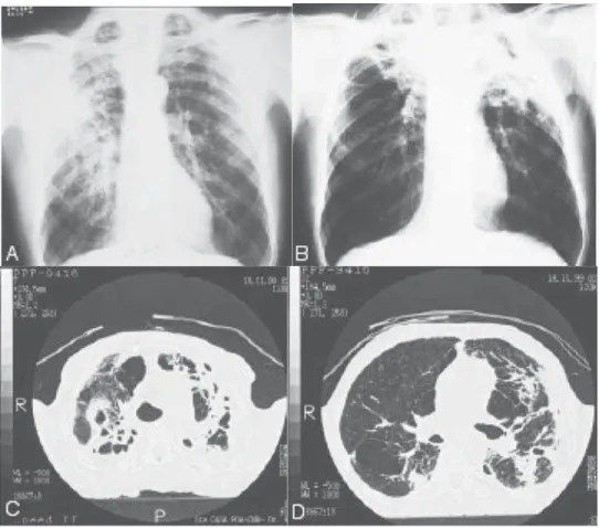
Imaging: Areas of consolidation of the axillary subsegments of the right lung, shown in Figure 2, as well as control of the evolution and the tomographic imaging.
Mycological evaluation: Immunodiffusion serology was positive for H. capsulatum, and the M band was present; immunodiffusion was negative for A. fumigatus; sputum staining was negative, and Grocott-Gomori silver methenamine staining of the biopsy sample was positive for H. capsulatum; the punch-biopsy sample of the lung revealed chronic inflammation in the lung parenchyma with accentuated fibrosis and a small focus of epithelioid granuloma with caseous necrosis; culture was positive for H. capsulatum.
Treatment and evolution: the patient was treated with itraconazole (200 mg/day); the patient regained his initial weight and was submitted to 18 months of treatment; the patient presented significant clinical improvement, and the antifungal treatment was suspended.
CASE 3 - A 53-year-old Caucasian male, resident of Sapucaia do Sul (RS), a security guard and pack-a-day smoker was hospitalized due to persistent fever, night sweats and weight loss (19 kg/3 months). Despite the fact that multiple sputum and bronchoalveolar lavage fluid smear tests were negative for acid-fast bacilli, the patient was submitted to empirical treatment for tuberculosis, to no effect.
Epidemiological history: The patient had been in contact with chicken droppings while working in an herb garden.
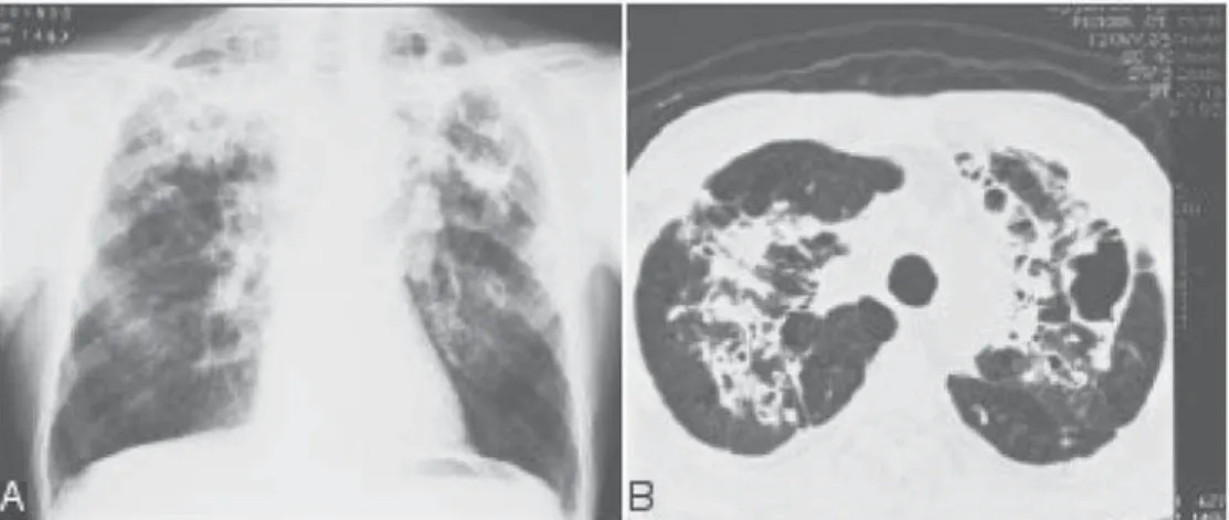
Imaging: Radiographic and tomographic aspects are shown in Figure 3.
Chronic pulmonary histoplasmosis mimicking tuberculosis 321
Mycological evaluation: Immunodiffusion serology was positive for H. capsulatum, and the M band was present; mycological examination of fresh sputum was negative; Grocott-Gomori silver methenamine staining of the sputum smear revealed small oval budding yeast-like organisms suggestive of H. capsulatum; sputum culture was negative for H. capsulatum on Sabouraud agar and positive for H. capsulatum on Mycosel®.
Treatment and evolution: The patient was given ketoconazole (400 mg/day) for a month, followed by itraconazole (100 mg/day) for nine months.
CASE 4 - A 38-year-old Caucasian male, a resident of Viamão, RS and a 10-cigarette-a-day smoker,
presented with a cough (including expectoration) and asthenia for three months.
Epidemiological history: Contact with a chicken coop. Imaging: Chest X-ray revealed fibronodular lesions, atelectasis and right-sided apical pleural thickening.
Mycological evaluation: Immunodiffusion serology was positive for H. capsulatum, and the M band was present; mycological test of fresh sputum was negative; Grocott-Gomori silver methenamine staining of the sputum smear revealed small oval budding yeast-like organisms suggestive of H. capsulatum; sputum culture was negative for H. capsulatum.
Treatment and evolution: Ketoconazole (200 Figure 2 - A) Chest X-ray showing areas of consolidation in the axillary subsegments of the right lung. B)
mg/day) was prescribed, and the patient was discharged. He did not return to the hospital.
Table 1 shows the diagnostic findings of the patients in this study, comparing them with the cases previously described in the literature. Table 2 shows demographic data, clinical profiles, treatment and evolution of the cases evaluated in the present
study, as well as of those previously described.
DISCUSSION
The first extensive study of CPH was conducted in tuberculosis sanatoriums involving patients suspected of being infected with tuberculosis.(6) Most
of the patients were initially submitted to treatment with tuberculostatic drugs prior to being diagnosed with mycosis, as was shown in 12 (92%) of the 13 cases of CPH with cavitation reported in another study.(7) We discovered that, in Brazil, the proportion
of patients submitted to empirical treatment for tuberculosis (50%) remains high, indicating that we tend to direct the diagnosis towards tuberculosis, which delays recognition of the clinical manifestation of mycosis, consequently allowing its progression.
The initial injury consists of interstitial pneumonitis, with frequent emphysematous air spaces, especially of the centrilobular or bullous type.(4) The colonization of these air spaces produces
fluids rich in fungal antigens that cause segmental pneumonitis due to bronchial dissemination. Later-stage injury is essentially characterized by a preexisting area of cavitation. Infected cavities tend to foster disease progression, with pulmonary fibrosis and exacerbation of respiratory insufficiency.(4) The
rapid change in the pattern of the lung parenchyma injury seen on the Case-2 chest X-ray is attributable Figure 3 - A) Chest X-ray showing poorly-defined opacities and possible cavities, predominantly in the
upper lobes; B) Computed tomography (CT) scan showing structural remodeling of both lungs, especially in the upper lobes, with areas of consolidation and atelectasis (mainly distributed along the axial interstices), as evidenced by bronchiectasis, cystoid cavities, parenchyma opacity and scarring-related emphysematous bullae. On the left side, cavities were larger and some contained septations or intracavitary vegetation. No ostensive lymph node enlargement was seen, although some lymph nodes were slightly more enlarged than usual.
TABLE 1
Mycological diagnosis
Case Sample GMS Culture IDh
LITERATURE
1(13) Sputum + + H, M
2(13) Sputum + + H, M
3(13) Sputum + + H, M
4(5) Sputum + + NF
BAL + +
Biopsy + +
5(5) Sputum + + M
Biopsy + +
PRESENT SERIES
6 Sputum - - M
(case 1) BAL +
-7
(case 2) Sputum - - M
Biopsy + +
8
(case 3) Sputum + + M
9
Chronic pulmonary histoplasmosis mimicking tuberculosis 323
on selective media during the first diagnostic investigation. Mycosel® and the similar product, Mycobiotic®, are selective media recommended for the isolation of dermatophytes. The inclusion of an antibiotic (chloramphenicol) and of an antifungal (cycloheximide) prevents the contamination of bacteria and fungi, allowing H. capsulatum, which has a slower growth rate, to be isolated.(10)
The recommended treatment for CPH is the administration of amphotericin B for severe cases (those requiring hospitalization), and itraconazole for mild and moderate cases.(11) Regardless of the
antifungal used, relapse may occur. In patients treated with itraconazole, especially if the drug is used for periods shorter than the currently recommended 12 to 24 months, relapse rates can range up to 15%.(11)
Patients in whom fungus ball is suspected and who present negative results on immunodiffusion tests may be submitted to serologic triage using the ELISA method for antigen detection.(12)
Only thirteen cases of CPH have been reported in Brazil.(5) Of these, five were CPH with cavitation: three
occurring in the state of Rio de Janeiro,(13) and two
in the state of Rio Grande do Sul.(5) These two states
TABlE 2
Chronic pulmonary histoplasmosis with cavitation in Brazil: clinical manifestations, comorbidities, treatment and evolution
Case Age, ET Symptoms Treatment Evolution; co-morbidities
Gender LITERATURE
1(13) 64, M No fever, productive cough Ketoconazole - 6m Epidermoid lung
Itraconazole - 7m carcinoma; Death Amphotericin B - 1g
2(13) 58, M No fever, productive cough Ketoconazole - 6m Death
Itraconazole - 22m
3(13) 74, M Yes productive cough, chest pain, Amphotericin B -1g No evolution
weight loss Itraconazole
4(5) 62, M No productive cough, hemoptysis, Ketoconazole - 7m fungus ball caused by fever, anorexia, dyspnea, Amphotericin B -1g A. fumigatus;
weight loss Ketoconazole - 30m postoperative death
5(5) 64, M Yes productive cough, fever, dyspnea, Ketoconazole - 18m Cure;
weight loss, night sweats Itraconazole - 13m fungus ball caused by A. fumigatus PRESENT SERIES
6 69, M Yes dyspnea, productive cough, fever, Itraconazole - 4m Cure;
(case 1) weight loss Itraconazole - 36m fungus ball caused by
A. fumigatus 7 63, M No dyspnea, productive cough, fever, Itraconazole - 18m Cure (case 2) anorexia, asthenia, weight loss
8 53, M Yes fever, night sweats, Ketoconazole - 1m Cure
(case 3) weight loss Itraconazole - 9m
ET: empirical treatment (for tuberculosis); M: male; m: months
to fibrotic retraction around the emphysematous bulla, which is a pattern characteristic of pulmonary histoplasmosis.(4)
Due to its high sensitivity, the immunodiffusion test is a good trial method for CPH, presenting positive results in virtually 100% of cases in which the diagnosis is later confirmed.(8) Diagnostic
confirmation is achieved through isolation of H. capsulatum in sputum or bronchoscopy samples in 60% to 85% of the cases if multiple samples are tested.(9) For definitive confirmation of the diagnosis,
clinical samples should be submitted to Grocott-Gomori silver methenamine staining and seeded on selective culture media.(8)
are recognizably endemic for histoplasmosis, presenting high rates of histoplasmin skin test positivity (93% and 89%, respectively),(14-15) and the
fungus can be easily isolated from the soil.(16-17) A
hypothetical diagnosis of CPH should be considered for patients with chronic obstructive pulmonary disease and exacerbation of symptoms whose epidemiologic history indicates the handling of soil contaminated with chicken droppings or bat guano. Serologic evaluation is extremely useful for diagnostic triage, as was observed in the present case study.
Cases of histoplasmosis preceding or concomitant to fungus ball have been reported, typically require immediate surgical treatment.(18) In most cases of CPH
with cavitation, pulmonary function has already been affected and, due to the presence of emphysematous bullae and fibrosis, and there are no cleavage plans for surgical excision. In such cases, surgery may accelerate the incidence of evident pulmonary insufficiency and mortality.(4) Itraconazole is a
treatment alternative.(19) Of the Brazilian patients, three
were diagnosed with fungus ball (Table 2), and two of these presented fungal colonization resulting from active histoplasmosis. Only one patient underwent surgery, which led to the death of that patient.(5)
We would like to highlight the fact that the presence of cavitary lesions in the upper lobes of patients with chronic obstructive pulmonary disease, especially those who present negative for acid-fast bacilli sputum smear microscopy and tuberculin skin test results and reside in endemic areas, should lead to a hypothetical diagnosis of CPH. In this group of patients, only 5% of the cases are likely to be infected with tuberculosis.(20) Hemoptysis in the presence of
late lesions or after the disease has been cured is indicative of Aspergillus colonization.
ACKNOWLEDGMENTS
The authors would like to thank Dr. Klaus Irion for reviewing the imaging and for providing the computed tomography scans of cases 1 and 2.
REFERENCES
1. Unis G, Roesch EW, Severo LC. Acute pulmonary histoplasmosis in the state of Rio Grande do Sul, Brazil. J Bras Pneumol 2005;31(1):52-9. Portuguese. 2. Unis G, Pegas KL, Severo LC. Pulmonary histoplasmoma in
Rio Grande do Sul. Rev Soc Bras Med Trop. 2005;38(1):11-4. Portuguese.
3. Unis G, Oliveira FM, Severo LC. Disseminated histoplasmosis in Rio Grande do Sul. Rev Soc Bras Med Trop. 2004;37(6):463-8. Portuguese.
4. Goodwin RA Jr, Owens FT, Snell JD, Hubbard WW, Buchanan RD, Terry RT et al. Chronic pulmonary histoplasmosis. Medicine (Baltimore). 1976;55(6):413-52. 5. Severo LC, Rizzon CF, Roesch EW, Oliveira FM, Porto NS. Chronic pulmonary histoplasmosis in Brazil: report of two cases with cavitation diagnosed by transthoracic needle biopsy. Rev Inst Med Trop S Paulo. 1997;39(5):293-7. 6. Furcolow ML, Brasher CA. Chronic progressive (cavitary)
histoplasmosis as problem in tuberculosis sanatoriums. Am Rev Tuberc.1956;73(5):609-19.
7. Loewen DF, Procknow JJ, Loosli CG. Chronic active pulmonary histoplasmosis with cavitation. A clinical and laboratory study of thirteen cases. Am J Med. 1960;28:252-80.
8. Wheat LJ. Laboratory diagnosis of histoplasmosis: update 2000. Semin Respir Infect. 2001;16(2):131-40. 9. Wheat LJ, Wass J, Norton J, Kohler RB, French MLV. Cavitary
histoplasmosis occurring during two large urban outbreaks. Analysis of clinical, epidemiologic, roentgenographic, and laboratory features. Medicine (Baltilmore). 1984;63(4):201-9. 1 0 . Unis G, Silva VB, Severo LC. Histoplasmose disseminada e SIDA. Importância do meio de cultivo para o espécime clínico-broncoscópico. Rev Soc Bras Med Trop. 2004;37 (3):234-7.
11. Wheat J, Sarosi G, McKinsey D, Hamill R, Bradsher R, Johnson P, et al. Practice guidelines for the management of patients with histoplasmosis. Infectious Diseases Society of America. Clin Infect Dis. 2000;30(4):688-95. 1 2 . Latgé JP. Aspergillus fumigatus and aspergillosis. Clin
Microbiol Rev. 1999;12(2):310-50.
13. Capone D, Wanke B, Monteiro PCF, Lazéra MS, Andrade GN, Valle ACF. Chronic pulmonary histoplasmosis in the state of Rio de Janeiro, Brazil. Mycopathologia. 1999; 145(2):75-9.
1 4 . Zembrzuski MM, Bassanesi MC, Wagner LC, Severo LC. I n q u é r i t o i n t r a d é r m i c o c o m h i s t o p l a s m i n a e paracoccidioidina em duas regiões do Rio Grande do Sul. Rev Bras Med Trop. 1996;29(1):1-3.
15. Fava SC, Fava Netto C. Epidemiologic surveys of histoplasmin and paracoccidioidin sensitivity in Brazil. Rev Inst Med Trop S Paulo. 1998;40(3):155-64. Portuguese. 16. Severo LC, Petrillo VF, Camargo JJ, Geyer GR, Porto NS.
Acute pulmonary histoplasmosis and first isolation of Histoplasma capsulatum from soil of Rio Grande do Sul, Brasil. Rev Inst Med Trop S Paulo. 1986;28(1):51-5. Portuguese.
17. Zancopé-Oliveira RM, Wanke B. Distribuição das fontes de infecção do Histoplasma capsulatum var. capsulatum em Rio da Prata - Município do Rio de Janeiro (RJ). Rev Inst Med Trop S Paulo. 1987;29(4):243-50.
18. Schwarz J, Baum GL, Straub M. Cavitary histoplasmosis complicated by fungus ball. Am J Med. 1961;31:692-700. 19. Campbell JH, Winter JH, Richardson MD, Shankland GS, Banham SW. Treatment of pulmonary aspergilloma with itraconazole. Thorax. 1991;46(11):839-41.