INTRODUCTION
Urologic surgery continues to evolve fo-cusing efforts on adequate treatment of patho-logic uropatho-logic conditions in a safe and minimally invasive manner (1).
Laparoscopic surgery has well defined benefits for the patient and has, over time, be-come accepted as a standard of care access strat-egy for the management of benign and malignant Urologic diseases.
Despite significant advances in laparo-scopic technique and technologies, laparolaparo-scopic Urologic surgery remains technically demanding. Unlike open surgery, at the end of laparoscop-ic extirpative procedures, the Urologist is often faced with the additional challenges of specimen retrieval and extraction.
Laparoscopic specimen entrapment and extraction occur at what is falsely considered the “end of the procedure”. During open surgery, after the specimen has been mobilized, the specimen is Despite significant advances in laparoscopic technique and technologies,
laparo-scopic Urologic surgery remains technically demanding regarding various surgical steps including the challenge of specimen retrieval and extraction, whether to in-stall a drainage system and the best option for wound closure. Laparoscopic speci-men entrapspeci-ment and extraction occurs at what is falsely considered the “end of the procedure”. During open surgery, after the specimen has been mobilized, the speci-men is simply lifted out of the larger incision which has been made to achieve the surgical objectives. In contrast, significant laparoscopic skill is required to entrap and safely extract laparoscopic specimens. Indeed, the Urologist and surgical team which are transitioning from open surgery may disregard this important part of the procedure which may lead to significant morbidity. As such, it is imperative that during laparoscopic procedures, the “end of the procedure” be strictly defined as the termination of skin closure and dressing placement. Taking a few minutes to focus on safe specimen entrapment and extraction will substantially reduce major mor-bidity. The following review focus on the technology and technique of specimen entrapment and extraction, on the matter of whether to install a drainage system of the abdominal cavity and the options for adequate closure of trocar site wounds. This article’s primary objectives are to focus on how to minimize morbidity while maintain the advantages of a minimally invasive surgical approach.
The final stage of the laparoscopic procedure: exploring
final steps
_______________________________________________
Ricardo A. Natalin, Fabio S. Lima, Thomé Pinheiro, Eugenio Vicari, Valdemar Ortiz, Cassio Andreoni,
Jaime Landman
Universidade Federal de Sao Paulo (RAN, FSL, TP, EV, VO, CA,) Sao Paulo, Brazil and Columbia University Medical Center (RAN, JL), New York, USA
ABSTRACT
ARTICLE
INFO
_______________________________________________________________ _____________________
Key words:
Laparoscopy; urology; kidney; outcomes
Int Braz J Urol. 2012; 38: 4-16
________________
Submitted for publication: March 21, 2011
________________
simply lifted out of the larger incision which has been made to achieve the surgical objectives. At this time the open surgical team is typically more relaxed, and may turn up the volume on what is commonly referred to as “closing music”.
In contrast, significant laparoscopic skill is required to entrap and safely extract laparoscop-ic specimens. Indeed, the Urologist and surglaparoscop-ical team which are transitioning from open surgery may disregard this important part of the proce-dure which may lead to significant morbidity. As such, it is imperative that during laparoscopic pro-cedures, the “end of the procedure” be strictly de-fined as the termination of skin closure and dress-ing placement.
The following review will focus on the technology and technique of specimen entrap-ment, extraction, and drainage of the abdominal cavity for safely exiting the abdomen. This arti-cle’s primary objectives are to focus on how to minimize morbidity while maintaining the advan-tages of a minimally invasive surgical approach.
Entrapment and retrieval devices
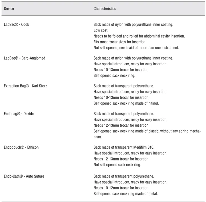
Important characteristics to be evaluated in a retrieval device are the sack permeability, re-sistance and sack stability inside the abdominal cavity (2). Clinical application of specimen retriev-al devices requires some characteristics in order to enable surgical manipulation and safety. The de-vice needs to be easily handled from trocar inser-tion, sack visibility, opening, closure and removal. A variety of different retrieval devices are commercially available, each one presenting par-ticular characteristics as show in Table-1.
Several technologic improvements have been made regarding the retrieval devices like the use of impermeable sack, used for both intact and fragmented specimen removal. The impermeable sack improves safety regarding port-site recur-rence as this rare but serious complication has been reported after organ retrieval without protec-tion. (3).
Cytologic washings from intact specimen retrieval devices sacks revealed that low-stage, low-grade tumors after minimal manipulation do not exfoliate cells into the retrieval sack.
Howev-er, higher grade and staged tumors may present different outcomes regarding cell exfoliation and should be properly treated (4).
Recently Ganpule et al. described a new entrapment and retrieval device, arguing it to be effective and with a lower cost, the Nadiad Bag. The bag is manually constructed by the use of a 5F ureteral catheter (Devon Industries, Banga-lore, India), a nylon thread and a polyethylene bag (Steribag, PCI, Kandivali, Mumbai, India); the bag is sealed at one end with an autoseal de-vice (Rainbow Manufacturers, Rajkot, India) and a tunnel is created around the open end of the bag to thread the ureteral catheter with the nylon thread. For specimen entrapment the device is in-serted through the working port; after specimen placement into the bag the ureteral catheter is re-moved and the nylon thread used to tight the bag. The authors found the device easy to make and to be deployed, effective in removing the surgi-cal specimen, with a low cost. However, attention should be made to the lack of permeability tests and stability tests and it should not be used for morcellation (5).
Alternative Entrapment Devices
Various approaches for benign specimen entrapment have been described in the literature. Raj et al. described an inexpensive alternative to specific specimen retrieval devices that achieves the same benefits but with very low cost. The new and low cost device utilizes a regular sterile latex glove with fingers extremities removed thus creat-ing a latex bag. This approach is a good alternative to specific retrieval devices that are usually expen-sive; however in the setting of oncologic disease treatment it should not be used due to the fragility of the sack and easy rupture with potential tumor seeding (6).
Table 1 - Laparoscopic specimen retrieval devices.
Device Characteristics
LapSac® - Cook Sack made of nylon with polyurethane inner coating. Low cost.
Needs to be folded and rolled for abdominal cavity insertion. Fits most trocar sizes for insertion.
Not self opened, needs aid of more than one instrument.
LapBag® - Bard-Angiomed Sack made of nylon with polyurethane inner coating. Have special introducer, ready for easy insertion. Needs 10-13mm trocar for insertion.
Self opened sack neck ring.
Extraction Bag® - Karl Storz Sack made of transparent polyurethane. Have special introducer, ready for easy insertion. Needs 10-13mm trocar for insertion.
Self opened sack neck ring made of nitinol.
Endobag® - Dexide Sack made of transparent polyurethane. Have special introducer, ready for easy insertion. Needs 12-13mm trocar for insertion.
Self opened sack neck ring made of plastic, without any spring mecha-nism.
Endopouch® - Ethicon Sack made of transparent Medifilm 810. Have special introducer, ready for easy insertion. Needs 12-13mm trocar for insertion.
Not self opened sack neck ring.
Endo-Cath® - Auto Suture Sack made of transparent polyurethane. Have special introducer, ready for easy insertion. Needs 10-12mm trocar for insertion.
Self opened sack neck ring made of metal.
low cost and easily performed by any laparo-scopic surgeon. However the authors have made an important observation of never using this ma-terial for morcellation because they are not leak-proof or strong enough (7).
These alternative entrapment devices pre-sented another important concern related to its primary purpose: they were not designed to be used with this indication; however they may be
used as prototype of new devices that will be spe-cially designed for surgical and medical purpose.
Specimen Retrieval
and if there is any other incision needed during surgery that could be used for this reason. The common extirpative Urologic surgical procedures include simple and radical nephrectomy, partial nephrectomy, nephroureterectomy, radical and simple prostatectomy, lymphadenectomy, adre-nalectomy, orchiectomy, and extraction of uri-nary calculi.
We have previously reported a laparo-scopic technique for large burden kidney stone, without the need of port site extension. A laparo-scopic pyelolithotomy is performed in a standard manner and after the stone is removed from the renal pelvis, it is allocated inside an Endocatch device (U.S. Surgical; Norwalk, CT) and an ultra-sonic lithotripter is used for stone fragmentation through a regular nephroscope, inside the perito-neal cavity. Stone fragments are removed along with the fluid aspiration. This technique is effec-tive in completely remove the calculi, maintain-ing the procedure in a complete minimally inva-sive approach, with no increased complication rates (8).
Options at the termination of extirpative Urologic procedures include intact extraction and specimen morcellation. With intact extraction, specimen removal can be achieved by trocar site extension, connecting existing trocar site inci-sions, or by incising prior abdominal scars or cre-ating a new incision. Transverse abdominal inci-sions are commonly chosen by surgeons because they achieve good cosmetics with potentially less pain if compared to incisions of other orientations (9). The use of retrieval devices facilitates either technique as with the Endocatch (U.S. Surgical; Norwalk, CT) for intact removal or the LapSac (Cook Urological; Spencer, IN) for the morcel-lated one. In a prospective study comparing the patient’s impact from both retrieval techniques - intact and morcellated, Gettman et al. found no significant differences in long term quality of life evaluation (10).
During extirpative procedures for malig-nant disease, there remains significant contro-versy regarding the acceptability of morcella-tion. Although it has been used for a long time in Urology, there are limited reports regarding tumor seeding or complications which have resulted
from Urologic specimen morcellation (11-13). The decision to morcellate should be made in con-junction with the patient who must understand the risks and benefits of specimen morcellation.
Changes in skin incision have been also studied as alternative to a mini-laparotomy for large specimen retrieval. Casciola et al. described an umbilical trocar incision extension causing minimal aesthetic impact since this extension is kept hidden by the umbilical scar. The authors found this approach effective in surgical speci-mens with a great variability of shape and size, with or without the use of a laparoscopic retrieval device. The authors were able to retrieve consid-erable large specimens of up to 6 or 7 cm, main-taining the minimally invasive advantages of laparoscopic surgery (14).
Some patients have the final cosmetic re-sult as a major concern. To reduce abdominal scar-ification in women, specimen retrieval through a vaginal incision has been proposed in a reproduc-ible technique, with excellent patient acceptance and satisfaction and low morbidity. However this technique should not be performed in young nul-liparious women, patients with atrophic vaginitis, an extremely large specimen, vaginal infection or a vaginal prolapse, or in those in whom the cos-metic result is not a matter of concern (15,16).
Supporters of intact specimen retrieval within an impermeable sack argument the supe-riority of this method due to the simplicity of ex-tending a trocar-site incision or perform a Pfan-nenstiel incision, without compromising cosmetic or functional results. The intact specimen allows complete and more precise evaluation of tumor pathologic characteristics that may be adequately used for prognosis evaluation, guide oncologic follow-up, counseling and further adjuvant ther-apy (17,18).
Morcellation
proce-dure was soon surrounded by debates regarding safety and pathologic tissue examination.
Morcellated specimen retrieval is based on the sense of maintaining the minimally inva-sive characteristics of the laparoscopic procedure and the oncologic safety by performing it inside a sack, leak proof and strong enough to prevent perforation. The use of port-site wounds for or-gan morcellation and for the intact sack extrac-tion containing the morcellated tissue is the ratio-nale of this type of procedure. Other advantages besides the improved cosmetic result are an im-provement in post-operative recovery due to the smaller incision, minimal skin wound and a lower risk of incisional hernia because there is reduced port-site manipulation and trauma (10).
In order to perform safe morcellation pro-cedure, with reduced risk of tumor cell seeding it is necessary that the tissues are kept under direct or laparoscopic view with precise protection around the tissue, trocar and port-site through which the fragmented tissue will be retrieved. Before continu-ing with surgery, all equipment and surgical instru-ments, gowns and gloves need to be changed at the end of the morcellation procedure. Safe mor-cellation without tissue spillage or entrapment bag perforation have been achieved with the use of the LapSac or EndoCatch II sack. These sacks have proved to be made of impermeable materials pre-venting tissue and cell dissemination (20,21).
Besides all debate and critics around mor-cellation procedure, there have been a limited number of case reports with of seeding after this procedure. Possible contributor factors for these recurrences are the fact of not using a sack spe-cifically designed for morcellation and unrecog-nized microperforations in the sack. However this is a rare complication, with only a few reported cases in the literature (11). Another disadvantage is the impossibility of an adequate pathologic tu-mor staging that may impact on the ability to en-roll patients in clinical chemotherapy trials and adequate oncologic follow-up.
Oncologic disease staging may be achieved based on radiologic imaging since com-puted tomography or magnetic resonance images have good accuracy; however they may under- or overstage tumors in 5% to 35% of cases (22).
Long-term studies evaluating oncolog-ic results of morcellation have shown that this technique did not significantly impact the abil-ity to detect pT3 disease and that there are no significant differences on recurrence-free, cancer specific or overall survival. Recurrence rates are similar to intact specimen retrieval, with similar oncological outcomes (23).
In vitro evaluation of pathologic as-sessment of morcellated specimen revealed that staging information was similar to that obtained from intact specimen retrieval; however these data have not been reproduced in the clinical setting (24).
Clinical pathologic staging after morcel-lation may be improved by removing larger frag-ments through a small extension on skin incision (25). Important pathologic characteristics for prognosis such as microvascular invasion can also be evaluated in morcellated fragments.
In the setting of keeping the procedure in a minimally invasive approach, morcellation enables specimen retrieval without enlargement of skin incision, with less post-operative pain and lower risk of incisional-related complica-tions (26).
Port-site fascial closure
Deep port-site closure should comprise fascial reapproximation and deep subcutaneous suture in order to eliminate subcutaneous dead space, decreasing wound tension and maximizing skin edge eversion (27).
Hernia is a major concern in laparoscop-ic surgery since the trocar created wounds that are large enough to allow the bowel or omentum through it. Closure of fascial defects is quite dif-ficult and frequently incomplete due to the small length of the skin incision.
Various different techniques and devices have been developed to aid the port-site fascial closure. Table-2 shows a brief description of these techniques, divided into three major groups.
wall to the bowel and other viscera. Another advan-tage of maintaining the pneumoperitoneum is that it enables surgeon inspection of abdominal cavity through smaller ports regarding adequate fascial wound closure, haemostasis and that any viscera is implicated in the suture (28).
Improving fascial port-site wound closure
Simple and more economic methods of laparoscopic port-site wound closure have been de-scribed as the use of a Foley catheter for the closure of 10-12 mm wounds. The Foley catheter allows for abdominal wall traction with easy wound evalua-tion to avoid any trapped viscera, while the inflated balloon may prevent herniation of the omentum or bowel through port-site defect. This method showed no significant difference in operative time, postop-erative pain and complication rate if compared to traditional suturing closure, while has demonstrated to be easy to apply, not expensive, without the need for special training or having to handle a new in-strument (29).
Another technique that has been described for port-site closure is by using a hemostat clamp for suture guidance. The hemostat is used to grasp the peritoneum and rectus sheath of both incision edges under direct laparoscopic view followed by deflation of the pneumoperitoneum and standard suturing of the wound edges (30).
Port-site skin closure
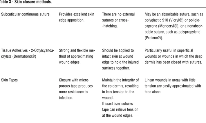
Port-site skin wounds may be closed in a wide variety of techniques, applying different kinds of sutures and materials (Table-3).
Buchweitz et al., in a prospective random-ized study, assessed the outcome of 5 mm laparo-scopic port site skin closure by three different tech-niques. The authors found that patient’s cosmetic satisfaction was higher with the use of transcutane-ously sutured wounds compared to the subcutane-ous suturing or the use of papertape closure. The authors attributed the improved cosmetics with the transcutaneous suture to the better coaptation of skin edges, enabling a higher quality scar result (31).
Table 2 - Laparoscopic fascial wound closure techniques.
Technique Characteristic Example
Closure assisted from inside abdomen (i.e., requiring two additional ports: one for the laparoscope and one for the grasper)
Instrument manipulation is done under direct visualization, allowing higher safety in avoiding visceral injuries.
Maciol needles; Grice needle; Use of ca-theter or spinal needles; The Endoclose device; The Gor-Tex device.
Closure assisted by the use of extracor-poreal instruments (i.e., needing only one additional port for the laparoscope).
Suture is performed extracorporeally under intra-abdominal direct visualization by the laparoscope. One of the most used techniques.
Carter-Thomason CloseSure System; Endo-Judge wound closure device; Tahoe Surgical Instrument Ligature de-vice; eXit Disposable Puncture Closure device; The Closure techniques using a 5 or 2mm trocar.
Closure performed with or without visual control.
Suture aided by a tactile sense, applicable during insufflation or after desufflation.
Table 3 - Skin closure methods.
Subcuticular continuous suture Provides excellent skin edge apposition.
There are no external sutures or cross--hatching.
May be an absorbable suture, such as polyglactic 910 (Vicryl®) or poligle-caprone (Monocryl®), or a nonabsor-bable suture, such as polypropylene (Prolene®).
Tissue Adhesives - 2-Octylcyanoa-crylate (Dermabond®)
Strong and flexible me-thod of approximating wound edges.
Should be applied to intact skin at wound edge to hold the injured surfaces together.
Particularly useful in superficial wounds or wounds in which the deep dermis has been closed with sutures.
Skin Tapes Closure with micro-porous tape produces more resistance to infection.
Maintain the integrity of the epidermis, resulting in less tension to the wound.
If used over sutures tape can relieve tension at the wound edges.
Linear wounds in areas with little tension are easily approximated with tape alone.
The use of skin adhesives associated to traditional suture provides extra closure support with an impermeable suture line, decreasing the need for postoperative care. It is especially inter-esting to use in pediatric patients since there is no need of postoperative suture removal (32).
Skin adhesives
Cyanoacrylate based glues are fast-acting adhesives formed by an association of a mono-mer and a plasticizer that forms a flexible bond presenting a breaking strength comparable to that of 5-0 monofilament suture and the intent of its use is to achieve good skin edges coaptation, just as it is done with the traditional sutures. It sets quickly, usually under 1 min, a characteristic that enables it an easy to use technology in small inci-sions such as traumatic skin lacerations and lapa-roscopic port-site wounds (33).
Advantages of the use of skin adhesives are the formation of a watertight barrier that al-low patients to shower any time after surgery, en-abling a more adequate recovering from surgery and high patient acceptance. Other possible
bene-fit that may rise from the use of these adhesives is a decrease in the use of needled sutures reducing personal needle exposure in the operating room.
Major disadvantages of the use of OCA are the need of a learning curve since it has some particularities for application as the need of a dry surface, with good edge-to-edge approximation. This technique is necessary in order to avoid the substance within the incision, since its presence will cause an intense foreign body reaction lead-ing to a not acceptable final skin cosmetics. Cost is another matter of concern to the use of these skin adhesives, when compared to the cost of tra-ditional sutures (33,34).
skin adhesives usage is the high cost, when com-pared to suture closure of port-site wounds - each vial of adhesive may cost three times or more the cost of a suture, estimated for a same length inci-sion (34,35).
Port-site Hernia formation
Hernia formation at the site of a laparo-scopic trocar is not frequent but is a deleterious complication since it is likely to require a new surgical procedure in order to correct the abdomi-nal wall defect (36,37).
Different characteristics may help to clas-sify an incisional hernia as the time from surgery that it occurred, if there is any content from ab-dominal cavity trapped inside of it, if there is any functional consequence as bowel obstruction or pain. These properties will guide surgeon’s deci-sion to observe or to recommend a new interven-tion in an elective or emergency setting (38).
Reasons contributing to the development of an incisional hernia at a laparoscopic trocar-port may be early suture disruption, skin or sub-cutaneous infection, patient malnutrition status, patient’s early return to daily activities or failure to adequately reapproximate fascial wound edges.
There is an inherent fascial weakness at the paraumbilical region, leading to a higher in-cidence on hernia formation. Midline trocars are also associated with a higher incidence of this complication, probably due to the fact that these trocars are usually of larger size and are actively manipulated during the surgical procedure. Um-bilical and midline trocars are also frequently used for organ retrieval, seriously influencing fascial tissue trauma and weakness, predisposing to hernia formation (37,39).
Lateral port-sites usually have a lower in-cidence on hernia formation, due to the presence of multiple abdominal wall muscular layers im-proving wound closure; still, frequently smaller size trocars are used at these sites, resulting in smaller wound defects (40).
It is recommended to end the pneumoperi-toneum completely before closing the trocar port wounds, in order to prevent intestine or omentum herniation due to gas pushing (37).
Trocar diameter is associated to the devel-opment of an incisional hernia at trocar wound, presenting a proportional risk to the trocar size. After laparosocopic surgery all 10 and 12 mm tro-car sites are best treated if properly closed, with adequate fascial wound edges suture and coapta-tion. Regarding the 5 mm trocar fascia closure, there has been some arguing on the matter, es-pecially in pediatric population (41). In general, authors advocate that all trocar wounds in the pe-diatric population should be closed, while in the adult patients they recommend closing all trocars larger than 5 mm (42,43).
Prophylatic Drainage
Prophylactic abdominal drainage at the beginning of the 1900’s had became a major con-cern and was not routinely recommended. Yates after an experimental study had stated in 1905 that “drainage of the peritoneal cavity is physi-cally and physiologiphysi-cally impossible” (44). Drain-age had presented so many complications that in 1919 Frank Hathaway wrote “Its day is past, and soon it will only be seen, where it should be, in a museum” in his article about abuse of drainage tubes (45).
There is enough evidence in the literature not to recommend prophylactic abdominal drain-age in all abdominal procedures, being unneces-sary or even harmful (46). In some cases, the use of drains may even be related to longer hospital stay and higher postoperative morbidity (47).
The rational for abdominal cavity drain-age is based on the fact that the presence of gas or fluid in the peritoneal cavity may disrupt inflam-matory reaction leading clinically to an increase of complications as pain and infection (48,49). Other reasons for drainage placement after a sur-gical procedure are any doubt about potential complication as difficulty in obtaining hemosta-sis or any vessel ligation, intestine suture that the surgeon was not totally confident with the result and even problematic or difficult urinary tract closure (50).
Urologic Laparoscopy Drainage
The improvement of techniques and de-velopment of new instruments along with the fact of laparoscopic expertise being achieved by many surgeons made the number of partial nephrecto-mies to grow and surgical indications expanded to larger and deeply located tumors, with a higher frequency of collecting system involvement (51). As well it enables the use of this technique for Radical Prostatectomy in the treatment of pros-tate cancer, with comparable results to the stan-dard retropubic open surgery (52). However with an increase in the frequency of this surgical ap-proach it is expected to find also an increase in the number of complications (53,54).
Laparoscopic surgery has been success-fully introduced in the treatment of urinary tract stone disease, with major applications for ureteral calculli and treatment of Ureteropelvic junction (UPJ) anomaly or caliceal diverticula (55,56).
The most frequent surgical complications after laparoscopic urologic procedures include bleeding, hematomas, urine leak, and infection. The use of a drainage system enables early di-agnosis of any of these conditions allowing fast intervention in order to adequately treat the
com-plication. Commonly urologic procedures that may require drainage are listed in Table-4.
Urine leak risk factors are associated to tumor size, tumor endophytic location and the need to open the collecting system during surgery. The majority of urine leak cases were successfully treated by prolonged drainage and drain manip-ulation. When non invasive treatment achieved unsuccessful urine leak resolution, treatment in-cluding ureteral stenting should be done being highly effective (57). It is only in cases where conservative maneuvers were not efficacious in achieving resolution that open repair or even ne-phrectomy may be necessary.
Drainage after Robotic Surgery
Indication for drainage after radical pros-tatectomy are usually tension at the urethrovesi-cal anastomosis, bladder neck deformity, a large median lobe, rectal injury, urinary bladder inju-ry, and need to redo urethrovesical anastomosis. Other potential complications related to abdomi-nal wall drain insertion are muscle or subcutane-ous hematoma, pain at the drain site, injury to the inferior epigastric vessels, and loss of part of the drain inside the abdominal cavity. Potential
Table 4 - Urologic laparoscopic drainage.
Procedure Drainage
Nephrectomy Needs drainage if there is doubt about bleeding or extensive linfonode dissection.
Partial Nephrectomy Always drain due to kidney’s resection bed and possible or needed opening of the collecting system.
Lymphadenectomy Drain if extensive dissection due to the higher risk of lymphocele.
Radical Prostatectomy Routinely require drainage to observe urethrovesical anas-tomosis. There is some debate when surgery is done with magnification and result in a watertight anastomosis.
Adrenalectomy Usually does not require drainage. Recommended if any con-cerns about bleeding.
urinary complications that may arise from drain omission after radical prostatectomy are usually the development of collection of urine, anasto-motic stricture due to urine leak, lymphoceles and urinary incontinence.
In a recent study Canes et al. evaluated the existence of an association between pelvic drain-age and postoperative complications on patients who underwent laparoscopic radical prostatecto-my. The authors found that drainage resulted in longer operative times and greater narcotic use if compared to undrained patients. Regarding other complications there was no increase of clinically detected urine leak, collection of urine, hemato-ma or lymphocele. Although routine pelvic drain-age is usually part of the radical prostatectomy procedure, these findings support the possibility of drain omission when a urethrovesical anas-tomosis is watertight during the intraoperative test (58). This approach reduces hospital stay and costs, and has been demonstrated to be safe with no rise in the complication rates (59).
The improved technology led to some debate whether a drain is really necessary af-ter partial nephrectomy. Robotic assisted partial nephrectomy demonstrated the ability to reduce blood loss, operative time and warm ischemia time when compared to pure laparoscopic partial nephrectomy (60). Closure of the collecting sys-tem may be improved through the use of robotic technology in laparoscopic renal surgery result-ing in a decrease of the urine leak frequency. Per-haps in the future the technologic improvement with safer and watertight closure of the collecting system will enable surgeons to elude the use of drains in those situations.
Open Passive Versus Closed Drainage
Skin wound and abdominal wall trajectory of Penrose drainage works as an entrance door for bacterial colonization and migration, in a higher frequency than when using closed suction drains (61,62).
Multiple drainage systems were developed to be use as a closed system, either with or without suction, with efficacy on removing fluid from the abdominal cavity after laparoscopic surgery (63).
A closed drainage system is achieved when the drain insertion is performed in a way to be water and air tight, precluding external contact to the drained cavity. It may be inserted and used in a passive way, allowing drainage mostly of liquid fluid material. When the material to be drained is a thicker fluid it is advisable to add a suction system in order to facilitate drainage.
Closed suction drains have been avoided by Urologists due to the potential risk of pro-longed urinary drainage that has been expressed in the statement “Penrose drains should be used in all patients because closed suction drains can perpetuate” and by perpetuating we should under-stand as a urinary fistula or even a delayed hemor-rhage after drain removal (64). However we have enough data to make this orientation differently since many have successfully used closed suction drains after urologic laparoscopic surgery, finding it effective, with no increased morbidity (65).
In a comparison of suction and non-suc-tion drains there was similar pain scores associat-ed to the period before or after drain removal. The removal procedure is usually more painful when the drain used is of suction type, probably due to soft early adherences of small bowel or omentum to the drain tube holes (66).
Data obtained from general surgery litera-ture have been able to clearly demonstrate that closed suction drains are associated with fewer complications when compared to an open passive drain as the Penrose one (67).
CONFLICT OF INTEREST
None declared.
REFERENCES
1. Rajan P, Turna B: New trends in minimally invasive urologi-cal surgery. Int Braz J Urol. 2009; 35: 514-20.
2. Rassweiler J, Stock C, Frede T, Seemann O, Alken P: Organ retrieval systems for endoscopic nephrectomy: a compara-tive study. J Endourol. 1998; 12: 325-33.
4. Ankem MK, Hedican SP, Pareek G, Waterman BJ, Moon TD, Selvaggi SM, et al.: Examination of laparoscopic retrieval bag washings for malignant cells after hand-assisted lapa-roscopic radical nephrectomy and intact specimen remov-al. Urology. 2006; 68: 50-2.
5. Ganpule AP, Gotov E, Mishra S, Muthu V, Sabnis R, Desai M: Novel cost-effective specimen retrieval bag in laparos-copy: Nadiad bag. Urology. 2010; 75: 1213-6.
6. Raj PK, Katris F, Linderman CG, ReMine SG: An inexpensive laparoscopic specimen retrieval bag. Surg Endosc. 1998; 12: 83.
7. Terai A, Ichioka K, Inoue K, Yoshimura K: A simple kidney entrapment technique using a zipped plastic bag during retroperitoneoscopic radical nephrectomy. BJU Int. 2005; 96: 683-4.
8. Collins S, Marruffo F, Durak E, Hruby G, Bergman A, Gupta M, et al.: Laparoscopic pyelolithotomy with intraperitoneal ultrasonic lithotripsy: report of a novel minimally invasive technique for intracorporeal stone ablation. Surg Laparosc Endosc Percutan Tech. 2006; 16: 435-6.
9. Tisdale BE, Kapoor A, Hussain A, Piercey K, Whelan JP: Intact specimen extraction in laparoscopic nephrectomy procedures: Pfannenstiel versus expanded port site inci-sions. Urology. 2007; 69: 241-4.
10. Gettman MT, Napper C, Corwin TS, Cadeddu JA: Laparo-scopic radical nephrectomy: prospective assessment of impact of intact versus fragmented specimen removal on postoperative quality of life. J Endourol. 2002; 16: 23-6. 11. Castillo OA, Vitagliano G: Port site metastasis and tumor
seeding in oncologic laparoscopic urology. Urology. 2008; 71: 372-8.
12. Fentie DD, Barrett PH, Taranger LA: Metastatic renal cell cancer after laparoscopic radical nephrectomy: long-term follow-up. J Endourol. 2000; 14: 407-11.
13. Castilho LN, Fugita OE, Mitre AI, Arap S: Port site tumor recurrences of renal cell carcinoma after videolaparoscopic radical nephrectomy. J Urol. 2001; 165: 519.
14. Casciola L, Codacci-Pisanelli M, Ceccarelli G, Bartoli A, Di Zitti L, Patriti A: A modified umbilical incision for specimen extraction after laparoscopic abdominal surgery. Surg En-dosc. 2008; 22: 784-6.
15. Gill IS, Cherullo EE, Meraney AM, Borsuk F, Murphy DP, Falcone T: Vaginal extraction of the intact specimen follow-ing laparoscopic radical nephrectomy. J Urol. 2002; 167: 238-41.
16. Ghezzi F, Raio L, Mueller MD, Gyr T, Buttarelli M, Franchi M: Vaginal extraction of pelvic masses following operative laparoscopy. Surg Endosc. 2002; 16: 1691-6.
17. Novick AC: Laparoscopic radical nephrectomy: specimen extraction. BJU Int. 2005; 95(Suppl 2): 32-3.
18. Kaouk JH, Gill IS: Laparoscopic radical nephrectomy: morcel-late or leave intact? Leave intact. Rev Urol. 2002; 4: 38-42.
19. Clayman RV, Kavoussi LR, Soper NJ, Dierks SM, Merety KS, Darcy MD, et al.: Laparoscopic nephrectomy. N Engl J Med. 1991; 324: 1370-1.
20. Wu SD, Lesani OA, Zhao LC, Johnston WK, Wolf JS Jr, Clayman RV, et al.: A multi-institutional study on the safety and efficacy of specimen morcellation after laparoscopic radical nephrectomy for clinical stage T1 or T2 renal cell carcinoma. J Endourol. 2009; 23: 1513-8.
21. Varkarakis I, Rha K, Hernandez F, Kavoussi LR, Jarrett TW: Laparoscopic specimen extraction: morcellation. BJU Int. 2005; 95(Suppl 2): 27-31.
22. Rabban JT, Meng MV, Yeh B, Koppie T, Ferrell L, Stoller ML: Kidney morcellation in laparoscopic nephrectomy for tumor: recommendations for specimen sampling and pathologic tumor staging. Am J Surg Pathol. 2001; 25: 1158-66.
23. Gabr AH, Gdor Y, Strope SA, Roberts WW, Wolf JS Jr.: Ap-proach and specimen handling do not influence oncological perioperative and long-term outcomes after laparoscopic radical nephrectomy. J Urol. 2009; 182: 874-80.
24. Landman J, Lento P, Hassen W, Unger P, Waterhouse R: Feasibility of pathological evaluation of morcellated kidneys after radical nephrectomy. J Urol. 2000; 164: 2086-9. 25. Landman J, Venkatesh R, Kibel A, Vanlangendonck R:
Mod-ified renal morcellation for renal cell carcinoma: laboratory experience and early clinical application. Urology. 2003; 62: 632-4; discussion 635.
26. Camargo AH, Rubenstein JN, Ershoff BD, Meng MV, Kane CJ, Stoller ML: The effect of kidney morcellation on op-erative time, incision complications, and postopop-erative analgesia after laparoscopic nephrectomy. Int Braz J Urol. 2006; 32: 273-9; discussion 279-80.
27. Toriumi DM, O’Grady K, Desai D, Bagal A: Use of octyl-2-cyanoacrylate for skin closure in facial plastic surgery. Plast Reconstr Surg. 1998; 102: 2209-19.
28. Shaher Z: Port closure techniques. Surg Endosc. 2007; 21: 1264-74.
29. Su WH, Cheng MH, Tsou TS, Cheung SM, Chang SP, Wang PH: Port wound closure assisted by Foley catheter: an eas-ier way to provide fascia security. J Obstet Gynaecol Res. 2009; 35: 725-31.
30. Rastogi V, Dy V: Simple technique for proper approxima-tion and closure of peritoneal and rectus sheath defects at port site after laparoscopic surgery. J Laparoendosc Adv Surg Tech A. 2001; 11: 13-6.
31. Buchweitz O, Wülfing P, Kiesel L: A prospective randomized trial of closing laparoscopic trocar wounds by transcuta-neous versus subcuticular suture or adhesive papertape. Surg Endosc. 2005; 19: 148-51.
33. Sebesta MJ, Bishoff JT: Octylcyanoacrylate skin closure in laparoscopy. J Endourol. 2003; 17: 899-903.
34. Matin SF: Prospective randomized trial of skin adhesive versus sutures for closure of 217 laparoscopic port-site incisions. J Am Coll Surg. 2003; 196: 845-53.
35. Sajid MS, Siddiqui MR, Khan MA, Baig MK: Meta-analysis of skin adhesives versus sutures in closure of laparoscopic port-site wounds. Surg Endosc. 2009; 23: 1191-7.
36. Callery MP, Strasberg SM, Soper NJ: Complications of laparo-scopic general surgery. Gastrointest Endosc Clin N Am. 1996; 6: 423-44.
37. Azurin DJ, Go LS, Arroyo LR, Kirkland ML: Trocar site hernia-tion following laparoscopic cholecystectomy and the signifi-cance of an incidental preexisting umbilical hernia. Am Surg. 1995; 61: 718-20.
38. Tonouchi H, Ohmori Y, Kobayashi M, Kusunoki M: Trocar site hernia. Arch Surg. 2004; 139: 1248-56.
39. Tisdale BE, Kapoor A, Hussain A, Piercey K, Whelan JP: In-tact specimen extraction in laparoscopic nephrectomy proce-dures: Pfannenstiel versus expanded port site incisions. Urol-ogy. 2007; 69: 241-4.
40. Bird VG, Au JK, Sandman Y, De Los Santos R, Ayyathurai R, Shields JM: Comparison of different extraction sites used during laparoscopic radical nephrectomy. J Urol. 2009; 181: 1565-70.
41. Spalding SC, Ponsky TA, Oristian E: A new Dual-hemostat technique to facilitate the closure of small laparoscopic trocar incisions. Surg Endosc. 2003; 17: 164-5.
42. Sanz-López R, Martínez-Ramos C, Núñez-Peña JR, Ruiz de Gopegui M, Pastor-Sirera L, Tamames-Escobar S: Incisional hernias after laparoscopic vs open cholecystectomy. Surg En-dosc. 1999; 13: 922-4.
43. Kulacoglu IH: Regarding: Small bowel obstruction and inci-sional hernia after laparoscopic surgery: should 5-mm trocar sites be sutured? J Laparoendosc Adv Surg Tech A. 2000; 10: 227-8.
44. Yates JL: An experimental study of the local effects of perito-neal drainage. Surg Gynecol Obstet. 1905; 1: 473-92. 45. Hathaway F: The Abuse of Drainage Tubes. Br Med J. 1918;
1: 718-20.
46. Petrowsky H, Demartines N, Rousson V, Clavien PA: Evidence-based value of prophylactic drainage in gastrointestinal sur-gery: a systematic review and meta-analyses. Ann Surg. 2004; 240: 1074-84; discussion 1084-5.
47. de Rougemont O, Dutkowski P, Weber M, Clavien PA: Ab-dominal drains in liver transplantation: useful tool or useless dogma? A matched case-control study. Liver Transpl. 2009; 15: 96-101.
48. Abbott J, Hawe J, Srivastava P, Hunter D, Garry R: Intraperi-toneal gas drain to reduce pain after laparoscopy: randomized masked trial. Obstet Gynecol. 2001; 98: 97-100.
49. Shen CC, Huang FJ, Hsu TY, Weng HH, Chang HW, Chang SY: A prospective, randomized study of closed-suction drainage after laparoscopic-assisted vaginal hysterectomy. J Am Assoc Gynecol Laparosc. 2002; 9: 346-52.
50. Tzovaras G, Liakou P, Fafoulakis F, Baloyiannis I, Zacharoulis D, Hatzitheofilou C: Is there a role for drain use in elective laparoscopic cholecystectomy? A controlled randomized trial. Am J Surg. 2009; 197: 759-63.
51. Simmons MN, Gill IS: Decreased complications of contem-porary laparoscopic partial nephrectomy: use of a standard-ized reporting system. J Urol. 2007; 177: 2067-73; discussion 2073.
52. Bove P, Asimakopoulos AD, Kim FJ, Vespasiani G: Laparo-scopic radical prostatectomy: a review. Int Braz J Urol. 2009; 35: 125-37; discussion 137-9.
53. Richstone L, Montag S, Ost M, Reggio E, Permpongkosol S, Kavoussi LR: Laparoscopic partial nephrectomy for hilar tu-mors: evaluation of short-term oncologic outcome. Urology. 2008; 71: 36-40.
54. Permpongkosol S, Link RE, Su LM, Romero FR, Bagga HS, Pavlovich CP, et al.: Complications of 2,775 urological laparo-scopic procedures: 1993 to 2005. J Urol. 2007; 177: 580-5. 55. Micali S, Moore RG, Averch TD, Adams JB, Kavoussi LR: The
role of laparoscopy in the treatment of renal and ureteral cal-culi. J Urol. 1997; 157: 463-6.
56. Nambirajan T, Jeschke S, Albqami N, Abukora F, Leeb K, Janetschek G: Role of laparoscopy in management of renal stones: single-center experience and review of literature. J En-dourol. 2005; 19: 353-9.
57. Meeks JJ, Zhao LC, Navai N, Perry KT Jr, Nadler RB, Smith ND: Risk factors and management of urine leaks after partial nephrectomy. J Urol. 2008; 180: 2375-8.
58. Canes D, Cohen MS, Tuerk IA: Laparoscopic radical prostatec-tomy: omitting a pelvic drain. Int Braz J Urol. 2008; 34: 151-8. 59. Sharma S, Kim HL, Mohler JL: Routine pelvic drainage not
required after open or robotic radical prostatectomy. Urology. 2007; 69: 330-3.
60. Benway BM, Bhayani SB, Rogers CG, Dulabon LM, Patel MN, Lipkin M, et al.: Robot assisted partial nephrectomy versus laparoscopic partial nephrectomy for renal tumors: a multi-institutional analysis of perioperative outcomes. J Urol. 2009; 182: 866-72.
61. Nora PF, Vanecko RM, Bransfield JJ: Prophylactic abdominal drains. Arch Surg. 1972; 105: 173-6.
62. Raves JJ, Slifkin M, Diamond DL: A bacteriologic study com-paring closed suction and simple conduit drainage. Am J Surg. 1984; 148: 618-20.
64. Naitoh J, Smith R: Complications of renal surgery. In: Com-plications of Urologic Surgery. Taneja SS, Smith RB, Ehrlich RM (ed.), Philadelphia: W.B. Saunders Co. 2001. pp. 299-325. 65. Chan DY, Marshall FF: Partial nephrectomy for centrally locat-ed tumors. Urology. 1999; 54: 1088-91; discussion 1091-2. 66. Raymond AP, Chan K, Deans R, Bradbury R, Vancaillie TG,
Ab-bott JA: A comparative, single-blind, randomized trial of pain associated with suction or non-suction drains after gyneco-logic laparoscopy. J Minim Invasive Gynecol. 2010; 17: 16-20.
67. Sánchez-Ortiz R, Madsen LT, Swanson DA, Canfield SE, Wood CG: Closed suction or penrose drainage after partial nephrec-tomy: does it matter? J Urol. 2004; 171: 244-6.