Rupture of ectopic renal arterial pseudoaneurysm after
percutaneous nephrolithotomy
_______________________________________________
Mingshuai Wang
1, Junhui Zhang
1, Nianzeng Xing
11 Department of Urology, Beijing Chao - Yang Hospital, Capital Medical University, Beijing, China
ABSTRACT
_______________________________________________________________________________________
845
RADIOLOGY PAGE
A 35-year-old female patient presented with swelling pain at left waist for 1 month. Left renal pelvis stones were found and standard percutaneous nephrolithotomy was successfully performed. Two weeks later, the patient suddenly suffered massive bleeding presented with gross hematuria. Rupture of ectopic renal artery pseudoaneurysm was identified by computed tomography and angiography of the renal artery. Emergency selective angioembolization of one branch of the artery was performed. To our knowledge, this is the first report of ruptured ectopic renal arterial pseudoaneurysm.
DESCRIPTION OF CASE
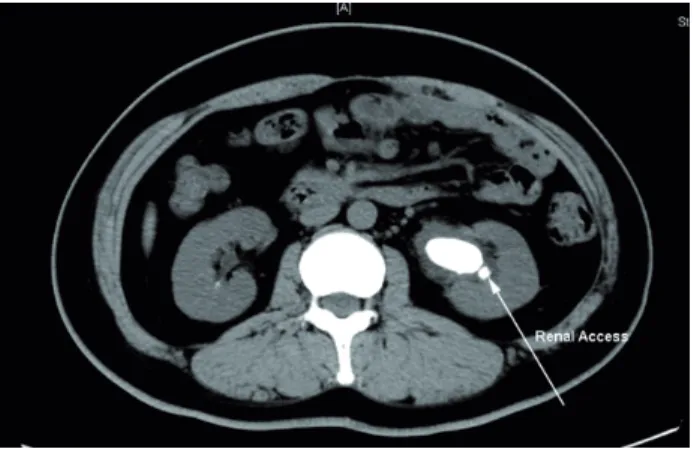
A 35-year-old female patient presented with back pain for 1 month. Plain computed to-mography (CT) scan showed a stone measuring 3.2*1.6cm and a smaller one located in the left renal pelvis (Figure-1).
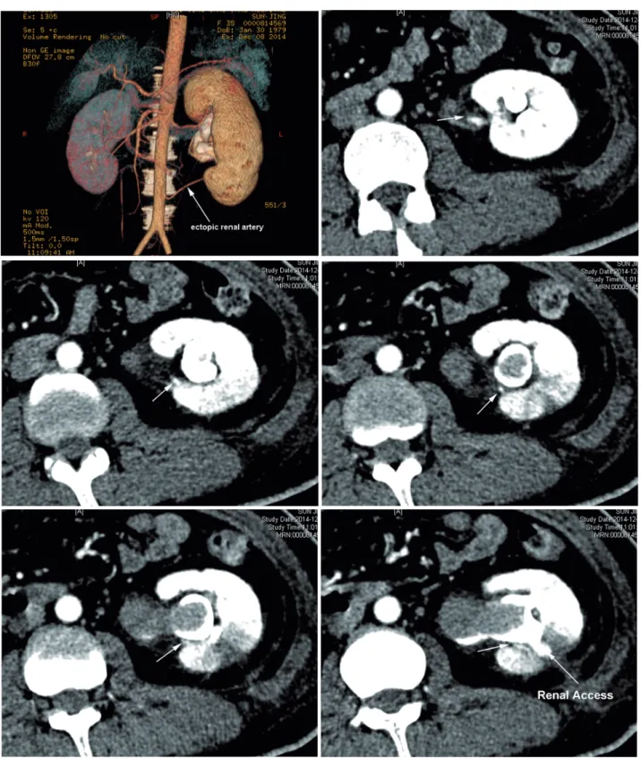
One experienced surgeon performed standard percutaneous nephrolithotomy. After general anesthesia, percutaneous renal access was obtained under ultrasound with an 18-gau-ge needle. Tract dilatation was accomplished using balloon dilator of 24F. The stone was fragmented utilizing an ultrasonic lithotripter through a rigid 24F nephroscope. A 20F ne-phrostomy tube was inserted after the successful completion of the procedure. The nephrostomy tube and urinary catheter were removed 1 week postoperatively. Unfortunately, 2 weeks after operation, the patient suddenly suffered mas-sive bleeding presented with gross hematuria. Her blood hemoglobin decreased to 7.2g/L. CT angiography identified an ectopic renal artery leading to a pseudoaneurysm which appeared to
Figure 1 - Left renal stone, measuring 3.2*1.6cm.
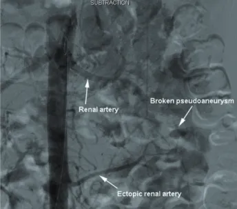
be in the pathway of the access tract (Figure-2). Emergency selective angioembolization of this branch was performed in conjunction with the angiogram confirming rupture and pseudoaneu-rysm of the ectopic branch artery (Figure-3).
Arterial pseudoaneurisms have occurred as consequence of extracorporeal shock wave li-thotripsy (1). Gavant et al. firstly described rup-ture of renal pseudoaneurysm as a complication of percutaneous nephrostomy (2). It was
repor-doi: 10.1590/S1677-5538.IBJU.2015.0017
IBJU| RADIOLOGY PAGE
846
IBJU| RADIOLOGY PAGE
847
ARTICLE INFO
Int Braz J Urol. 2016; 42: 845-7
_____________________
Submitted for publication: January 11, 2015
_____________________
Accepted after revision: February 22, 2016
REFERENCES
1. Lang EK, Earhardt V. Arterial pseudoaneurysm after extracorporeal shock wave lithotripsy. J Urol. 2005;173:1366. 2. Gavant ML, Gold RE, Church JC. Delayed rupture of
renal pseudoaneurysm: complication of percutaneous nephrostomy. AJR Am J Roentgenol. 1982;138:948-9.
_______________________ Correspondence address: Nianzeng Xing, MD
Professor of the Department of Urology Vice President of Beijing Chao - Yang Hospital
Gongti South Street, NO. 8, Chao - Yang District, Beijing, China, 100026
Fax: +86 010 8593-5241
Email: [email protected]
Figure 3 - Rupture of one branch of the ectopic renal artery angiography.
ted that pseudoaneurysm after percutaneous re-nal surgery was the most common angiographic finding (3). The access route to the stone has a major impact on the incidence of the com-plication, causing pseudo-aneurisms or arterio--venous fistulae. Because of the trajectory of the access-tract between arterial and venous chan-nels in the upper and mid-pole arterial-venous fistula-formation may occur. This trajectory is different for a subcostal and intercostal appro-ach, hence a different rate of attendant compli-cations (4). However, rupture of ectopic renal arterial pseudoaneurysm is very rare, so much care should be taken for this kind of patient.
CONFLICT OF INTEREST
None declared.
3. Richstone L, Reggio E, Ost MC, Seideman C, Fossett LK, Okeke Z, et al. First Prize (tie): Hemorrhage following percutaneous renal surgery: characterization of angiographic findings. J Endourol. 2008;22:1129-35.