Endovascular treatment of abdominal aortic aneurysm
in patient with horseshoe kidney: a case report
Tratamento endovascular de aneurisma de aorta abdominal
em paciente com rim em ferradura: relato de caso
José Manoel da Silva Silvestre1, Gustavo Teixeira Fulton Schimit2, Wander Eduardo Sardinha3, Guilherme da Silva Silvestre4, Guilon Otávio Santos Tenório5, Fernando Barbosa Trevisan5
Abstract
Horseshoe kidney is one of the most common urologic anomalies, present in about 0.12% of patients with abdominal aortic aneurysm. Conventional surgical repair is associated with technical diiculties that probably increase morbidity and mortality but can be avoided with endovascular treatment. We report the case of a 64-year-old patient presenting with horseshoe kidney and abdominal aortic aneurysm and successfully treated with endovascular repair.
Keywords: abdominal aortic aneurysm; congenital abnormalities; blood vessel prosthesis implantation.
Resumo
O rim em ferradura é uma das anomalias urológicas congênitas mais comuns e está presente em cerca de 0,12% dos pacientes com aneurisma de aorta abdominal. O reparo cirúrgico convencional está associado a diiculdades técnicas que provavelmente aumentam a morbidade e a mortalidade, mas que podem ser evitadas com o tratamento endovascular. Relatamos um caso de um paciente de 64 anos com rim em ferradura e aneurisma de aorta abdominal, que foi submetido ao reparo endovascular do aneurisma com sucesso.
Palavras-chave: aneurisma de aorta abdominal; anormalidades congênitas; implante de prótese vascular.
R E P O R T
1 Universidade Estadual de Londrina – UEL, Londrina, PR, Brazil 2 Hospital Universitário Regional do Norte do Paraná – Londrina, PR, Brazil
3 Universidade Estadual de Londrina – UEL, Departamento de Clínica Cirúrgica, Londrina, PR, Brazil 4 Universidade do Oeste do Paraná – Cascavel, PR, Brazil
5 Hospital Universitário Regional do Norte do Paraná, Londrina, PR, Brazil
Financial support: None.
Conlicts of interest: No conlicts of interest declared concerning the publication of this article. Submitted:29.08.11.Accepted: 14.11.12.
INTRODUCTION
Horseshoe kidney is a very frequent congenital abnormality, with an incidence of one in every 600 to 800 individuals1. According to Ferko et al.,
horseshoe kidneys are found in every 200 cases of patients with abdominal aortic aneurysms (AAA) treated surgically2.
Resection of an AAA associated with horseshoe
kidney involves two basic problems: irst, renal
parenchyma are present, often covering the aneurysm
and making its exposure more dificult; second, aneurysm resection is usually very dificult because
the lesion is present in anomalous arteries. In this scenario, endovascular therapy emerges as a very attractive procedure for the treatment of AAAs in patients with horseshoe kidneys, especially because of its less aggressive nature and increased effectiveness in this type of patient.
Notwithstanding, in the literature reviewed, including both Brazilian and international papers, the investigation of endovascular approaches to the treatment of AAAs has been limited to case reports or small case series. In the present journal, the problem has only been addressed with a focus on conventional surgical treatment3.
The objective of the present study was to report the case of a patient presenting with horseshoe kidneys and subjected to endovascular treatment of an AAA. We discuss the clinical importance of an individualized treatment approach, based on the latest evidence available for each different type or manifestation of this congenital anomaly.
CASE DESCRIPTION
A 64-year-old male patient presented at our vascular surgery service seeking treatment for an
infrarenal AAA. The patient had no symptoms and informed that the aneurysm had been diagnosed 5 years before during an ultrasound examination performed for other reasons. Over this period, the patient had been followed through regular exams that showed constant increase of aneurysm diameter. Medical history included hypertension, diabetes mellitus, and myocardial revascularization in 1987 and 2004 using the great saphenous vein and the mammary artery as grafts.
On physical examination, the patient was
normotensive; abdominal examination findings
were poor due to obesity. Longitudinal scars were observed in the lower limbs (bilateral saphenectomy). Right posterior tibial artery pulse was absent upon palpation. Laboratory tests revealed normal glycemia and creatinine levels.
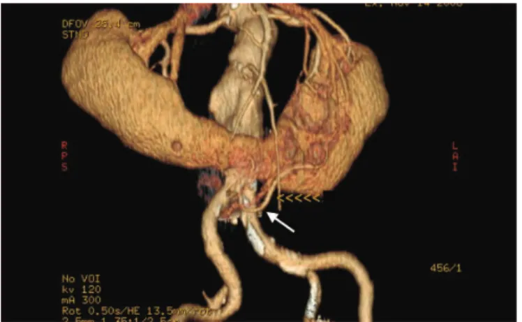
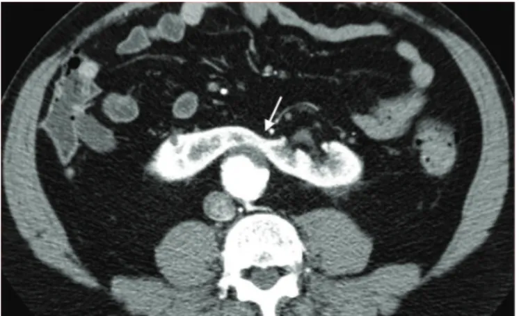
Computed tomographic (CT) angiography was carried out because of the continuous growth of the aneurysm and revealed a fusiform aneurysm of the infrarenal aorta, with a maximal cross-sectional diameter of 50 mm, and horseshoe kidneys with an isthmus of parenchymal tissue. About 20 to 30% of the kidney, in the isthmus region, was irrigated by an accessory renal artery arising from the aortoiliac junction (Figures 1 and 2). The exam also revealed
signiicant stenosis (60%) at the origin of the left
renal artery.
Because of the continuous growth of the aortic aneurysm, elective treatment was indicated. In December 2008, the patient underwent angioplasty and stent placement in the left renal artery, with success. In the same procedure, selective angiography of the accessory renal artery was performed and
conirmed the indings previously obtained with
CT angiography. Based on the favorable anatomy
of the aneurysm and taking into consideration
the dificulties involved in conventional surgical
correction of infrarenal aneurysms, the team decided to adopt an endovascular approach, with occlusion of the accessory renal artery 2 months after angioplasty of the left renal artery. The endovascular procedure was performed with bilateral femoral access, using a bifurcated prosthesis with distal anchorage on the common iliac arteries.
Follow-up CT angiographic indings evidenced
an adequately implanted endograft, well adapted, with no signs of leakage (Figure 3). An area of renal infarction was observed in the isthmus region, corresponding to approximately 20% of the total renal parenchyma (Figure 4). Angioplasty of the left renal artery did not show evidence of stenosis. Serial investigations revealed a gradual reduction in aneurysm diameter.
Throughout the follow-up period, the patient presented asymptomatic, with well-controlled blood pressure by the use of medications and normal
laboratory indings. At present, the patient undergoes
clinical and CT angiographic exams yearly.
DISCUSSION
In patients with horseshoe kidney, two distinct kidneys are located on either side of the body and anteriorly connected to the spine by an isthmus of
either parenchymal or ibrous tissue.4 Horseshoe
kidneys are a very common congenital urologic abnormality, affecting about 0.12% of patients who require surgical correction of aortic aneurysms5.
In most cases, horseshoe kidney is associated with normal renal function, and patients are asymptomatic. The anomaly can take different forms, with arterial and venous variations, as well as different presentations of the renal parenchyma
Figure 2. Cross-sectional view of computed tomographic angiography showing renal isthmus composed of parenchymal tissue (arrow).
and excretory organs, sometimes posing dificulties
or even preventing conventional surgical correction of AAAs6.
Horseshoe kidneys are usually connected by their lower poles, often between the vena cava and
the aorta. The isthmus can be composed of ibrous
tissue or, more commonly, of viable, vascularized parenchymal tissue7,8.
Several classiications have been proposed to
categorize the different manifestations of horseshoe kidneys according to variations in blood supply. The
classiication system proposed by Eisendrath et al.4,9
is the most widely used and divides indings into ive types:
• Type I: one renal artery to each side of the horseshoe kidney. Corresponds to 20% of cases;
• Type II: one renal artery to each side of the aorta and an aortic branch to the isthmus aorta. Corresponds to 30% of cases. This is the type of the case here described;
• Type III: two renal arteries to each side of the horse
-shoe kidney and an aortic branch to the isthmus. Corresponds to 15% of cases;
• Type IV: two renal arteries to each side of the horse
-shoe kidney, associated with one or more branches arising from the iliac arteries. Corresponds to 15% of cases; and
• Type V: multiple renal arteries arising from the aorta, mesenteric and iliac arteries. Corresponds to 20% of cases.
The greatest challenge in the surgical treatment of patients with infrarenal AAAs associated with
horseshoe kidneys is inding an access route to the
aneurysm, as well as an adequate approach to treat accessory renal arteries6,10.
In the few cases presenting with a fibrous isthmus, the best approach is to section the structure. Notwithstanding, in patients with a functional
isthmus, this approach should be avoided as a result of potential technical complications, such as chronic renal disease that could affect the graft. Ureters are anteriorly positioned, close to the isthmus, and may be present in duplicate11. During surgery, extreme
care should be taken not to injure these abnormally positioned ureters.
There are two main approaches to the treatment of accessory renal arteries during the correction of
AAAs. The irst one consists of reimplanting all
accessory renal arteries12,13. The second approach is
renal artery ligation, in an attempt to prevent future segmental renal infarction. This latter approach has the advantage of being faster and less aggressive14,15.
The irst reports of endovascular treatment of
AAAs in patients with horseshoe kidneys date back to 1997, either alone or combined with the occlusion of smaller-caliper accessory renal arteries2,16-21. Some
studies have assessed the clinical and laboratory results of the occlusion of accessory renal arteries in patients subjected to endovascular treatment of AAAs. Aquino et al. showed that, even though segmental renal infarction was observed in 21% of the cases, transient hypertension occurred in only one of 24 patients subjected to accessory renal artery occlusion using an aortic endograft22. Kaplan et al.
reported similar results18 and concluded that, in cases
where renal arteries do not present stenosis, where accessory arteries smaller than 3 mm are present, and where there is no history of renal disease, occluding
these arteries will not have signiicant effects on
renal perfusion.
These indings seem to suggest that the importance
of accessory renal arteries has been overestimated. This fact, combined with the rare cases of AAA in patients with horseshoe kidneys, and also with the high morbidity associated with conventional surgery,
makes endovascular treatment extremely attractive for these patients.
According to Ruppert et al., in patients with horseshoe kidneys types I and II according to
Eisendrath’s classiication, as is the case of our
patient, endovascular repair should be preferred whenever the procedure is technically feasible taking into consideration aneurysm anatomy. Accessory arteries with a diameter below 3 mm can be occluded. In cases showing accessory arteries larger than 3 mm, selective angiography should be performed to determine the amount of parenchyma irrigated by the
vessel. As a result, in patients with Eisendrath types
III and IV, the decision to adopt an endovascular approach will depend on each individual evaluation. Finally, in patients with type V horseshoe kidney and an acceptable operative risk, conventional surgical repair is still the treatment of choice23.
It is important to emphasize that larger caliper accessory arteries, when occluded, may lead to the occurrence of type II endoleak and increase the risk
of rupture, as reported by White et al.24 Some selected
reports can be found in the literature describing patients with accessory arteries with minimum collateral circulation subjected to coil embolization before endovascular treatment of the aneurysm23.
CONCLUSION
Over the last decade, endovascular treatment of AAAs in patients with horseshoe kidneys was rarely discussed in both national and international publications, and there is no consensus regarding the best treatment approach in these cases.
The present study concludes that the endovascular treatment of AAAs in patients with horseshoe kidneys is safe and effective. In cases with horseshoe
kidneys types I and II according to Eisendrath’s classiication, with nondominant accessory arteries
smaller than 3 mm, endovascular treatment can be considered the treatment of choice, as these patients
will beneit from a less invasive procedure. In all
other cases and types, treatment should be chosen based on individual evaluations.
REFERENCES
1. Campbell MF. Urology. 3rd ed. Philadelphia: WB Saunders; 1970. p. 448.
2. Ferko A, Krajina A, Jon B, Lesko M, Voboril Z. Juxtarenal aortic aneurysm associated with a horseshoe kidney: transfemoral endoluminal repair. Arch Surg. 1997;132:316-7. PMid:9125035. http://dx.doi.org/10.1001/archsurg.1997.01430270102021
3. Bonamigo TP, Tornquist FA, Furlan NM. Aneurisma da aorta abdominal e a presença de rim em ferradura. Cir Vasc Angiol. 1999;16: 59-64.
4. Eisendrath DN, Phifer FM, Culver HB. Horseshoe kidney. Ann Surg. 1925;82:735-64. PMid:17865363 PMCid:1400255. http:// dx.doi.org/10.1097/00000658-192511010-00009
5. Artioukh DY, Wake PN, Edwards PR, Moody AP. Problems of abdominal aortic aneurysm associated with horseshoe kidney. Eur J Vasc Endovasc Surg. 1997;14:75-8. http://dx.doi.org/10.1016/ S1078-5884(97)80230-4
6. Faggioli G, Freyrie A, Pilato A, et al. Renal anomalies in aortic surgery: contemporary results. Surgery. 2003;133:641-6. PMid:12796732. http://dx.doi.org/10.1067/msy.2003.156
7. Gutierrez R. Operative technique for division of renal isthmus in horseshoe kidney. Am J Surg 1942;55:762-7. http://dx.doi. org/10.1016/S0002-9610(42)90212-5
8. Donati A, Bartolomeo R, Turinetto B, et al. Abdominal aortic aneurysm and horseshoe kidney. J Cardiovasc Surg. 1980;21:632.
9. Faris I, Buxton B. Aneurysm surgery. Edinburgh: Churchill Livingstone; 1995. p. 175-80.
10. Stroosma OB, Koostra G, Schurink GWH. Management of aortic aneurysm in the presence of a horseshoe kidney. Br J Surg 2001;88:500-9. PMid:11298616. http://dx.doi. org/10.1046/j.1365-2168.2001.01718.x
11. Taylor DC, Sladen JG, Maxwell T. Aortic surgery and horseshoe kidney: a challenge surgical problem. Can J Surg. 1987;30:431 PMid:3664411.
12. Kasirajan K, O’Hara PJ. Renal ectopia and renal fusion in patients requiring abdominal aortic operations. In: Ernst CB, Stanley JC, editors. Current therapy in vascular surgery. St Louis: Mosby; 2001. p. 257-61.
13. Cronenwett JL, Krupski WC, Rutherford RB. Abdominal aortic and iliac aneurysms. In: Rutherford RB, editor. Vascular surgery. 5th ed.
14. Hollier LH. [comment in discussion]. In: Aquino RV, Rhee RY, Muluk SC, Tzeng EY, Nita-Missig C, Makaroun MS. Exclusion of accessory renal arteries during endovascular repair of abdominal aortic aneurysms. J Vasc Surg. 2001;34:878-84.
15. Edwards WH. [comment in discussion]. In: Kaplan DB, Kwon CC, Marin ML, Hollier LH. Endovascular repair of abdominal aortic aneurysms in patients with congenital renal vascular anomalies. J Vasc Surg. 1999;30:407-16. http://dx.doi.org/10.1016/ S0741-5214(99)70067-4
16. Dorfner R, hurnher S, Prokesch R, Youssefzadeh S, Holzenbein T, Lammer J. Spiral ct during selective accessory renal artery angiography: assessment of vascular territory before aortic stent-grafting. Cardiovasc Interv Radiol. 1998;21:179-82. http://dx.doi. org/10.1007/s002709900239
17. Loftus IK, Thompson MM, Fishwick G, Boyle JR, Bell PRF. Endovascular repair of aortic aneurysms in the presence of a horseshoe kidney. J Endovasc Surg. 1998;5:278-81. http://dx.doi. org/10.1583/1074-6218(1998)005<0278:EROAAI>2.0.CO;2
18. Kaplan DB, Kwon CC, Marin ML, Hollier LH. Endovascular repair of abdominal aortic aneurysms in patients with congenital renal vascular anomalies. J Vasc Surg. 1999;30:407-16. http://dx.doi. org/10.1016/S0741-5214(99)70067-4
19. Lee WA, Rubin GD, Arko F, Hill BB, Zarins CK. Endovascular stent graft repair of an infrarenal abdominal aortic aneurysm with a horseshoe kidney. Circulation. 2001;103:2126-7. PMid:11319206. http://dx.doi.org/10.1161/01.CIR.103.16.2126
21. Teijink JAW, Odink HF, Bendermacher B, Welten RJTJ, Veldhuijzen Van Zanten GO. Ruptured aaa in a patient with a horseshoe kidney: emergent treatment using talent acute aneurysm repair kit. J Endovasc Surg. 2003;10:240-3.
22. Aquino RV, Rhee RY, Muluk SC, Tzeng EY, Nita-Missig C, Akaroun MS. Exclusion of accessory renal arteries during endovascular repair of abdominal aortic aneurysms. J Vasc Surg. 2001;34:878-84. PMid:11700490. http://dx.doi.org/10.1067/mva.2001.118814 23. Ruppert V, Umscheid T, Rieger J, et al. Endovascular aneurysm
repair: treatment of choice for abdominal aortic aneurysm coincident with horseshoe kidney? hree case reports and review of literature. J Vasc Surg. 2004;40:367-70. PMid:15297835. http:// dx.doi.org/10.1016/j.jvs.2004.04.014
24. White RA, Donayre C, Walot I, Stewart M. Abdominal aortic aneurysm rupture following endoluminal graft deployment: report of a predictable event. J Endovasc her. 2000;7:257-62. http://dx.doi.org/10.1583/1545-1550(2000)007<0257:AAARF E>2.3.CO;2
Correspondence Gustavo Teixeira Fulton Schimit Av. Presidente Castelo Branco, 469 – Jardim Presidente CEP 86061-335 – Londrina (PR), Brazil E-mail: [email protected]
Author information JMSS is an associate professor of Angiology and Vascular Surgery at the Department of Surgical Practice of Universidade Estadual de Londrina (UEL). GTFS is a resident physician in angioradiology and endovascular surgery at Hospital Universitário Regional do Norte do Paraná. WES is an adjunct professor of Angiology and Vascular Surgery at the Department of Surgical Practice of Universidade Estadual de Londrina (UEL). GSS is a medical student at Universidade do Oeste do Paraná. GOST and FBT are resident physicians in vascular surgery at Hospital Universitário Regional do Norte do Paraná.