Value of CT angiography in reducing the risk of hemorrhage
associated with mini-percutaneous nephrolithotomy
_______________________________________________
Xiang-Jun Meng
1,2, Qi-Wu Mi
2, Tao Hu
3, Wei-De Zhong
1,41 Southern Medical University , Guangzhou, Guangdong, P.R. China; 2 Department of Urology, Dongguan
people’s Hospital, Dongguan 523059, Guangdong, P.R. China; 3 Department of Radiology, Dongguan people’s Hospital, Dongguan 523059, Guangdong, P.R. China; 4 Department of Urology, Guangzhou first people’s Hospital 510180, Guangzhou, Guangdong, P.R. China
ABSTRACT
ARTICLE
INFO
______________________________________________________________ ______________________
Purpose: To evaluate the clinical value of computed tomography angiography (CTA) in re-ducing the risk of hemorrhage associated with mini-percutaneous nephrolithotomy (PCNL). Materials and Methods: A total of 158 patients with renal or ureter stones who had un-dergone mini-percutaneous nephrolithotomy were retrospectively enrolled into this study from May of 2011 to April of 2014. Group 1 (65 patients) underwent computed tomography angiography, and Group 2 (93 patients) underwent non-contrast CT. The clinical charac-teristics of the patients and hemorrhagic complications were recorded. The hematologic complications (transfusion rate, and preoperative and postoperative hemoglobin values) were assessed.
Results: There were no statistically significant differences in age, body mass index(BMI), stone diameter, operative time, stone-free rate, and hospital stay between the 2 groups. In group 2, 1 patient (1.1%) developed a renal arteriovenous fistula and was treated with embolus therapy. In addition, Group 2 showed significantly drop in hemoglobin (3.6 g/dL vs. 2.4 g/dL, respectively; P <0.001) and more transfusions (9.7% vs. 1.5%, respectively; P <0.05) compared with Group 1.
Conclusion: The study showed that patients who underwent computed tomography angio-graphy prior to percutaneous nephrolithotomy had lower drop of hemoglobin and needed less transfusions. These findings may suggest that the use of computed tomography angio-graphy may reduce the risk of bleeding during percutaneous nephrolithotomy.
Key words:
Nephrostomy, Percutaneous; Urinary Tract; Calculi;Tomography, X-Ray Computed; Angiography
Int Braz J Urol. 2015; 41: 690-6
_____________________
Submitted for publication: June 21, 2014
_____________________
Accepted after revision: October 23, 2014
INTRODUCTION
Percutaneous nephrolithotomy has become an effective and valued procedure in the treatment of kidney and ureter stones. Although PCNL is a mi-nimally invasive technique, it carries the potential risk of complications.
The complication rates associated with PCNL range from 29% to 83% (1-3). In the percutaneous nephrolithotomy Global Study analysis, the
We report our experience in reducing the risk of hemorrhage associated with mini-percuta-neous nephrolithotomy with the use of pre-proce-dural computed tomography angiography.
MATERIALS AND METHODS
From May of 2011 to April of 2014, 158 pa-tients with renal or ureter stones were retrospectively enrolled into 2 groups, and they underwent a mini--percutaneous nephrolithotomy. In Group 1 (65
pa-tients), the patients underwent a preoperative com-puted tomography angiography (Figure-1), and in Group 2 (93 patients), the patients underwent non--contrast CT (Figure-2). Surgery was performed by a surgeon with 10 years of experience after the com-puted tomography angiography examination. Pa-tients were excluded if they had comorbidities which might affect bleeding risk such as diabetes, hyper-tension, high serum creatinine (Cr >200 umol/L). Du-ring follow-up, that ranged from 1 to 19 months, 1 case was lost to follow-up.
Figure 1 - Computed tomography angiography.
Figure 2 - Non-contrast computed tomography.
C) Frontal view; D) Posterior view. The access site is indicated by the red arrow. RA=renal artery; SSA=superior segmental artery; SASA=superior anterior segmental artery; IASA=inferior anterior segmental artery; ISA=inferior anterior segmental artery; PSA=posterior segmental artery.
The study protocol was approved by the ins-titutional review board, the patients in this manus-cript has given written informed consent to publish these case details.
Computed tomography angiography Protocol
A multidetector spiral CT (Brilliance iCT, Philips Medical Systems, Cleveland, Ohio) was used (65 patients), and CTA was performed with the follo-wing parameters: standard modality, gantry rota-tion 0.4s, pitch 0.915, KV 120, mAs 300, collima-tion 128*0.625, matrix 512*512, thickness 0.9mm, increment-0.45mm, and Dose Length Product(DLP) 802.3mGy*cm.
A contrast agent (Ultravist, 370mgI/mL was used via the CT high-pressure syringe, with a mode-rate injection mode-rate(4~5 mL/s) and dose(1.0~1.5 mL/ kg). Arterial, venous, and urographic phases were acquired after a bolus tracking test.
Images were post-processed on an indepen-dent workstation (Extended Brilliance Workspace V4.5, Philips Medical Systems, Cleveland,Ohio) to obtain multiplanar reconstructions (MPRs), maxi-mum intensity projections (MIPs), as well as surface and volume renderings (Figure-1).
The computed tomography angiography was reviewed carefully by two radiologistes indi-vidually who were blinded to this study. Disagree-ments were resolved by two radiologists in a final consensus reading.
Surgical Protocol
In Group 1, before the procedure, the CT an-giography was analyzed carefully by two radiologist who obtained renal vasculature map. The selection of an access site far away from larger vessels was performed by the radiologist, and the urologist per-formed the puncture in view of the selected access site(s). In Group 2, the urologist used non-contrast CT to select access site(s) and perform the puncture.
All patients underwent the placement of an ipsilateral ureteral catheter under general anesthesia and were turned to a full prone position. In Group 1, based on the computed tomography angiography, access calyces were identified. In Group 2, renal ac-cess was obtained basing on non-contrast CT. The puncture was performed with fluoroscopic guidance
by the urologist in view of the distribution of the stones. Dilation was done with serial manual dilators up to a size of 20-Fr, and a 20-Fr Amplatz sheath (COOK medical inc., Bloomington, USA) was posi-tioned in the target renal calyx. Wolf nephroscope (11.5Fr) was used in the mini-percutaneous nephro-lithotomy, and the stones were fragmented with an pneumatic lithotripsy device (Hawk medical inc., Shenzhen, China).
No residual fragments were defined as stone--free. The outcome was confirmed by performing a B-ultrasound or abdominal plain film examination on the third postoperative day.
Hemoglobin level was measured on preo-perative day and on the first postopreo-perative day, or when severe acute bleeding occurred. A blood trans-fusion was required when hemoglobin level ≤7g/dL or a progressive drop in the hemoglobin level was observed (≤10g/dL).
Statistical analysis
The statistical analysis was performed using SPSS 17.0 (SPSS, Chicago, IL, USA). The data are reported as the mean plus or minus the standard de-viation (SD) or as the median and range. Continuous variables were compared using the Wilcoxon rank sum test, and nominal variables were analyzed using the Pearson chi square test. A P value <0.05 was considered statistically significant.
RESULTS
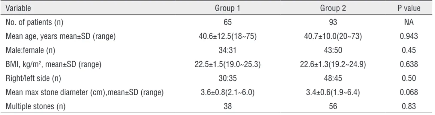
The patient demographics and stone cha-racteristics are reported in Table-1. The two Groups were comparable in terms of age, the male to female ratio, BMI(body mass index), side and stone diame-ter. There were no significant differences in the ope-ration time, stone-free rate hospital stay and access tract number (number of renal calix punctured per patient) between the two Groups (Table-2).
Three cases in Group 1 suffered urinary sep-sis (fever<39ºC) and two cases in Group 2. A pro-longed urinary leak from the flank was observed in one case in Group 1, which spontaneously stopped without requiring a double J stent positioning.
A greater drop in the hemoglobin level (P<0.001) was reported in group 2 (3.6g/dL) than in Group 1(2.4g/dL).
In Group 2, 9.7% of the patients (n=9) re-quired a blood transfusion, and 1 patient deve-loped a renal arteriovenous fistula and required embolus therapy. However, in Group 1, only 1.5% of the patients (n=9) suffered a severe hemorrhage and required a blood transfusion
postoperative-ly. In Group 1, a significant decrease in the blood transfusion rate was found compared with that in Group 2 (P=0.039; Table-3).
DISCUSSION
Percutaneous nephrolithotomy is less in-vasive than open surgery, and is associated with high success rates. Today, percutaneous nephro-lithotomy has become an effective and valued procedure for treating kidney and ureter stones. Although percutaneous nephrolithotomy is a minimally invasive technique, it carries out the potential risk of complications.
Table 1 - Patient demographics and stone characteristics\operative details and outcomes in the two Groups.
Variable Group 1 Group 2 P value
No. of patients (n) 65 93 NA
Mean age, years mean±SD (range) 40.6±12.5(18~75) 40.7±10.0(20~73) 0.943
Male:female (n) 34:31 43:50 0.45
BMI, kg/m2, mean±SD (range) 22.5±1.5(19.0~25.3) 22.6±1.3(19.2~24.9) 0.638
Right/left side (n) 30:35 48:45 0.50
Mean max stone diameter (cm),mean±SD (range) 3.6±0.8(2.1~6.0) 3.4±0.6(1.9~6.4) 0.068
Multiple stones (n) 38 56 0.83
Table 2 - Operative details and outcomes in the two groups.
Variable Group 1 Group 2 P value
Operation time(min) mean±SD 75.5±28.4 77.6±23.0 0.604
Stone-free rate(%) 87.7 88.2 0.60
Mean hospital stay (d) (SD) 8.1±1.7 8.3±1.5 0.279
Access number 1.2±0.5 1.3±0.6 0.241
Table 3 - Bleeding Complications.
Variable Group 1 Group 2 P value
Drop in hemoglobin (g/dL) (mean) 2.4 3.6 0.000
Blood transfusion (%) 1.5 9.7 0.039
Arteriovenous fistula(n) 0 1 0.40
The complication rates associated with PCNL have been reported to range from 29% to 83% (1-3), and in a recent analysis, the compli-cation rate was 15% (5). Complicompli-cations related to percutaneous nephrolithotomy include: infection, bleeding, pelvic perforation, urinary fistulas and perforations of adjacent organs. Bleeding is one of the most significant complications.
In the percutaneous nephrolithotomy Glo-bal Study analysis, the bleeding complication rate and transfusion rate were 9.4% and 7.0%, respec-tively (4). In the percutaneous nephrolithotomy study reported by Zehri and coworkers, the overall blood transfusion rate was 14.2% (6). In our study, 9.7% of the patients in Group 2 required a blood transfusion and only 1 patient in Group 1.
The preoperative factors that were reported to be predictors of blood loss in percutaneous ne-phrolithotomy include age, hypertension, diabetes mellitus (DM), ipsilateral pyelonephritis, the serum creatinine level, stone localization and burden, previous ipsilateral renal stone surgery and extra-corporeal shock wave lithotripsy and the degree of hydronephrosis and stone type (7-9). The operati-ve factors were the operation time, puncture calyx and tract number (9). An analysis suggested that BMI may increase the risk of bleeding (10), but other studies reported that BMI was not associated with the complication rates (11-13).
Rosette et al. reported that dilation was a risk factor for bleeding complications, with balloon di-lation significantly increasing the risk of bleeding compared with telescopic/serial dilation. In their stu-dy, bleeding was reported in 9.4% of the patients with balloon dilation compared with 6.7% with te-lescopic/serial dilation; the sheath size may have in-fluenced the transfusion rates. The transfusion rate was only 1.1% for the smallest sheath but was 12.0% for the largest sheath (5). We believe that the use of smaller sheaths could help reduce bleeding.
Bleeding is usually caused by a renal vascu-lar injury, which may occur at any step of the per-cutaneous nephrolithotomy. A renal vascular injury may lead to several consequences, including arterio-venous fistula, pseudoaneurysm, hemorrhage, hypo-tension, and loss of renal function (14).
Most vascular injuries manifest as hematu-ria. Bleeding from venous vessels can be managed
by simple maneuvers, such as placing the patient in the supine position, positioning a nephrostomy ca-theter, and clamping the nephrostomy catheter (15). Arterial lesions may lead to severe acute bleeding. Some types of severe bleeding, such as arteriovenous fistulas and pseudoaneurysms, can be persistent and require specific treatments. The incidence of arteriovenous fistulas or pseudoa-neurysms associated with percutaneous nephroli-thotomy is approximately 0.8% (16). In Group 2, 1 patient (1.1%) developed a renal arteriovenous fistula. The treatment is more troublesome if the bleeding occurs during the course of the percuta-neous nephrolithotomy.
Duplex US and computed tomography angiography are often used to diagnose vascular injuries. Hyperselective renal embolization is con-sidered to be the most appropriate technique for treating renal vascular injuries, with a high suc-cess rate and a low complication rate (15). Renal angiography is used to simultaneously diagnose and treat arteriovenous fistula or pseudoaneu-rysms (17). In our study, the patient who develo-ped a renal arteriovenous fistula was treated with hyperselective renal artery embolus therapy.
Although there are many maneuvers to control bleeding, which may lead the loss of renal function, the prevention of any bleeding should be a major focus during percutaneous nephroli-thotomy. To decrease the incidence of hemorrha-gic complications, the mini-percutaneous techni-que has been applied widely. In a recent study, B-mode combined with color Doppler ultrasound guidance in percutaneous nephrolithotomy avoi-ded renal vascular injury, and reduced the risk of bleeding occurrence (18). Penbegül reported that percutaneous nephrolithotomy with ultrasound guidance could be safely performed in children (19). Although the use of ultrasound guiding puncture could avoid vessel lesions, we think it is necessary to select calyces preoperatively.
vas-cular structure (20). Raman et al. reported that the sensitivity of computed tomography angio-graphy for demonstration of the renal artery was 98.5% (21). Computed tomography angiography has the advantage of evaluating not only the main vessels but also renal tumors and stones with only one test (22).
Currently, non-contrast CT has already been shown to be useful in pre-operative plan-ning for percutaneous nephrolithotomy. Al-though non-contrast CT (Figure-2) may be able to identify the posterior calyx, it cannot display renal vasculature, and reduce vascular compli-cations. In this study, computed tomography angiography was performed preoperatively (Fi-gure-1) and provided a map of the renal vascu-lature, renal artery, segmental artery and larger vessel branches were displayed accurately in Group 1. By reading the computed tomography angiography, we selected access site(s) far away from larger vessels, and the risk of hemorrhage was reduced. In Group 1, no patient developed a renal arteriovenous fistula and required embo-lus therapy, and only 1 patient required a blood transfusion.
In this study, there were several limita-tions. First, the number of enrolled patients was rather small, and further studies are needed to verify our results. Second, there were selection bias in our study due to the retrospective na-ture. Third, there were some disadvantages of computed tomography angiography, such as the need for intravenous contrast (that affects renal function in patients with renal insufficiency), significant radiation exposure and higher cost.
CONCLUSIONS
In conclusion, the study showed that pa-tients who underwent computed tomography angiography prior to mini-percutaneous nephro-lithotomy had lower drop of hemoglobin and nee-ded less transfusions. The use of computed tomo-graphy angiotomo-graphy may be a viable alternative to decrease the likelihood of bleeding complica-tions during mini-percutaneous nephrolithotomy.
ABBREVIATIONS
CT = Computed tomography
CTA = Computed tomography angiography
PCNL = Percutaneous nephrolithotomy
BMI = Body mass index
DM = Diabetes mellitus
ACKNOWLEDGEMENTS
We thank the medical staff (Department of Urology, Dongguan People’s Hospital) for supporting the study, Professors Xiaolin Zheng (Department of Radiology, Dongguan People’s Hospital) for provi-ding CT materials, and the patients who kindly vo-lunteered their time to participate in the study.
FUNDINGS
This project was supported by Science and Technological Program (201110515001078 and 2012105102027) for Dongguan’s Higher Education, Science and Research, and Health Care Institutions.
CONFLICT OF INTEREST
None declared.
REFERENCES
1. Dindo D, Demartines N, Clavien PA. Classification of surgical complications: a new proposal with evaluation in a cohort of 6336 patients and results of a survey. Ann Surg. 2004;240:205-13.
2. Assmy AM, Shokeir AA, Nahas AR, Shoma AM, Eraky I, El-Kenawy MR, et al. Outcome of percutaneous nephrolithotomy: effect of body mass index. Eur Urol. 2007;52:199-204.
3. Michel MS, Trojan L, Rassweiler JJ. Complications in percutaneous nephrolithotomy. Eur Urol. 2007;51:899-906; discussion 906.
5. de la Rosette J, Assimos D, Desai M, Gutierrez J, Lingeman J, Scarpa R, et al. The Clinical Research Office of the Endourological Society Percutaneous Nephrolithotomy Global Study: indications, complications, and outcomes in 5803 patients. J Endourol. 2011;25:11-7.
6. Zehri AA, Biyabani SR, Siddiqui KM, Memon A. Triggers of blood transfusion in percutaneous nephrolithotomy. J Coll Physicians Surg Pak. 201;21:138-41.
7. Turna B, Nazli O, Demiryoguran S, Mammadov R, Cal C. Percutaneous nephrolithotomy: variables that influence hemorrhage. Urology. 2007;69:603-7.
8. Srivastava A, Singh KJ, Suri A, Dubey D, Kumar A, Kapoor R, et al. Vascular complications after percutaneous nephrolithotomy: are there any predictive factors? Urology. 2005;66:38-40.
9. Akman T, Binbay M, Sari E, Yuruk E, Tepeler A, Akcay M, et al. Factors affecting bleeding during percutaneous nephrolithotomy: single surgeon experience. J Endourol. 2011;25:327-33.
10. Bagrodia A, Gupta A, Raman JD, Bensalah K, Pearle MS, Lotan Y. Impact of body mass index on cost and clinical outcomes after percutaneous nephrostolithotomy. Urology. 2008;72:756-60.
11. Sergeyev I, Koi PT, Jacobs SL, Godelman A, Hoenig DM. Outcome of percutaneous surgery stratified according to body mass index and kidney stone size. Surg Laparosc Endosc Percutan Tech. 2007;17:179-83.
12. Olbert PJ, Hegele A, Schrader AJ, Scherag A, Hofmann R. Pre- and perioperative predictors of short-term clinical outcomes in patients undergoing percutaneous nephrolitholapaxy. Urol Res. 2007;35:225-30.
13. Koo BC, Burtt G, Burgess NA. Percutaneous stone surgery in the obese: outcome stratified according to body mass index. BJU Int. 2004;93:1296-9.
14. Kukreja R, Desai M, Patel S, Bapat S, Desai M. Factors affecting blood loss during percutaneous nephrolithotomy: prospective study. J Endourol. 2004;18:715-22.
15. Martin X, Murat FJ, Feitosa LC, Rouvière O, Lyonnet D, Gelet A, et al. Severe bleeding after nephrolithotomy: results of hyperselective embolization. Eur Urol. 2000;37:136-9.
16. Lahme S, Bichler KH, Strohmaier WL, Götz T. Minimally invasive PCNL in patients with renal pelvic and calyceal stones. Eur Urol. 2001;40:619-24.
17. Rastinehad AR, Andonian S, Smith AD, Siegel DN. Management of hemorrhagic complications associated with percutaneous nephrolithotomy. J Endourol. 2009;23:1763-7.
18. Lu MH, Pu XY, Gao X, Zhou XF, Qiu JG, Si-Tu J. A comparative study of clinical value of single B-mode ultrasound guidance and B-mode combined with color Doppler ultrasound guidance in mini-invasive percutaneous nephrolithotomy to decrease hemorrhagic complications. Urology. 2010;76:815-20.
19. Penbegül N, Tepeler A, Sancaktutar AA, Bozkurt Y, Atar M, Yildirim K, et al. Safety and efficacy of ultrasound-guided percutaneous nephrolithotomy for treatment of urinary stone disease in children. Urology. 2012;79:1015-9.
20. Türkvatan A, Ozdemir M, Cumhur T, Olçer T. Multidetector CT angiography of renal vasculature: normal anatomy and variants. Eur Radiol. 2009;19:236-44.
21. Raman SS, Pojchamarnwiputh S, Muangsomboon K, Schulam PG, Gritsch HA, Lu DS. Utility of 16-MDCT angiography for comprehensive preoperative vascular evaluation of laparoscopic renal donors. AJR Am J Roentgenol. 2006;186:1630-8.
22. Kawamoto S, Montgomery RA, Lawler LP, Horton KM, Fishman EK. Multidetector CT angiography for preoperative evaluation of living laparoscopic kidney donors. AJR Am J Roentgenol. 2003;180:1633-8.
_______________________ Correspondence address: