Forming a stone in pelviureteric junction obstruction: cause
or effect?
_______________________________________________
Theodora Stasinou
1, Andreas Bourdoumis
2, Junaid Masood
31 South Manchester University Hospitals NHS Foundation Trust, Manchester, UK; 2 North Manchester General Hospital, Acute Pennine Hospitals NHS Trust, Manchester, UK; 3 Homerton University Hospital NHS Foundation Trust, London, UK
ABSTRACT
ARTICLE
INFO
______________________________________________________________ ______________________
Objectives: To investigate a possible causal relationship for stone formation in pelvi-ureteric junction obstruction and to outline management options.
Materials and Methods: A literature search and evidence synthesis was conducted via electronic databases in the English language using the key words pelviureteric junction obstruction; urolithiasis; hyperoxaluria; laparoscopic pyeloplasty; flexible nephros-copy; percutaneous nephrolithotomy, alone or in combination. Relevant articles were analysed to extract conclusions.
Results: Concomitant pelviureteric junction obstruction (PUJO) and renal lithiasis has been reported only scarcely in the literature. Although PUJO has been exten-sively studied throughout the years, the presence of calculi in such a patient has not received equal attention and there is still doubt surrounding the pathophysiology and global management.
Conclusions: Metabolic risk factors appear to play an important role, enough to justify metabolic evaluation in these patients. Urinary stasis and infection are well known factors predisposing to lithiasis and contribute to some extent. The choice for treat-ment is not always straightforward. Managetreat-ment should be tailored according to de-gree of obstruction, renal function, patient symptoms and stone size. Simultaneous treatment is feasible with the aid of minimally invasive operative techniques and lapa-roscopy in particular.
Keywords:
Pelviureteric Junction
Obstruction; Calculi; Urolithiasis; Nephrostomy, Percutaneous
Int Braz J Urol. 2017; 43: 13-9
_____________________
Submitted for publication: September 14, 2015
_____________________
Accepted after revision: May 28, 2016
_____________________
Published as Ahead of Print: September 14, 2016
INTRODUCTION
Pelviureteric junction obstruction (PUJO) is well described in the literature as far as diag-nosis and treatment are concerned. Yet, there is much controversy regarding stone formation and management in these patients. PUJO was first des-cribed as a syndrome by Dietl in 1864 (1)and the ensuing fibrotic changes were demonstrated by Allen TD in 1970 (2). Subsequently, it was proven that if left untreated the narrow junction
lead to obstruction by compression and kinking at the junction (8, 9). Concomitant lithiasis of the urinary tract is not uncommon and whether it co--exists as a separate entity or is the result of a nar-row renal outflow tract is still debated. The preva-lence of lithiasis in patients with malformations of the kidney is described as higher than that of the general population (10). In a retrospective re-view of 1639 paediatric patients during a 45 year period at the Mayo Clinic, the prevalence was 70-fold that of the aged matched population (11, 12). This seems to be also true for the adult population (13). In an early series, David and Lavengood (14) reported concomitant lithiasis in 16% of patients undergoing open pyeloplasty, whereas others re-ported an incidence of up to 20% (15). PUJO in horseshoe kidneys is described as high as 35% (16) and Lampel et al. suggested that at least 14% of stones treated in such patients were associated with a narrow pelviureteric junction (17).
We have conducted a literature search in three-3-electronic databases (Medscape/E-medi-cine, Pub Med, EmBase) using the following key words: pelviureteric junction obstruction; uroli-thiasis; hyperoxaluria; laparoscopic pyeloplasty; flexible nephroscopy; percutaneous nephrolitho-tomy, alone or in combination. We isolated arti-cles in the English language, relevant to research and/or reports of concomitant lithiasis on a back-ground of PUJO. Overall, 17 articles were identi-fied, mostly case series and presentation of surgi-cal techniques. Only two reports (11, 13) focused on identifying any underlying pathophysiological changes in paediatric populations, while one fur-ther study examined the metabolic factors in renal stones coinciding with PUJO (18).
Pathogenesis of calculi in PUJO
There are few reports in the literature that examine the significance and/or correlation of the ultrastructural changes in the narrow pelviureteric junction with the incidence of renal calcul (11-13). In one such retrospective analysis, all patients had histologic evidence of tissue changes (increased fibrosis) associated with anatomical obstruction, similar to those originally described by Allen TD for true congenital PUJO (13). In theory, an im-pacted stone at the pelviueteric junction is likely
ab-Table 1 - Metabolic risk factors for stone formation in patients with PUJO (percentages correspond to those that were metabolically evaluated).
Risk factors Study Series
Hyperoxaluria Hypercalciuria Hyperuricosuria Hypocitraturia
Hussman et al. (13) N/A 61%(observation group) and 17%(struvite group)
11% (observation group) and 8%(struvite
group)
22%(observation group) and 8% (struvite group)
Hussman et al. (11) N/A 36% (observation
group) and 17% (struvite group)
14% (observation group) and 17%(struvite group)
9% ( non-struvite only)
Matin and Streem (18) 24% vs 12% in control
33% vs 12% in control 29% vs 8% in control 19% vs 27% in control
normality. In contrast, subsequent medical mana-gement in the treatment arm (n=24) yielded only 17% stone recurrence rate. In the struvite group, 43% of recurrent calculi occurred in the con-tralateral kidney. Long term antibiotic treatment appeared to be beneficial with regards to stone recurrence in this group. The same authors sub-sequently reviewed a paediatric population with similar characteristics in retrospect, and found a recurrence rate of 68% in long term follow-up, with comparable results as for the metabolic fac-tors found in adults, further supporting the con-cept of an underlying metabolic etiology (11).
In their prospective observational study, Matin and Streem evaluated 47 patients with con-genital PUJO for factors predisposing to lithiasis (18). Of the 21 patients with stones, 67% presented identifiable metabolic risk factors vs. 38% of the 26 control patients with PUJO and no stones. The incidence was not unlike that found in stone form-ing populations (18, 20). The composition of such stones was found to be calcium oxalate in 93% of patients, with or without calcium phosphate as an additional mineral. The authors acknowledge small number of patients in the study (n=47), but pertain to the prospective design of the evalua-tion, to conclude that metabolic evaluation is re-quired in the treatment plan of concurrent PUJO and renal calculi. The same conclusion is also pro-duced by Hussman (Table-1) (13). Hyperoxaluria and hypercalciuria have been confirmed as
hav-ing positive correlation with PUJO and lithiasis in respective series of paediatric patients (21, 22). Summary of the metabolic risk factors identified during these studies is presented in Table-1. In the retrospective study by Bernado et al., 90 patients with PUJO who underwent endopyelotomy and simultaneous stone removal were compared with 80 patients without obstruction who underwent only stone extraction. The authors argue against metabolic factors as a prerequisite, since 71.4% of patients without PUJO were found to have a metabolic abnormality that predisposed to urinary stones, as opposed to 19% with obstruction (23). The authors concluded that correction of the ana-tomic obstruction facilitates the drainage of urine, thus decreasing the incidence of recurrent urinary stone formation.
Overall, the available evidence point to-ward a combination of factors that seem to be res-ponsible for lithiasis in PUJO other than urinary stasis caused by the obstruction. An undiagnosed metabolic abnormality and probably genetic pre-disposition are likely, while urinary tract infection and pH appear to play a part as well. It is therefore important to consider these possibilities and in-clude respective appropriate measures in the for-mulation of a global treatment strategy.
Management options
answering two important questions, that of when and how to treat. The significance of the exact location and number of the calculi in the pelvi-calyceal system is not adequately described in the majority of the studies. An initial period of obser-vation seems reasonable for asymptomatic stones of less than 5mm in greatest dimension, accom-panied by regular follow-up of the degree of obs-truction and renal function (24). With increasing symptoms, stone size, deteriorating renal function and/or recurrent infections, active treatment be-comes necessary. Minimally invasive procedures should be preferred where available. While open, laparoscopic and lately robotically assisted pyelo-plasty constitute established treatment options for PUJO, no such consensus exists for treating the stone. For our proposed algorithm in Figure-1, we suggest that PUJO is an already established diag-nosis at the time of choice of treatment, preferable by nuclear renogram studies that demonstrate
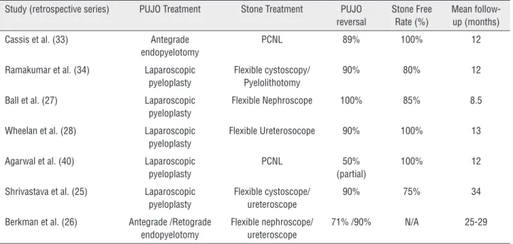
obs-truction as part of the pre-operative assessment. In the modern era of endoscopic stone surgery simultaneous treatment appears feasible, even in cases with multiple stones and difficult anatomy, i.e. calyceal stones, where retrograde flexible in-struments seem to be very useful (25-28). Table-2 provides a summary of the existing evidence in endoscopic management of such cases. A recen-tly published review by Skolarikos et al. and also laparoscopic series support this concept (29-34). Endoscopic combined intrarenal surgery (ECIRS) could also be an option, although the evidence is lacking. For staghorn and struvite stones in parti-cular, it is prudent to take caution and ensure peri--and postoperative antibiotic cover guided by uri-ne culture and local sensitivity patterns (35, 36). The duration and programming of follow-up is yet to be determined, but should include a history of symptoms, routine renal biochemistry and radio-nuclide imaging, i.e. MAG-3 renogram as a
mum (37, 38). We also recommend performing a thorough metabolic work-up, similar to that pro-posed for recurrent stone formers, both before and after definitive treatment, in order to identify the metabolic stone formers and formulate an appro-priate preventive strategy (39).
CONCLUSIONS
Stone disease in pelviureteric junction ob-struction is associated with an underlying metabolic disorder in up to a third of patients. Metabolic risk factors appear to play an important role, enough to justify metabolic evaluation of such patients. Urinary stasis and infection are well known fac-tors predisposing to lithiasis and also appear to be contributory factors. The choice for treatment is not always straightforward and relies on several factors, including organization of the department with dedi-cated stone clinic services, availability of appropria-te equipment to carry out complex endourological surgery and experience in postoperative follow-up and complication management. Upon verification of PUJO with nuclear functional imaging studies, further intervention should be tailored according to degree of obstruction, renal function, patient symp-toms and stone burden. Simultaneous treatment is
feasible with the aid of minimally invasive operative techniques and laparoscopic approach in particular appears to be the most promising solution (Table-2). Robotically-assisted laparoscopy is rapidly growing in the field and appears promising (40). Metabolic evaluation should be an integral part of initial eva-luation as well as follow-up and form the basis for future preventative planning against recurrences.
CONFLICT OF INTEREST
None declared.
REFERENCES
1. Dietl J: Wandernde nieren and deren einklemmung. Wien Med Wochenschr. 1864; 14: 153-66.
2. Allen TD. Congenital ureteral strictures. J Urol. 1970;104:196-204.
3. Whitaker RH. Methods of assessing obstruction in dilated ureters. Br J Urol. 1973;45:15-22.
4. Koff SA, Hayden LJ, Cirulli C, Shore R. Pathophysiology of ureteropelvic junction obstruction: experimental and clinical observations. J Urol. 1986;136:336-8.
5. Hanna MK, Jeffs RD, Sturgess JM, Barkin M. Ureteral structure and ultrastructure. Part II. Congenital ureteropelvic junction obstruction and primary obstructive megaureter. J Urol. 1976;116:725-30.
Table 2 - Summary of results following simultaneous PUJO correction and stone treatment.
Study (retrospective series) PUJO Treatment Stone Treatment PUJO reversal
Stone Free Rate (%)
Mean follow-up (months)
Cassis et al. (33) Antegrade
endopyelotomy
PCNL 89% 100% 12
Ramakumar et al. (34) Laparoscopic pyeloplasty
Flexible cystoscopy/ Pyelolithotomy
90% 80% 12
Ball et al. (27) Laparoscopic
pyeloplasty
Flexible Nephroscope 100% 85% 8.5
Wheelan et al. (28) Laparoscopic pyeloplasty
Flexible Ureterosocope 90% 100% 13
Agarwal et al. (40) Laparoscopic pyeloplasty
PCNL 50%
(partial)
100% 12
Shrivastava et al. (25) Laparoscopic pyeloplasty
Flexible cystoscope/ ureteroscope
90% 75% 34
Berkman et al. (26) Antegrade /Retograde endopyelotomy
Flexible nephroscope/ ureteroscope
6. Hanna MK. Some observations on congenital ureteropelvic junction obstruction. Urology. 1978;12:151-9.
7. Notley RG. Electron microscopy of the upper ureter and the pelvi-ureteric junction. Br J Urol. 1968;40:37-52. 8. Perlberg S, Pfau A. Management of ureteropelvic junction
obstruction associated with lower polar vessels. Urology. 1984;23:13-8.
9. Rutchik SD, Resnick MI. Ureteropelvic junction obstruction and renal calculi. Pathophysiology and implications for management. Urol Clin North Am. 1998;25:317-21. 10. Gambaro G, Fabris A, Puliatta D, Lupo A. Lithiasis in
cystic kidney disease and malformations of the urinary tract. Urol Res. 2006;34:102-7.
11. Husmann DA, Milliner DS, Segura JW. Ureteropelvic junction obstruction with concurrent renal pelvic calculi in the pediatric patient: a long-term followup. J Urol. 1996;156:741-3.
12. Rickwood AM, Reiner I. Urinary stone formation in children with prenatally diagnosed uropathies. Br J Urol. 1991;68:541-2.
13. Husmann DA, Milliner DS, Segura JW. Ureteropelvic junction obstruction with a simultaneous renal calculus: long-term followup. J Urol. 1995;153:1399-402.
14. David HS, Lavengood RW Jr. Ureteropelvic junction obstruction in nephrolithiasis. An etiologic factor. Urology. 1975;5:188-90.
15. Clark WR, Malek RS. Ureteropelvic junction obstruction. I. Observations on the classic type in adults. J Urol. 1987;138:276-9.
16. Smith JE, Van Arsdalen KN, Hanno PM, Pollack HM. Extracorporeal shock wave lithotripsy treatment of calculi in horseshoe kidneys. J Urol. 1989;142:683-6.
17. Lampel A, Hohenfellner M, Schultz-Lampel D, Lazica M, Bohnen K, Thürof JW. Urolithiasis in horseshoe kidneys: therapeutic management. Urology. 1996;47:182-6. 18. Matin SF, Streem SB. Metabolic risk factors in patients
with ureteropelvic junction obstruction and renal calculi. J Urol. 2000;163:1676-8.
19. Scardino PT. Obstruction at the ureteropelvic junction. In: Bergman H (ed.). The Ureter, 2nd Edition, Chapter. 33, Springer, New York. 1981; pp. 697.
20. Johri N, Cooper B, Robertson W, Choong S, Rickards D, Unwin R. An update and practical guide to renal stone management. Nephron Clin Pract. 2010;116:c159-71. 21. Tekin A, Tekgul S, Atsu N, Ergen A, Kendi S. Ureteropelvic
junction obstruction and coexisting renal calculi in children: role of metabolic abnormalities. Urology. 2001;57:542-5.
22. García-Nieto V, Navarro JF, Luis-Yanes MI, López-Méndez M, García-Rodríguez V. Hypercalciuria in pediatric patients with ureteropelvic junction obstruction is of genetic origin. Scand J Urol Nephrol. 2007;41:144-8.
23. Bernardo NO, Liatsikos EN, Dinlenc CZ, Kapoor R, Fogarty JD, Smith AD. Stone recurrence after endopyelotomy. Urology. 2000;56:378-81.
24. Koh LT, Ng FC, Ng KK. Outcomes of long-term follow-up of patients with conservative management of asymptomatic renal calculi. BJU Int. 2012;109:622-5.
25. Srivastava A, Singh P, Gupta M, Ansari MS, Mandhani A, Kapoor R, et al. Laparoscopic pyeloplasty with concomitant pyelolithotomy--is it na effective mode of treatment? Urol Int. 2008;80:306-9.
26. Berkman DS, Landman J, Gupta M. Treatment outcomes after endopyelotomy performed with or without simultaneous nephrolithotomy: 10-year experience. J Endourol. 2009;23:1409-13.
27. Ball AJ, Leveillee RJ, Patel VR, Wong C. Laparoscopic pyeloplasty and flexible nephroscopy: simultaneous treatment of ureteropelvic junction obstruction and nephrolithiasis. JSLS. 2004;8:223-8.
28. Whelan JP, Wiesenthal JD. Laparoscopic pyeloplasty with simultaneous pyelolithotomy using a flexible ureteroscope. Can J Urol. 2004;11:2207-9.
29. Skolarikos A, Dellis A, Knoll T. Ureteropelvic obstruction and renal stones: etiology and treatment. Urolithiasis. 2015;43:5-12. 30. Wang X, Li S, Liu T, Guo Y, Yang Z. Laparoscopic
pyelolithotomy compared to percutaneous nephrolithotomy as surgical management for large renal pelvic calculi: a meta-analysis. J Urol. 2013;190:888-93.
31. Stravodimos KG, Giannakopoulos S, Tyritzis SI, Alevizopoulos A, Papadoukakis S, Touloupidis S, et al. Simultaneous laparoscopic management of ureteropelvic junction obstruction and renal lithiasis: the combined experience of two academic centers and review of the literature. Res Rep Urol. 2014;6:43-50.
32. Agarwal A, Varshney A, Bansal BS. Concomitant percutaneous nephrolithotomy and transperitoneal laparoscopic pyeloplasty for ureteropelvic junction obstruction complicated by stones. J Endourol. 2008;22:2251-5.
33. Cassis AN, Brannen GE, Bush WH, Correa RJ, Chambers M. Endopyelotomy: review of results and complications. J Urol. 1991;146:1492-5.
34. Ramakumar S, Lancini V, Chan DY, Parsons JK, Kavoussi LR, Jarrett TW. Laparoscopic pyeloplasty with concomitant pyelolithotomy. J Urol. 2002;167:1378-80.
35. Gonen M, Turan H, Ozturk B, Ozkardes H. Factors affecting fever following percutaneous nephrolithotomy: a prospective clinical study. J Endourol. 2008;22:2135-8.
36. Gutierrez J, Smith A, Geavlete P, Shah H, Kural AR, de Sio M, et al. Urinary tract infections and post-operative fever in percutaneous nephrolithotomy. World J Urol. 2013;31:1135-40.
38. Inagaki T, Rha KH, Ong AM, Kavoussi LR, Jarrett TW. Laparoscopic pyeloplasty: current status. BJU Int. 2005;95:102-5.
39. Skolarikos A, Straub M, Knoll T, Sarica K, Seitz C, Petřík A, et al. Metabolic evaluation and recurrence prevention for urinary stone patients: EAU guidelines. Eur Urol. 2015;67:750-63.
40. Atug F, Castle EP, Burgess SV, Thomas R. Concomitant management of renal calculi and pelvi-ureteric junction obstruction with robotic laparoscopic surgery. BJU Int. 2005;96:1365-8.
_______________________ Correspondence address: