Oxygen desaturation in healthy subjects undergoing the
incremental shuttle walk test*
Dessaturação em indivíduos saudáveis submetidos ao incremental shuttle walk test
Daniel Machado Seixas, Daniela Miti Tsukumo Seixas, Monica Corso Pereira, Marcos Mello Moreira, Ilma Aparecida Paschoal
Abstract
Objective: To determine the probability of oxygen desaturation in healthy individuals undergoing the incremental
shuttle walk test (ISWT). Methods: We enrolled 83 healthy subjects: 55 males (including 1 smoker) and 28 females. We determined pre-ISWT FEV1, FEV6, HR and SpO2, as well as post-ISWT HR and SpO2. Results: Mean values overall were as follows: age, 35.05 ± 12.53 years; body mass index, 24.30 ± 3.47 kg/m2; resting HR, 75.12 ± 12.48 bpm; resting SpO2, 97.96 ± 1.02%; FEV1, 3.75 ± 0.81 L; FEV6, 4.45 ± 0.87 L; FEV1/FEV6 ratio, 0.83 ± 0.08 (no restriction or obstruction); incremental shuttle walk distance, 958.30 ± 146.32 m; post-ISWT HR, 162.41 ± 18.24 bpm; and post-ISWT SpO2, 96.27 ± 2.21%. In 11 subjects, post-ISWT SpO2 was higher than was pre-ISWT SpO2. In 17 subjects, there was a 4% decrease in SpO2 after the ISWT. There were no statistically significant differences between the groups with and without post-ISWT oxygen desaturation in terms of age, gender, FEV1, FEV6, FEV1/FEV6, pre-ISWT SpO2, incremental shuttle walk distance, HR, or percentage of maximal HR. In the individuals with post-ISWT oxygen desaturation, the body mass index was higher (p = 0.01) and post-ISWT SpO2 was lower (p = 0.0001). Conclusions: Healthy individuals can present oxygen desaturation after the ISWT. Using the ISWT to predict subtle respiratory abnormalities can be misleading. In healthy subjects, oxygen desaturation is common after the ISWT, as it is during any intense physical activity.
Keywords: Heart function tests; Respiratory function tests; Body mass index; Oximetry
Resumo
Objetivo: Determinar a probabilidade de dessaturação arterial em indivíduos saudáveis submetidos ao incremental
shuttle walk test (ISWT). Métodos: Foram estudados 83 indivíduos saudáveis, dos quais 55 eram homens (1 deles fumante) e 28 eram mulheres. Foram determinados VEF1 e VEF6 antes da realização do ISWT, assim como FC e SpO2 antes e depois do ISWT. Resultados: As médias gerais foram as seguintes: idade, 35,05 ± 12,53 anos; índice de massa corporal, 24,30 ± 3,47 kg/m2; FC em repouso, 75,12 ± 12,48 bpm; SpO
2 em repouso, 97,96 ± 1,02%; VEF1, 3,75 ± 0,81 L; VEF6, 4,45 ± 0,87 L; relação VEF1/VEF6, 0,83 ± 0,08 (sem restrição ou obstrução); distância percorrida no ISWT, 958,30 ± 146,32 m; FC pós-ISWT, 162,41 ± 18,24 bpm e SpO2 pós-ISWT, 96,27 ± 2,21% Em 11 indivíduos, houve um aumento da SpO2 após o ISWT, ao passo que em 17 houve uma queda de 4%. Não houve diferença estatística entre os grupos com e sem dessaturação após o ISWT no tocante às variáveis idade, gênero, VEF1, VEF6, VEF1/VEF6, SpO2 basal, distância percorrida no ISWT, FC e porcentagem da FC máxima. Nos indivíduos que apresentaram dessaturação, o índice de massa corporal foi maior (p = 0,01) e a SpO2 pós-ISWT foi menor (p = 0,0001). Conclusões: Indivíduos saudáveis podem apresentar dessaturação após o ISWT. O uso do ISWT para prever a presença de problemas respiratórios sutis pode ser enganador. Em indivíduos saudáveis, a dessaturação é um evento comum após o ISWT, assim como o é durante a atividade física intensa.
Descritores: Testes de função cardíaca; Testes de função respiratória; Índice de massa corporal; Oximetria.
*Study carried out at the State University at Campinas, Campinas, Brazil.
Correspondence to: Ilma Aparecida Paschoal. Cidade Universitária “Zeferino Vaz”, Disciplina de Pneumologia, FCM-UNICAMP, Distrito de Barão Geraldo, CEP 13083-888 Campinas, SP, Brasil.
Tel. 55 19 3521-7948. E-mail: [email protected] Financial support: None.
Introduction
Although patients with interstitial pulmonary fibrosis or pulmonary arterial hypertension can have normal SpO2 at rest, some will show oxygen desaturation after submaximal exercise.
(1,2) End-exercise PaO
2 decreases after maximal
exercise, and submaximal steady-state exercise was found to be an important measure of disease severity in interstitial pulmonary fibrosis.(3,4) Lama
et al. demonstrated that patients with usual interstitial pneumonia who developed oxygen desaturation during and after a six-minute walk test (6MWT)—a ≥ 4% decrease in oxygen saturation from baseline (Δsat ≥ 4%)—were more than four times more likely to die during follow-up.(5) The
abovementioned findings led us to hypothesize that a decrease in oxygen saturation during self-paced walking (a submaximal exercise test) is a meaningful measure of disease status in patients with scleroderma. Our results showed that, in the multiple logistic regression analysis, the variable Δsat ≥ 4% was significantly associated with age, dyspnea, and two other variables related to pulmonary involvement, i.e., FVC < 80% of the predicted value (as assessed by spirometry) and positivity for the Scl-70 antibody, which is a marker of pulmonary disease in scleroderma. However, the statistical model applied to the data did not indicate which of the dependent variables analyzed (Δsat ≥ 4% or the distance walked) was better at predicting pulmonary disease. Nevertheless, Δsat ≥ 4% appeared to be able to provide more information on this issue than did the distance walked.(6)
Another study conducted by our research group and involving patients with systemic lupus erythematosus showed that those with post-6MWT Δsat ≥ 4% (as assessed by pulse oximetry) showed a significant reduction in the six-minute walk distance (6MWD), which was 443 m in the group of patients who developed oxygen desaturation and 497 m in that of those who did not (p = 0.0291). However, both 6MWDs were well above the lower limit of the normal range. In addition, when compared with the patients who did not develop oxygen desaturation, those who did had a higher post-6MWT HR (p = 0.0170), a lower MEP (p = 0.0282), a lower MIP (p = 0.0504), and a restrictive pattern of lung disease (as determined by spirometry). These findings suggest that oxygen desaturation is more sensitive in detecting the presence of respiratory abnormalities in patients
with systemic lupus erythematosus than is the 6MWD (unpublished data). Therefore, oxygen desaturation during submaximal exercise seems to be a more sensitive endpoint to detect subtle respiratory abnormalities in pulmonary diseases and in systemic diseases that affect respiration.
Maximal exercise tests, such as cardiopulmonary exercise tests, can provide important information on the integration between the cardiovascular and respiratory systems during exercise. Although cardiopulmonary exercise tests have been extensively used in order to investigate various lung diseases, they are complex tests that require expensive equipment.
The incremental shuttle walk test (ISWT) is a maximal exercise test that provides data that correlate well with measurements made during incremental cardiopulmonary exercise testing, being simpler and less expensive than the latter. The fact that even patients with few symptoms and mild pulmonary involvement can present with a decrease in oxygen saturation in a submaximal test led to us inquire how oxygen saturation would behave during a maximal walk test, such as the ISWT. Could it improve the sensitivity of the 6MWT in detecting respiratory impairment? With the objective of exploring this hypothesis, we decided to perform the ISWT in healthy individuals in order to determine the probability of oxygen desaturation at the end of the test.
Methods
This was a cross-sectional study involving healthy subjects who attended a private fitness center in the city of Campinas, Brazil. All of the enrolled subjects attended the fitness center in order to keep physically active and in good health, and none were professional athletes. They were invited to participate in the study and agreed to perform the tests included in the protocol.
The study was approved by the Research Ethics Committee of the Universidade Estadual de Campinas (Unicamp, State University at Campinas) Hospital de Clínicas, located in the city of Campinas, Brazil, and all participants gave written informed consent.
A digital peak flow meter (Koko Peak Pro 6; Ferraris Cardiorespiratory PDS Healthcare Products Inc., Louisville, CO, USA) was used in order to determine pre-ISWT FEV1 and FEV6. The FEV1/ FEV6 ratio was calculated by the device and was displayed on the screen. Each subject performed at least three forced expiratory maneuvers after maximal inspiratory maneuvers, the best maneuver being automatically chosen by the peak flow meter. Pre-ISWT HR and oxygen saturation were determined with a pulse oximeter equipped with a finger probe (Nonin Medical, Inc., Plymouth, MN, USA). Fingernail polish, if worn by the subject, was removed before testing. In order to guarantee an accurate assessment of oxygen saturation, the principal investigator checked that the pulse oximeter showed an acceptable pulse signal and that the oximeter light was green and pulsing in synchrony with the HR before the beginning of the tests.
The protocol used for the ISWT was the 12-level version proposed by Singh et al.(7) for functional
capacity evaluation in patients with COPD. All participants were tested under standardized conditions by the same investigator. The walking course was level, being 10 m in length. It was delimited by two cones inset 0.5 m from either end to avoid the need for abrupt changes in
direction. The speed at which subjects walked was dictated by an audio signal played on a portable microcomputer. Each participant received a standardized explanation (“you should walk at a steady pace, aiming to turn around whenever you hear the signal; you should continue to walk until you feel that you are unable to maintain the required speed”). At the first level of the test, the calculated walking speed was 0.5 m/s and the number of expected shuttles was 3; at the twelfth level, walking speed was 2.37 m/s and 14 shuttles were expected. All subjects were carefully observed during the test so that they would not exceed their exercise limit.
Oxygen saturation was also measured immediately after the end of the ISWT. For the purpose of data analysis, oxygen desaturation was defined as a ≥ 4% decrease in oxygen saturation (SpO2) from baseline (Δsat = oxygen saturation
at rest − oxygen saturation immediately after the
ISWT). This 4% decrease in oxygen saturation was validated in studies of exercise-induced hypoxemia during maximal exercise tests in athletes.
(8) Maximum incremental shuttle walk distance
(ISWD) was defined as the maximum distance that the subjects were able to walk during the ISWT.
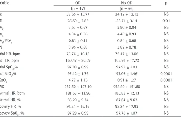
Table 1 - Comparison between the two groups (with and without oxygen desaturation).a
Variable OD No OD p
(n = 17) (n = 66)
Age 38.65 ± 13.77 34.12 ± 12.13 NS
BMI 26.59 ± 3.85 23.71 ± 3.14 0.01
FEV1 3.53 ± 0.67 3.80 ± 0.84 NS
FEV6 4.34 ± 0.56 4.48 ± 0.93 NS
FEV1/FEV6 0.83 ± 0.11 0.84 ± 0.08 NS
LLN 3.95 ± 0.68 3.82 ± 0.78 NS
Initial HR, bpm 73.76 ± 10.16 75.47 ± 13.06 NS
Final HR, bpm 160.47 ± 20.59 162.91 ± 17.72 NS
Initial SpO2,% 97.88 ± 0.99 97.99 ± 1.03 NS
Final SpO2,% 93.12 ± 1.76 97.08 ± 1.46 0.0001
Δ SpO2 4.77 ± 1.15 0.91 ± 1.27 0.0001
ISWD 956.50 ± 127.10 958.80 ± 151.80 NS
Maximal HR, bpm 181.53 ± 13.96 185.88 ± 12.13 NS
Maximal HR, % 88.29 ± 9.34 87.64 ± 9.62 NS
Recovery HR, % 91.24 ± 15.16 92.24 ± 17.93 NS
Recovery SpO2, % 97.29 ± 0.99 97.70 ± 1.07 NS
A ≥ 4% decrease in oxygen saturation was used in order to divide the study population into two groups. The Anderson-Darling test was applied to the measured variables and the demographic characteristics of the two groups to determine their distribution. Variables with normal distribution were analyzed with the Student’s t-test. Variables with non-normal distribution were analyzed with the Wilcoxon test. Categorical data were compared by the chi-square test or Fisher’s exact test. The statistical analysis was performed with the SAS software, version 8 (SAS Institute, Inc., Cary, NC, USA). Differences were considered significant at p < 0.05.
Results
Eighty-three individuals who attended a fitness center in the city of Campinas, Brazil, were invited and agreed to participate in the study. Of those, 55 were male and 28 were female. Only 1 was a smoker. The mean age was 35.05 ± 12.53 years, the median being 32 years. The mean body mass index (BMI) was 24.30 ± 3.47 kg/m2, the median
being 24.5 kg/m2. The mean resting HR was 75.12
± 12.48 bpm, the median being 73 bpm. The mean pre-ISWT SpO2 was 97.96 ± 1.02%, the median being 98%. The mean FEV1 was 3.75 ± 0.81 L, the median being 3.65 L. The mean FEV6 was 4.45 ± 0.87 L, the median being 4.38 L. The mean FEV1/FEV6 ratio was 0.83 ± 0.08, the median being 0.82. In all enrolled subjects, measured FEV6 was above the lower limit of the predicted FVC (as determined by the equations devised for the Brazilian population), and all subjects had a FEV1/FEV6 ratio ≥ 0.8. These findings allow the assumption that none of the participants had restrictive or obstructive lung disease (Table 1).
The mean ISWD was 958.30 ± 146.32 m, the median being 1,020 m. The mean post-ISWT HR was 162.41 ± 18.24 bpm, the median being 166 bpm. The mean post-ISWT SpO2 was 96.27 ± 2.21%. In 11 subjects, post-ISWT oxygen saturation values were higher than pre-ISWT
oxygen saturation values. In 17 subjects, oxygen saturation had decreased by ≥ 4% by the end of the test. In 2 subjects, SpO2 was < 92% (Table 2). The study population was divided into two groups on the basis of the presence of a post-ISWT oxygen desaturation ≥ 4%. No differences were found between the two groups regarding age, gender, FEV1, FEV6, FEV1/FEV6, initial oxygen saturation (pre-ISWT SpO2), ISWD, pre-ISWT HR, post-ISWT HR, or percentage of maximal HR (Table 1). The BMI was significantly higher in those who developed oxygen desaturation (p = 0.01), and post-ISWT SpO2 was significantly different between the two groups (p < 0.0001).
Discussion
In 66 subjects, post-ISWT oxygen saturation values were quite similar to pre-ISWT oxygen saturation values, a finding that was expected because of the intensity of the exercise performed. In 11 subjects, post-ISWT SpO2 values were higher than pre-ISWT SpO2 values. This finding is not unusual, given that physical activity improves ventilation and alveolar recruitment. However, 17 (20.7%) of the 83 individuals in the study sample showed a significant drop in oxygen saturation during the ISWT (Δsat ≥ 4%). This was an unexpected finding, and there is little information in the literature regarding what happens with oxygen saturation after the ISWT in healthy subjects.
Exercise-induced hypoxemia in athletes is arbitrarily defined as a decrease in PaO2 of approximately 7.5 mmHg,(7) an SaO
2 below 95%, or
both; extreme cases will show an SaO2 of less than 88%.(9) Oxygen uptake increases during exercise in
order to meet the needs imposed by an increased metabolic rate and correlate with work intensity until all subjects achieve maximal oxygen uptake.
(10) Each step of oxygen transport from ambient air
to the cells can limit whole-body oxygen uptake, and circulation has been considered the most important factor limiting maximal oxygen uptake during large muscle mass exercise.
A decrease in PaO2 and SaO2 is quite common during maximal ergometer rowing.(11) In such
subjects, cardiac output can exceed 30 L/min; under such circumstances, the ability to renew alveolar air and maintain high oxygen partial pressures, the diffusion resistance to oxygen at the alveolar-capillary membrane, the reduction in red blood cell transit time in the pulmonary capillary,
Table 2 - Proportions of individuals who reached more than 85% or 85% or less of the predicted maximal HR in the two groups.a
Maximal HR OD No OD
> 85% 12 (71) 47 (71)
≤ 85% 5 (29) 19 (29)
Total 17 (100) 66 (100)
pulmonary impedance, a situation that can also impair the renewal of alveolar air.
Although it has been described as an incremental field walking test that produces a symptom-limited maximal performance, the ISWT cannot be compared with the maximal exercise tests cited above. Nevertheless, 17 healthy subjects showed oxygen desaturation after the ISWT in the present study. The only significant difference between the individuals who developed oxygen desaturation and those who did not was that the BMI was higher in the former. This finding is consistent with the hypothesis that the lungs, during physical activities that cause the HR to get closest to the maximal HR expected for a given subject, are unable to arterialize the fast flowing blood, especially in those subjects whose needs are amplified because of a higher body mass. Although Durand et al.(16)
found no differences in height, weight, or lung volume between athletes who developed oxygen desaturation and those who did not, it can be argued that the proportions of those variables are more important than their absolute values.
Our findings and the data from the literature lead to a worrisome possibility: oxygen desaturation during aerobic activity is probably more common than previously thought and can pose a threat to high-performance athletes that has been systematically overlooked. Although the occurrence of oxygen desaturation is acknowledged in studies that date back to the second half of the last century, none of those studies elaborated on the potential harmful effects of such periods of intermittent hypoxemia.
We are unaware of any evaluation protocol for high-performance aerobic training that includes the determination of exercise-induced oxygen desaturation. Given that sudden death is relatively common in athletes, screening for oxygen desaturation seems justified.
Sudden cardiac death (SCD) is considered the leading cause of death in young athletes. The true incidence of SCD is unknown and highly underestimated. The studies reporting the highest incidence estimated that up to 110 deaths occur each year in young athletes, which is equivalent to 1 death every 3 days in the United States.
(17) The available evidence points to a structural
cardiac abnormality as the underlying cause of SCD. Hypertrophic cardiomyopathy and coronary artery anomalies account for approximately 25% and 14%, respectively, of all SCDs in the and the increased probability of ventilation/
perfusion mismatch are critically important to oxygen uptake.(12)
The prevalence of exercise-induced hypoxemia seems to be as high as 50%.(13) Exercise intensity
determines the degree of hypoxemia.(14) Oxygen
desaturation is also more pronounced during whole-body exercise, such as rowing or running, than during leg exercise, and leg exercise is more capable of inducing hypoxemia than is arm exercise.
(14) This suggests that the amount of muscle mass
involved in the exercise influences the development of oxygen desaturation.
In 1984, Dempsey et al.(15) studied the incidence
United States.(18) Arrhythmogenic right ventricular
cardiomyopathy/dysplasia is a cardiac disease characterized by myocardial necrosis followed by fibrofatty replacement. These altered myocardial areas constitute the anatomical substrate for reentry circuits that propitiate the onset of ventricular arrhythmias.(19) This last condition can be
particularly significant in the context of the present study. Oxygen desaturation during intense physical activity can cause repeated episodes of hypoxic pulmonary vascular constriction and pulmonary hypertension. The walls of the right ventricle can suffer during these episodes, to the point of myocardial necrosis, fibrofatty replacement being the expected consequence of this kind of stress. Another common cause of SCD is Brugada syndrome,(20) which is characterized by an ST-segment
elevation in the right precordial electrocardiogram leads followed by a negative T wave. The worldwide prevalence of Brugada syndrome is estimated at 1-5 per 10,000 population, although it is higher in Southeast Asia.(21) Brugada syndrome is traditionally
thought of as a primary electrical cardiac disease arising in myocardium that is otherwise structurally normal. However, magnetic resonance imaging, positron emission tomography, and pathological evaluation of biopsy specimens have identified structural abnormalities in many patients with a diagnosis of Brugada syndrome, including fibrofatty replacement of the right ventricular free wall and fibrotic disruption of the right bundle branch.
It is established in the literature that Brugada syndrome is the result of an autosomal dominant mutation in the SCN5A gene on chromosome 3, resulting in a loss of function sodium channel abnormality.(22) It has become increasingly clear
that ion channel gene expression is highly dynamic and can respond to many environmental stimuli.
(23) Hypoxemia is possibly one of these stimuli.
Therefore, a genetic predisposition to cardiac arrhythmia does not preclude the superimposition of hypoxemia causing the sudden deaths of young people or athletes.
The data in the present study and the accumulated knowledge regarding oxygen desaturation during physical activity raise the hypothesis that hypoxemia during exercise can be dangerous and suggest that it is advisable to include a screening test for oxygen desaturation in the evaluation protocols for endurance athletes. Further studies are needed in order to explore this hypothesis.
In conclusion, because of the possibility of oxygen desaturation in healthy individuals undergoing the ISWT, the use of the ISWT to predict the presence of subtle respiratory abnormalities undetected by submaximal tests such as the 6MWT can be misleading. The finding that oxygen desaturation is common in healthy subjects undergoing the ISWT adds to the knowledge that oxygen desaturation during intense physical activity is quite common and can have deleterious effects.
References
1. Hallstrand TS, Boitano LJ, Johnson WC, Spada CA, Hayes JG, Raghu G. The timed walk test as a measure of severity and survival in idiopathic pulmonary fibrosis. Eur Respir J. 2005;25(1):96-103. http://dx.doi.org/10.1183/09031 936.04.00137203 PMid:15640329
2. Morales-Blanhir JE, Palafox Vidal CD, Rosas Romero Mde J, García Castro MM, Londo-o Villegas A, Zamboni M. Six-minute walk test: a valuable tool for assessing pulmonary impairment. J Bras Pneumol. 2011;37(1):110-7. http://dx.doi.org/10.1590/S1806-37132011000100016 PMid:21390439
3. King TE Jr, Tooze JA, Schwarz MI, Brown KR, Cherniack RM. Predicting survival in idiopathic pulmonary fibrosis: scoring system and survival model. Am J Respir Crit Care Med. 2001;164(7):1171-81. http://dx.doi.org/10.1164/ ajrccm.164.7.2003140 PMid:11673205
4. King TE Jr, Schwarz MI, Brown K, Tooze JA, Colby TV, Waldron JA Jr, et al. Idiopathic pulmonary fibrosis: relationship between histopathologic features and mortality. Am J Respir Crit Care Med. 2001;164(6):1025-32. http:// dx.doi.org/10.1164/ajrccm.164.6.2001056 PMid:11587991 5. Lama VN, Flaherty KR, Toews GB, Colby TV, Travis WD,
Long Q, et al. Prognostic value of desaturation during a 6-minute walk test in idiopathic interstitial pneumonia. Am J Respir Crit Care Med. 2003;168(9):1084-90. http:// dx.doi.org/10.1164/rccm.200302-219OC PMid:12917227 6. Villalba WO, Sampaio-Barros PD, Pereira MC, Cerqueira
EM, Leme CA Jr, Marques-Neto JF, et al. Six-minute walk test for the evaluation of pulmonary disease severity in scleroderma patients. Chest. 2007;131(1):217-22. http:// dx.doi.org/10.1378/chest.06-0630 PMid:17218579 7. Singh SJ, Morgan MD, Scott S, Walters D, Hardman
AE. Development of a shuttle walking test of disability in patients with chronic airways obstruction. Thorax. 1992;47(12):1019-24. http://dx.doi.org/10.1136/ thx.47.12.1019 PMid:1494764 PMCid:PMC1021093 8. Prefaut C, Durand F, Mucci P, Caillaud C. Exercise-induced
arterial hypoxaemia in athletes: a review. Sports Med. 2000;30(1):47-61. http://dx.doi.org/10.2165/00007256-200030010-00005 PMid:10907757
9. Dempsey JA, Wagner PD. Exercise-induced arterial hypoxemia. J Appl Physiol. 1999;87(6):1997-2006. PMid:10601141
11. Nielsen HB, Madsen P, Svendsen LB, Roach RC, Secher NH. The influence of PaO2, pH and SaO2 on maximal oxygen uptake. Acta Physiol Scand. 1998;164(1):89-7. http://dx.doi. org/10.1046/j.1365-201X.1998.00405.x PMid:9777029 12. Nielsen HB. pH after competitive rowing: the lower
physiological range? Acta Physiol Scand. 1999.;165(1):113-4. http://dx.doi.org/10.1046/j.1365-201x.1999.00485.x PMid:10072104
13. Powers SK, Dodd S, Lawler J, Landry G, Kirtley M, McKnight T, et al. Incidence of exercise induced hypoxemia in elite endurance athletes at sea level. Eur J Appl Physiol Occup Physiol. 1988;58(3):298-302. http://dx.doi.org/10.1007/ BF00417266 PMid:3220070
14. Nielsen HB. Arterial desaturation during exercise in man: implication for O2 uptake and work capacity. Scand J Med Sci Sports. 2003;13(6):339-58. http://dx.doi.org/10.1046/ j.1600-0838.2003.00325.x PMid:14617055
15. Dempsey JA, Hanson PG, Henderson KS. Exercise-induced arterial hypoxaemia in healthy human subjects at sea level. J Physiol. 1984;355:161-75. PMid:6436475 PMCid:PMC1193484
16. Durand F, Mucci P, Préfaut C. Evidence for an inadequate hyperventilation inducing arterial hypoxemia at submaximal exercise in all highly trained endurance athletes. Med Sci Sports Exerc. 2000;32(5):926-32. http://dx.doi. org/10.1097/00005768-200005000-00008 PMid:10795782 17. Casa DJ, Guskiewicz KM, Anderson SA, Courson RW,
Heck JF, Jimenez CC, et al. National athletic trainers’
association position statement: preventing sudden death in sports. J Athl Train. 2012;47(1):96-118. PMid:22488236 PMCid:PMC3418121
18. Maron BJ. Sudden death in young athletes. N Engl J Med. 2003;349(11):1064-75. http://dx.doi.org/10.1056/ NEJMra022783 PMid:12968091
19. Bauce B, Daliento L, Frigo G, Russo G, Nava A. Pregnancy in women with arrhythmogenic right ventricular cardiomyopathy/dysplasia. Eur J Obstet Gynecol Reprod Biol. 2006;127(2):186-9 http://dx.doi.org/10.1016/j. ejogrb.2005.10.011 PMid:16337730
20. Brugada P, Brugada J. Right bundle branch block, persistent ST segment elevation and sudden cardiac death: a distinct clinical and electrocardiographic syndrome. A multicenter report. J Am Coll Cardiol. 1992;20(6):1391-6. http://dx.doi. org/10.1016/0735-1097(92)90253-J
21. Antzelevitch C. Brugada syndrome. Pacing Clin Electrophysiol. 2006;29(10):1130-59. http://dx.doi.org/10.1111/j.1540-8159.2006.00507.x PMid:17038146 PMCid:PMC1978482 22. Walker J, Calkins H, Nazarian S. Evaluation of cardiac
arrhythmia among athletes. Am J Med. 2010;123(12):1075-81. http://dx.doi.org/10.1016/j.amjmed.2010.05.008 PMid:20870195 PMCid:PMC3010317
23. Hilber K. Skeletal myocyte plasticity: basis for improved therapeutic potential? Curr Opin Pharmacol. 2008;8(3):327-32. http://dx.doi.org/10.1016/j.coph.2008.01.007 PMid:18329336 PMCid:PMC2957812
About the authors
Daniel Machado Seixas
Professor of Physical Education. State University at Campinas, Campinas, Brazil.
Daniela Miti Tsukumo Seixas
Physician. State University at Campinas, Campinas, Brazil.
Monica Corso Pereira
Physician. State University at Campinas, Campinas, Brazil.
Marcos Mello Moreira
Respiratory Diseases Service. State University at Campinas, Campinas, Brazil.
Ilma Aparecida Paschoal