ORIGINAL
RES
EAR
CH
Correspondence to: Rodrigo Cappato – Universidade de Pernambuco, Campus Petrolina, BR 203 Km 2 S/N – Vila Eduardo – CEP: 56300-000 – Petrolina (PE), Brasil – E-mail: [email protected]
Presentation: jun. 2012 – Accepted for publication: jan. 2013 – Financial support: none – Conflict of interest: nothing to declare – Approval from the Ethics Committee nº 202/09.
ABSTRACT | The purpose of this study was to analyze
the influence of high-heeled shoes on the quadriceps electromyographic activity (EMG) during the sit-to-stand task. Ten healthy females (20.2±3.0 years) and 10 females with patellofemoral pain syndrome (PFPS) (21.3±3.4 years) participated in this study. The subjects performed a stan-dardized sit-to-stand task under 3 conditions: barefoot, wearing sneakers and wearing 10 cm high-heeled shoes. The electromyographic (EMG) activity was recorded from the vastus medialis obliquus (VMO), vastus lateralis (VL) and rectus femoris (RF) muscles during the tasks us-ing simple differential surface electrodes connected to an EMG system. To compare data between groups and tasks, the ANOVA test with repeated measures and the Tukey
post hoc test were applied (p<0,05). Results demonstrated higher EMG activity for the VMO muscles during stand and sit tasks performed with high-heeled shoes in the con-trol group. In the PFPS group, an increased EMG activity for the VL muscle during the stand task was observed, and the VMO:VL ratio decreased with the use of high heels. Results show that the use of high-heeled shoes can
Influence of high-heeled shoes on the quadriceps
electromyographic activity in women with and
without patellofemoral pain syndrome during the
sit-to-stand task
Inluência do calçado de salto alto na atividade eletromiográica do músculo quadríceps em
mulheres com e sem síndrome da dor femoropatelar durante a tarefa de levantar e sentar
Inluencia del calzado de tacón alto en la actividad electromiográica del músculo
cuádriceps en mujeres con y sin síndrome de dolor patelofemoral durante la tarea de
levantarse y sentarse
Laísla da Silva Paixão Batista1, Valéria Mayaly Alves de Oliveira1, Lucas Pereira Lopes de Souza2, Ana Carolina Rodarti Pitangui3, Rodrigo Cappato de Araújo4
Study conducted at the Laboratório de Pesquisa em Reabilitação Musculoesquelética e Saúde da Mulher (LAPRESM), of Universidade de Pernambuco(UPE) – Petrolina (PE), Brazil.
1Physical therapy student at UPE – Petrolina (PE), Brazil. 2Physical therapist by UPE – Petrolina (PE), Brazil.
3PhD and Adjunct Professor of the Physical Therapy course in the Masters Program of Adolescent Medicine at UPE – Recife (PE), Brazil. 4PhD, Adjunct Professor of the Physical Therapy course and of the Associate Postgraduate Program in Physical Education and the
Masters Program of Adolescent Medicine at UPE – Recife (PE), Brazil.
further increase the EMG activity of the VL muscle than the VMO in women with PFPS, a fact that may contrib-ute to the increased joint imbalance and worsened PFPS. Therefore, the results suggest that this type of footwear should be avoided by women with PFPS.
Keywords | patellofemoral pain syndrome; knee; shoes.
INTRODUCTION
he use of high-heeled shoes became a frequent habit in the past few years1-3, being an essential accessory to
the female aesthetics. However, the prolonged use can trigger biomechanical changes in the lower limb, and it may contribute for the development of musculoskeletal disorders of the knee2. One of the most frequent
occur-rences at the clinic is the patellofemoral pain syndrome (PFPS), characterized as a difuse pain in the anterior, peripatellar or retropatellar regions4-7, which worsens
with tasks such as crouching, kneeling, going up and down the stairs8,9.
he etiological factors of PFPS are not completely clear, and its origin may be proximal, related to changes in the mechanism of the hip, or distal, with changes in the structure and movement of the ankle and subtalar joints. Besides, the poor patellar alignment and insta-bility, caused by the imbalance of static and dynamic stabilizing structures, have been suggested as the main causal factors10,11.
Another proposed causal factor is the imbalance of forces and the time of activation between the vastus medialis obliquus (VMO) and the vastus lateralis (VL), which could lead to a not properly aligned patella, and consequently it would cause pain in this joint, especially during tasks that require its use, such as crouching and
going up or down the stairs. herefore, the electromyo-graphic (EMG) activity of the VMO and VL muscles has been investigated during the performance of difer-ent functional activities and therapeutic exercises12-18, in
order to conirm or deny this hypothesis.
Besides, in literature it is possible to observe the search to better understand PFPS and the possible factors that may inluence the balance of stabilizing forces. By observ-ing the higher incidence of PFPS among females, and knowing that the use of high-heeled shoes could trig-ger changes in the musculoskeletal system in response to biomechanical changes in the lower limbs3, researchers
developed studies that assessed the efect of high heels in the muscular activity3, as well as in biomechanics and
kinetics variables of the lower limb19,20.
Ho et al.19 veriied that the high-heeled shoes increase
the stress of the patellofemoral joint due to the increased knee extensor moment, while Simonsen et al.20
demon-strated that high heels cause the knee abduction moment to increase. According to these authors, the changes in the internal moments both in the sagittal and in the frontal planes would be related to the increased EMG activity of the extensor muscles of the knee, and with the greater stress over the patellofemoral joint, resulting from changes in the patellar movement.
Many studies2,3,23 have demonstrated that the use of
high heels leads to the increased EMG activity of the de Tukey (p<0,05). Os resultados demonstraram maior atividade
EMG do músculo VMO, no grupo Controle, durante as tarefas de levantar e sentar utilizando o salto alto. No grupo SDFP, foi obser-vado aumento da atividade EMG do VL na tarefa de levantar do banco e diminuição da razão VMO:VL com o uso do salto alto. Os resultados mostraram que o uso do salto alto pode provocar um aumento da atividade do VL em relação ao VMO em mulhe-res com SDFP, fato esse que pode colaborar para o mau alinha-mento patelar e agravaalinha-mento da SDFP. Portanto, os resultados sugerem que esse tipo de calçado deve ser evitado por mulheres com SDFP.
Descritores | síndrome da dor patelofemoral; joelho; sapatos.
RESUMEN | El objetivo del estudio fue analizar la influencia del calzado de tacón alto en la actividad electromiográfica (EMG) del músculo cuadríceps durante la tarea de sentarse y le-vantarse. Participaron de este estudio 10 voluntarias asintomáti-cas con 20,2±3,0 años y 10 voluntarias con síndrome de dolor pa-telofemoral (SDPF) con 21,3±3,4 años. Las voluntarias ejecutaron
la tarea de sentarse y levantarse en tres diferentes condiciones: descalzas, con zapatillas y con calzado de tacón de diez centíme-tros. La actividad EMG del vasto medial oblícuo (VMO), vasto la-teral (VL) y recto femoral (RF) fue registrada durante la ejecución de las tareas por medio de electrodos de superficie diferenciales conectados al electromiógrafo. Para la comparación entre gru-pos y tareas fue utilizada la prueba ANOVA con medidas repeti-das y post test de Tukey (p<.05). Los resultados demostraron más actividad EMG del músculo VMO, en el grupo Control durante las tareas de levantarse y sentarse utilizando el tacón alto. En el gru-po SDPF fue observado un aumento de la actividad EMG del VL en la tarea levantarse del banco y disminución de la relación VMO:VL con el uso de tacón alto. Los resultados mostraron que el uso de tacón alto puede causar un aumento de la actividad del VL en relación al VMO en mujeres con SDPF, un hecho que puede colaborar para un mal alineamiento patelar y una agrava-ción del SDPF. Por lo tanto, los resultados sugieren que ese tipo de calzado debe ser evitado por mujeres con SDFP.
Palabras clave | síndrome de dolor patelofemoral; rodilla; zapatos.
VMO and VL muscles, but without causing changes or imbalance between these muscles. However, most of these studies have been conducted with asymptomatic volunteers, so it is not possible to deine if this type of shoes could contribute with the imbalance of extensor muscles of the knee in women with PFPS. he knowl-edge about the possible inluences of the high heels on symptomatic subjects is very relevant, since it can di-rectly inluence the decision making of the profession-als involved in the rehabilitation process; therefore, they could include the restriction or suspension of high heels for women with PFPS.
So, the objective of this study was to investigate, by means of the surface electromyography, the inlu-ence of high-heeled shoes on the activity of the VMO, VL and rectus femoris (RF) muscles during the sit-to-stand task, with a bench, in asymptomatic women and those with PFPS. Considering that high heels can cause changes in the saggital plane, and especially in the fron-tal plane, the hypothesis of this study is that this type of shoe can cause changes in the activity and the balance of VMO and VL muscles.
METHODOLOGY
Sample
In this study, 20 volunteers were selected and divided into 2 groups with the same number of participants: the irst one was comprised of women who presented symptoms of PFPS (PFPS group), and the second one had asymptomatic women (Control group). he an-thropometric data of the volunteers are presented in Table 1. Also, there was no sample loss during the study.
In the PFPS group were included those volunteers who presented with symptoms of the dysfunction, such as previous pain in the anterior or retropatel-lar region in at least three of the following activities:
prolonged sitting, running, going up or down the stairs, crouching, kneeling and the isometric con-traction of the quadriceps; presence of at least three clinical signs observed at the functional assessment (increased Q angle, medialization of the patella, ex-cessive subtalar pronation, sensitivity to patellar facet palpation, external tibial torsion)18; marking at least
three for pain in the Visual Analogue Scale22. In the
Control group, volunteers who did not present with history of pain, surgery trauma or lesion in the lower limb were included. Both groups consisted of women who wear shoes size 36 (Brazil). Besides, with the objective to ensure homogeneity among the groups, the intention was to pair volunteers according to age, body mass, height and weekly frequency of wear-ing high heels. his study was submitted to and ap-proved by the Ethics Committee of Universidade de Pernambuco, protocol 202/09.
Instruments
he myoelectric signals of the RF, VMO and VL were captured by means of electrodes that were active sin-gle diferential surface electrodes with gain of 20 times, composed of 2 parallel rectangular bars of pure silver (10x2x1 mm, 10 mm distance between bars), from Datahominis Tecnologia Ltda. (Uberlândia, Brazil). he reference electrode (tweezer type) was placed in the distal portion of the tibia.
In order to obtain the EMG registration, three channels from the Myosystem Br-1 (Datahominis Tecnologia Ltda.) were used. he equipment had elec-trical grounding and common simultaneous acquisition for channels, 10 Hz to 5 Hz band ilters, 3 ampliication stages, impedance of the 10 GΩ channels in diferen-tial mode, common mode rejection ratio of 92 dB, 16 bits and dynamic resolution rendering, -10 V to +10 V amplitude range and analog digital converter. he vi-sualization and the processing of signals were per-formed with the Myosistem Br-1, version 3.5. he raw signal was used to derive the EMG amplitude values obtained through the calculation of the Root Mean Square (RMS). Data were collected at 4000 H, and bandpass digital ilters of 15,500 Hz. he RMS values were normalized by the mean value of EMG ampli-tude in three maximum voluntary isometric contrac-tions (MVIC) of knee extension. Besides, the analysis of the proportion of muscle activation (VMO and VL) was performed and deined by the ratio (VMO/VL) of the normalized RMS values.
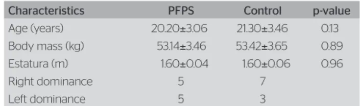
Table 1. Mean (standard-deviation) of the variables: age, body mass, height and dominance of the assessed groups
Characteristics PFPS Control p-value
Age (years) 20.20±3.06 21.30±3.46 0.13
Body mass (kg) 53.14±3.46 53.42±3.65 0.89
Estatura (m) 1.60±0.04 1.60±0.06 0.96
Right dominance 5 7
Left dominance 5 3
Procedures
After physical evaluation, the order of tasks was sorted out and trichotomy and skin cleansing were performed. Afterwards, the surface electrodes were ixated with adhe-sive plaster to the RF, VL and VMO muscles. he electro-de of the RF muscle was placed according to the guielectro-delines by SENIAM23, and the ixation of electrodes on the VMO
and VL muscles were in accordance with the procedures described by Grossi et al.18. he dominant limb was the
criterion of muscle choice assessed by the Control group, whilst in the group with PFPS the EMG assessment was performed by the afected limb, or the most afected limb, in the case of bilateral PFPS. In order to determine the dominant limb, a ball was placed in front of the volunteers, and they were asked to kick it. he limb chosen to kick the ball was considered as the dominant one.
Afterwards, three MVIC of the leg extension mus-cles were performed, and the volunteers were sitting on an extension chair, with the lower limb support locked, maintaining hip and knee lexion at 90º24. he
volun-teers were asked to perform three MVIC against the support for four seconds, with a two minute interval between each contraction. After the last MVIC, there was a period of ten minutes to rest until the beginning of the tasks: standing up and sitting on the bench.
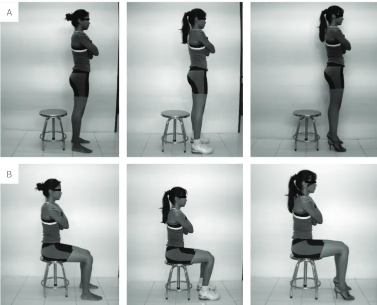
Volunteers were placed sitting on a bench that allowed height regulation, so that all of them would keep the knees lexed at 90º (Figure 1), controlled with the use of a universal goniometer, keeping the feet aligned at a distance similar to the shoulder width. Besides, the arms should be crossed in a way that the hands could touch the opposite shoulder, to avoid movements and compensations from the upper limbs. In all of the executions, the EMG collection and the
A
B
metronome were simultaneously activated. Volunteers were recommended to wait until the second visual and sound warning (after 1.8 second) to start the task, and then they should complete it by the second visual and sound warn-ing (1.8 second), and keep the position for three seconds. For the EMG analysis, the gap between 1.8 and 3.6 seconds was chosen, since it represented the muscle activity rate per-formed during the amplitude of the knee joint movement between 90º and 180º. Before the performance of tasks, vol-unteers were allowed to practice and get familiar with the training, which enabled the correct performance of the task.
For the sitting down task, the volunteers were rec-ommended to do the inverse movement in relation to the previously described task, respecting the same posi-tion of upper and lower limbs and time of execuposi-tion.
Both tasks were performed three times, with a two minute interval between them. Each step was performed in three situations: wearing a 10 cm high-heeled shoe; with sneakers that had a 1 cm elevation from the sole at the midtarsus region in relation to the metatarsus and forefoot; and barefoot. For each shoe change, the volunteers had a ten minute period to adapt to the new shoe, during which they stood up and took some steps. he time of 1.8 second to perform the tasks was determined according to the data from the study by Ikeda et al.25, which indicated the mean
time of execution of 1.86 second, for young people, for the task to stand up from a chair.
Statistical analysis
All statistical tests were performed with the software SPSS, version 16.0. At irst, the normality of data was checked by means of the Shapiro-Wilk test. In order to analyze the in-luence of the diferent types of shoes in the EMG activity of the studied muscles, the ANOVA test was applied with repeated measures, as well as the Tukey post hoc test, while for the intergroup comparisons the unpaired t test was used. All of the statistical tests considered a 5% signiican-ce level. Besides, in variables with p<0.05, by means of the Winpepi software, version 10.8, values of Cohen’s d were calculated to assess the magnitude of the efect. Cohen’s
d values lower than 0.2 indicated low magnitude efect, and higher values meant high magnitude efect.
RESULTS
Standing from the bench
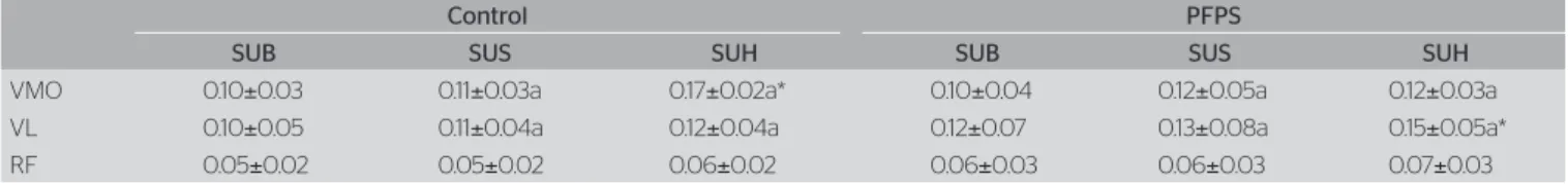
It was not possible to observe statistical diferences between the activities of the three muscles during this task with the barefoot volunteers in the Control group (p≥0.08; d≤0.98) and in the PFPS group (p≥0.20; d≤0.90). However, it was observed that the use of sneakers and high heels provided higher EMG activity in the VMO and VL muscles when compared to the RF muscle in the Control group (p<0.01; d>1.89) and in the PFPS group (p<0.01; d>1.45). he cal-culation of Cohen’s d value was higher than 0.80, and this size was considered as high magnitude efect.
In the Control group, the use of high heels increased the VMO activity in relation to the conditions of being barefoot (p=0.01; d=2.74) and with sneakers (p=0.03; d=2.35), but no statistical diference was observed for VL (p≥0.10; d≤0.52) and RF (p≥0.07; d≤0.50) (Table 2). Cohen’s d index demonstrated a high magnitude efect in relation to the use of high heels and the increased VMO activity. No changes were shown in the VMO:VL ratio in the Control group (p≥0.28; d≤0.30) (Table 3).
For the PFPS group, no statistical changes were ob-served in the VMO (p≥0.06; d≤0.59) and RF activities (p≥0.28; d≤0.66). However, the use of high heels caused the VL activity to increase in relation to the condition of being barefoot (p=0.01; d=0.72) (Table 2) and led to the decreased ratio VMO:VL in relation to the task of standing up from the bench barefoot (p=0.03; d=-0.52) (Table 3). In this case, it was possible to observe a medium magnitude efect.
Sitting on the bench
In both groups, it was not possible to observe diferenc-es in the activitidiferenc-es of the three muscldiferenc-es in the conditions
Table 2. Mean of the normalized RMS values during the activities of standing up from the bench barefoot (SUB), wearing sneakers (SUS) and wearing high heels (SUH)
Control PFPS
SUB SUS SUH SUB SUS SUH
VMO 0.10±0.03 0.11±0.03a 0.17±0.02a* 0.10±0.04 0.12±0.05a 0.12±0.03a
VL 0.10±0.05 0.11±0.04a 0.12±0.04a 0.12±0.07 0.13±0.08a 0.15±0.05a*
RF 0.05±0.02 0.05±0.02 0.06±0.02 0.06±0.03 0.06±0.03 0.07±0.03
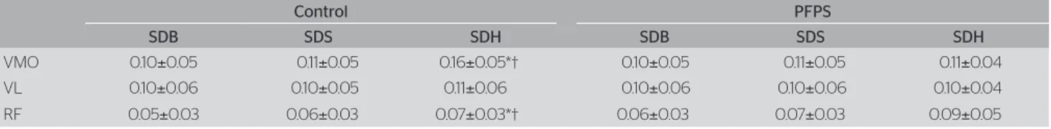
of being barefoot (control: p≥0.08; d≤0.68; PFPS: p≥0.21; d≤0.35), with sneakers (control: p≥0.08; d≤0.53; PFPS: p≥0.20; d≤0.47) and with high heels (control: p≥0.08; d≤0.66; PFPS: p≥0.54; d≤0.30) (Table 4).
In the Control group, results demonstrated a signii-cant increase in the VMO activity when wearing high heels in relation to the conditions of being barefoot (p=0.01; d=1.20) and with sneakers (p=0.03; d=1.00), as well as the increased RF activity with high heels in relation to the conditions of being barefoot (p=0.01; d=0.66) and with sneakers (p=0.03; d=0.33). he Cohen’s d index dem-onstrated a high magnitude efect for VMO and an efect that ranged from low and mean magnitude for RF. here was no signiicant change in the activity of the VL muscle (p≥0.12; d≤0.66) (Table 4). No changes were observed in the VMO:VL ratio in the Control group (p≥0.30; d≤0.30).
In relation to the PFPS group, no statistical difer-ences were identiied for this task in any of the analyzed situations (p>0.35; d≤0.34) (Tabela 4). However, the use of high heels caused the VMO:VL ratio to decrease in relation to the task of sitting down barefoot (p=0.04; d=-0.25) (Table 3).
Intergroup analysis
he intergroup analyses did not show statistical dife-rences in the EMG amplitude of the VMO,VL and RF muscles during the task of sitting on the bench in any
of the tested situations (p≥0.28; d≥0.49) (Table 4). In the task of standing up from the bench there were no signiicant diferences in the EMG values of the three muscles in any of the tested situations (p≥0.30; d≥0.51) (Table 2). he values of the VMO:VL ratio did not pre-sent signiicant diferences between the groups, both for the task of standing up (p≥0.42; d≥0.37) and the task of sitting down (p≥0.63; d≥0.32) (Table 3).
DISCUSSION
he results showed that the use of high-heeled shoes and sneakers interfered in the EMG activity of all the muscles in healthy women. In women from the PFPS group, no signiicant diferences were registered as to the intensity of EMG activity in the RF muscle; how-ever, the use of high heels caused changes in the ac-tivities of the VMO and VL muscles, especially in the VMO:VL ratio.
In a previous study, Edwards et al.2 assessed
the inluence of diferent heels in the EMG activ-ity of the VM and VL muscles of healthy women. In this study, it was observed that a 1 cm heel was not enough to cause changes in the EMG activity of the VM and VL muscles. On the other hand, the 5 cm heel led to the increased EMG activity for both muscles. However, no changes in the VM:VL ratio were found, suggesting that the heel does not cause imbalance between them. Even though in the pres-ent study, during the task of standing up, only the signiicant increase of the VMO muscle has been ob-served, it was possible to check that this fact did not inluence the VMO:VL ratio. his inding corrobo-rates the study by Edwards et al.2, demonstrating that
in the Control group there was no inluence of the high heels on the VMO:VL ratio. Likewise, the use of sneakers, which would be similar to the 1 cm heel, did not inluence this ratio.
Table 3. Mean of VMO:VL ratio values during the tasks of sitting down and standing up for both groups
VMO:VL VMO:VL
Control PFPS
SUB 1.18±0.33 1.19±0.40#
SUS 1.17±0.56 1.12±0.27
SUH 1.25±0.73 1.02±0.23#
SDB 1.15±0.33 1.15±0.40ǂ
SDS 1.14±0.33 1.15±0.40
SDH 1.16±0.52 1.05±0.39ǂ
#Statistical diference between estatística entre SUB and SUS (p=0.03); ǂStatistical diference between SDB and SDH (p=0.04)
Table 4. Mean of RMS normalized values during the activities of sitting down barefoot (SDB), wearing sneakers (SDS) and wearing high heels (SDH) for both groups
Control PFPS
SDB SDS SDH SDB SDS SDH
VMO 0.10±0.05 0.11±0.05 0.16±0.05*† 0.10±0.05 0.11±0.05 0.11±0.04
VL 0.10±0.06 0.10±0.05 0.11±0.06 0.10±0.06 0.10±0.06 0.10±0.04
RF 0.05±0.03 0.06±0.03 0.07±0.03*† 0.06±0.03 0.07±0.03 0.09±0.05
On the other hand, during the sitting down task, it was possible to observe a signiicant increase in the ac-tivities of the RF and VMO muscles. Anderson et al.26
observed that, during the performance of the crouching task, the activity of the RF muscle increased while the knee lexion also increased, and that the VMO muscle also increases its activity in order to keep a proper pa-tellar alignment. Besides, added to the fact that the in-creased knee lexion in a closed kinetic chain (CKC) is responsible for the increased activity of the knee extensor group, several authors3,27,28 have reported the increased
external knee lexion moment provided by the ankle inclination. hese two factors contribute with the in-creased knee extensor moment and the higher stress of the patellofemoral joint19.
Finally, despite being an eccentric task, unlike the concentric task analyzed by Edwards et al.2, it
was still possible to observe the concordance as to the balance aspect between VMO and VL, since the efect of high heels in the VMO:VL ratio was not observed among asymptomatic women. However, Edwards et al.2 observed that high heels caused a
signiicant increase in the activity of the VL muscle, which was not observed in the Control group of this study for both the tested tasks.
his divergence in relation to the VL activity can be justiied by the methodological diferences between the studies. his one used a 10 cm high-heeled shoe, while Edwards et al.2 used a wooden device to simulate a 5 cm
high-heeled shoe. Besides, the wooden device that sim-ulated the high heel had a broader base, while in this study the shoes had thin heels. his could inluence the position of the feet, since Foster et al.29 demonstrated
that a 9.5 cm heel signiicantly increases the plantarlex-ion angles of the ankle and inversplantarlex-ion of the foot. his condition may have required from the volunteers some diferent strategies in order to keep the balance during the execution of tasks, and may have caused changes in the balance of forces not only in the sagittal plane, but also in the other planes.
Diverging from the results of the Control group, in the volunteers with PFPS the high-heeled shoes caused the decrease of the VMO:VL ratio in both tasks. his fact may be related to the increased external knee adduction moment due to the use of high heels2,21.
With the objective to confront the external adduction moment, the quadriceps muscle, by contracting, gener-ates an internal abduction moment20. However, a major
increase in the internal moment caused by the muscles of the lateral side o the knee could also increase the
lateral slide of the patella2. So, the increased activity of
the VL muscle should be followed by the simultaneous increase of the VMO, in order for the balance of forces to occur and to avoid patella lateralization.
Indeed, the volunteers from the Control Group presented signiicant VMO increase, maintain-ing the VMO:VL ratio. However, the volunteers from the PFPS group presented only signiicant increase in the VL muscle, which consequently led to the de-creased VMO:VL ratio. Some authors30 suggest that
such decrease in the VMO:VL ratio is a consequence of a neuromuscular imbalance, which could be caused by disorders of the neurophysiological mechanism. his is because the presence of pain or signals of inlamma-tion in the knee joint have been pointed out as being responsible for causing an inhibition of the quadriceps muscle, and the VM muscle is the most afected one31.
his inhibition mechanism of the VMO muscle could justify the diferences found between the Control and PFPS groups in this study. Besides, some
stud-ies32,33 have suggested that people with PFPS may
present with decreased capacity to slow down or resist the valgus external movement during functional tasks. hus, due to changes in the balance of forces of abduc-tor muscles and hip external rotaabduc-tors, the femur could excessively adduce during functional tasks, with weight discharge, leading to the increased dynamic valgus32,
and this could lead to the lateral slide of the patella32-34.
However, to analyze this issue it is necessary to as-sess the activity of the muscles that work in the hip joint, as well as to perform biomechanics and kinetics evaluations in the sagittal plane, and especially in the frontal plane. In relation to the intergroup comparisons, this study did not show statistic diferences. his dem-onstrate that maybe the most important aspect of this dysfunction is not to assess possible changes in the lev-els of muscle activity between diferent subjects, but to consider the proportion of activation of diferent mus-cles in the same subject.
ratio, which suggests that its use may lead to the imbal-ance of the patella stabilizing forces. Finally, the higher activity of the VMO and VL muscles in relation to RF during the task of standing up from the bench wearing high heels indicates that na action of patellar stabiliza-tion was necessary, thus conirming the stabilizing role of the VMO and VL muscles.
Even though it was a small group and two speciic tasks were analyzed, the results in this study provide initial relevant information. However, this study pres-ents some limitations, such as the small sample, the lack of kinetic and biomechanical analyses of the movement, which limits some conclusions. On the other hand, numberless questions need to be answered, thus open-ing possibilities for future studies to analyze the inlu-ence of the use and the time of use of diferent types of shoes for the activation of the stabilizing muscles of the knee and their correlation with the worsening of signs and symptoms of PFPS; then, it would be possible to create evidence that support or not the interruption of the use of shoes in patients with PFPS.
CONCLUSIONS
he results demonstrated the the use of high-heeled shoes provides diferent responses between groups, causing the increased VL activity in relation to VMO in women with PFPS. he decreased VMO:VL ratio suggests that the high heels may be an aggravating fac-tor for the muscular imbalance of the stabilizers of the patellofemoral joint in women with PFPS.
REFERENCES
1. Barton CJ, Coyle JA, Tinley P. The efect of heel lifts on trunk muscle activation during gait: a study of young healthy females. J Electromyogr Kinesiol. 2009;19(4):598-606.
2. Edwards L, Dixon J, Kent JR, Hodgson D, Whittaker V. Efect of shoe heel height on vastus medialis and vastus lateralis electromyographic activity during sit to stand. J Orthop Surg Res. 2008;3:2-7.
3. Yoon JY, An DH, Yoo WG, Kwon YR. Diferences in activities of the lower extremity muscles with and without heel contact during stair ascent by young women wearing high-heeled shoes. J Orthop Sci. 2009;14:418-22.
4. Brindle TJ, Mattacola CG, McCrory JL. Electromyographic changes in the gluteus medius during stair ascent and descent in subjects with anterior knee pain. Knee Surg Sports Traumatol Arthrosc. 2003;11:244-51.
5. Fagan V, Delahunt E. Patellofemoral pain syndrome: a review on the associated neuromuscular deficits and current treatment options. Br J Sports Med. 2008;42(10):789-95.
6. Pulzatto F, Say KG, Siqueira AC, Santos GM, Grossi DB, Oliveira AS, et al. Step height influence on backward step-up exercise: an electromyographic study in healthy individuals and in those with patellofemoral pain syndrome Acta Ortop Bras. 2005;13(4):168-70.
7. Witvrouw E, Werner S, Mikkelsen C, Van Tiggelen D, Vanden Berghe L, Cerulli G. Clinical classification of patellofemoral pain syndrome: guidelines for nonoperative treatment. Knee Surg Sports Traumatol Arthrosc. 2005;13(2):122-30.
8. Boling MC, Bolgla LA, Mattacola CG, Uhl TL, Hosey RG. Outcomes of a weight bearing rehabilitation program for patients diagnosed with patellofemoral pain syndrome. Arch Phys Med Rehabil. 2006;87(11):1428-35.
9. Powers CM. The influence of altered lower-extremity kinematics on patellofemoral joint dysfunction: a theoretical perspective. J Orthop Sports Phys Ther. 2003;33:639-46.
10. Davis IS, Powers CM. Patellofemoral pain syndrome: proximal, distal and local factors, na international retreat, April 30-May 2, 2009, Fells Point, Baltimore, MD. J Orthop Sports Phys Ther. 2010;40(3):A1-16.
11. Baker V, Bennell K, Stillman B, Cowan S, Crossley K. Abnormal knee joint position sense in individuals with patellofemoral pain syndrome. J Orthop Res. 2002;20(2):208-14.
12. Tang SF, Chen CK, Hsu R, Chou SW, Hong WH, Lew HL. Vastus medialis obliquus and vastus lateralis activity in open and closed kinetic chain exercises in patients with patellofemoral pain syndrome: an electromyographic study. Am J Phys Med Rehabil. 2002;81(9):684-90.
13. Barak Y, Ayalon M, Dvin Z. Spectral EMG changes in vastus medialis muscle following short range of motion isokinetic training. J Electromyogr Kinesiol. 2006;16(5):379-83.
14. Herrington L, Pearson S. Does Level of load afect relative activation levels of vastus medialis oblique and vastus lateralis? J Electromyogr Kinesiol. 2006;16(4):379-83.
15. Alves FSM, Oliveira FS, Junqueira CHBF, Azevedo BMS, Dionísio VC. Analysis of electromyographic patterns during standard and declined squats. Rev Bras Fisioter. 2009;13(2):164-72.
16. Dionísio VC, Almeida GL, Duarte M, Hirata RP. Kinematic, Kinetic and EMG parameters during downward squatting. J Electromyogr Kinesiol. 2008;18(1):134-43.
17. Earls JE, Schmitz RJ, Arnold BL. Activation of the VMO and VL during dynamic mini-squat exercises with and without isometric hip adduction. J Electromyogr Kinesiol. 2001;11(6):381-86.
18. Grossi DB, Felicio LR, Simões R, Coqueiro KRR, Monteiro-Pedro V. Electromyographic activity evaluation of the patella muscles during squat isometric exercise in individuals with patellofemoral pain syndrome. Rev Bras Med Esporte. 2005;11(3):159-63.
19. Ho KY, Blanchette MG, Powers CM. The influence of heel height on patellofemoral joint kinetics during walking. Gait Posture. 2012;36(2):271-5
20. Simonsen EB, Svendsen MB, Norreslet A, Baldvinsson HK, Heilskov-Hansen T, Larsen PK, et al. Walking on high heels changes muscle activity and the dynamics of human walking significantly. 2012;28:20-8.
22. Cowan SM, Bennell KL, Hodges PW, Crossley KA, McConnell J. Delayed onset of electromyographic activity of vastus medialis obliquus relative to vastus lateralis in subjects with patellofemoral pain syndrome. Arch Phys Med Rehabil. 2001;82:183-98.
23. Hermens HJ, Freiks B, Disselhorst-Klug C, Rau G. Development of recommendations for SEMG sensors and sensors placement procedures. J Electromyogr Kinesiol. 2000;10:361-74.
24. Grossi DB, Felício LR, Silvério GWP. Início da atividade elétrica dos músculos estabilizadores da patela em indivíduos com SDFP. Acta Ortop Bras. 2009;17(5):297-99.
25. Ikeda ER, Schenkman ML, Riley PO, Hodge WA. Influence of age on dynamics of rising from a chair. Phys Ther. 1991;71(6):473-81.
26. Anderson R, Courtney C, Carmeli E. EMG analysis of the vastus medialis/vastus lateralis muscles utilizing the unloaded narrow and wide stance squats. J Sports Rehabil. 1998;7:236-47.
27. Opila-Correia KA. Kinematics of High-Heeled Gait. Arch Phys Med Rehabil. 1990;71:304-9.
28. Cowan SM, Hodges PW, Bennell KL, Crossley KM. Altered vastii recruitment when people with patellofemoral pain syndrome complete a postural task. Arch Phys Med Rehabil. 2002;83:989-95.
29. Foster A, Blanchette MG, Chou YC, Powers CM. The influence of heel height on frontal plane ankle biomechanics: implications for lateral ankle sprains. Foot Ankle Int. 2012;33(1):64-9.
30. Fonseca ST, Cruz ABC, Lima SS, Seixas AFAM. Análise eletromiográfica dos músculos vasto medial oblíquo e vasto lateral em exercícios usados no tratamento da síndrome da dor patelofemoral. Rev Fisiot Univers SP.2001;8(1):1-10.
31. Torry MR, Decker MJ, Viola RW, O’Connor DD, Steadman JR. Intraarticular knee joint efusion induces quadriceps avoidance gait patterns. Clin Biomech. 2000;15:147-59.
32. Nakagawa TH, Muniz TB, Baldon RDM, Dias Maciel C, Menezes Reif RB, Serrao FV. The efect of additional strengthening of hip abductor and lateral rotator muscles in patellofemoral pain syndrome: a randomized controlled pilot study. Clin Rehabil. 2008;22: 1051-60.
33. Nakagawa TH, Moriya ETU, Dias C, Serrao FV. Frontal Plane Biomechanics in Males and Females With and Without Patellofemoral Pain. Med Sci Sports Exerc. 2012. DOI: 10.1249/MSS.0b013e318256903a [in press]. 34. Powers CM. The influence of altered lower-extremity kinematics on