Response of
Rhodococcus erythropolis
strain IBB
Po1to toxic organic solvents
Mihaela Marilena Stancu
Institute of Biology Bucharest of Romanian Academy, Bucharest, Romania.
Submitted: May 29, 2014; Approved: March 11, 2015.
Abstract
Recently, there has been a lot of interest in the utilization of rhodococci in the bioremediation of pe-troleum contaminated environments. This study investigates the response of Rhodococcus erythropolisIBBPo1cells to 1% organic solvents (alkanes, aromatics). A combination of microbiol-ogy, biochemical, and molecular approaches were used to examine cell adaptation mechanisms likely to be pursued by this strain after 1% organic solvent exposure.R. erythropolisIBBPo1was found to utilize 1% alkanes (cyclohexane, n-hexane, n-decane) and aromatics (toluene, styrene, ethylbenzene) as the sole carbon source. Modifications in cell viability, cell morphology, membrane permeability, lipid profile, carotenoid pigments profile and 16S rRNA gene were revealed in R. erythropolisIBBPo1cells grown 1 and 24 h on minimal medium in the presence of 1% alkanes (cyclo-hexane, n-(cyclo-hexane,n-decane) and aromatics (toluene, styrene, ethylbenzene). Due to its environmen-tal origin and its metabolic potential, R. erythropolis IBBPo1 is an excellent candidate for the bioremediation of soils contaminated with crude oils and other toxic compounds. Moreover, the carotenoid pigments produced by this nonpathogenic Gram-positive bacterium have a variety of other potential applications.
Key words:Rhodococcus, solvents, morphology, lipids, carotenoids.
Introduction
The existence of contaminated areas with highly toxic organic solvents is a clear indication of the lack of biologi-cal systems that have the ability to efficiently degrade these compounds. Research in recent years has focused on the search for solvent-tolerant bacteria that have the catabolic potential necessary to remove comprehensively these toxic compounds (Torreset al., 2011). Many of the organic sol-vents are relatively stable and toxic to bacteria because they are able to bind to the cell membrane (Torreset al., 2011). When organic solvents accumulate in the cell membrane, its integrity is affected, resulting in loss of function as a per-meability barrier, as a protein and reaction matrix and as an energy transducer leading concomitantly to damages of the cellular metabolism, growth inhibition, and, finally leading to cell death (Isken and de Bont, 1998; Heipieperet al., 2007). The large genomes ofRhodococcusstrains, their re-dundant and versatile catabolic pathways, their ability to uptake and metabolize hydrophobic compounds, to form
biofilms, to persist in adverse conditions, as well as the availability of recently developed tools for genetic engi-neering in rhodococci make them suitable industrial micro-organisms for biotransformations and the biodegradation of many toxic compounds (Larkinet al., 2006; Martínkováet al., 2009). Furthermore, these bacteria are capable to pro-duce several carotenoid pigments (e.g.,b-carotene,g -caro-tene, chlorobactene) with different medical, industrial, and nutritional applications (Taoet al., 2006). Some organic solvent tolerance mechanisms in rhodococci have been pro-posed (e.g., induction of general stress regulon, production of organic solvent emulsifying or deactivating enzymes, active solvent efflux pumps) (Torreset al., 2011). In re-sponse to toxic organic solvents cell morphology alter-ations and filamentous growth were also observed in some solvent-tolerant bacteria (Torreset al., 2011). The variety of mechanisms that could confer adaptation to organic sol-vents implies that bacterial solvent tolerance cannot be pro-vided by a single mechanism (Heipieper et al., 2007).
DOI: http://dx.doi.org/10.1590/S1517-838246420140462
Send correspondence to M.M. Stancu. Institute of Biology Bucharest of Romanian Academy, 296 Splaiul Independentei, P.O. Box 56-53, 060031 Bucha-rest, Romania. E-mail: [email protected].
Taking into account the interest raised by the utilization of rhodococci in the bioremediation of petroleum contami-nated environments, this study investigates the response of
Rhodococcus erythropolisIBBPo1cells to 1% organic sol-vents (alkanes, aromatics). Cyclohexane,n-hexane,n -dec-ane, toluene, styrene, and ethylbenzene with different log
POW values (logarithm of the partition coefficient in
octanol-water mixture) have been used as the sole carbon source in minimal medium. The possible cell adaptation mechanisms pursued by this strain after 1% organic solvent (alkanes, aromatics) exposure was studied by following the changes in the cell viability, cell morphology, membrane permeability, lipid profile, carotenoid pigments profile, and 16S rRNA gene.
Materials and Methods
Bacterial strain
The strain used in the present study was R. erythropolisIBBPo1(KF059972.1), which has been previ-ously isolated in our laboratory from Poeni crude oil-con-taminated soil sample (Stancu, 2014). The analysis of the 16S rRNA gene sequence located strain IBBPo1 (KF059972.1) within the genus Rhodococcus, showing 95% similarity to otherRhodococcusstrains from the pub-lic databases (GenBank/DDBJ/EMBL).
Response ofR. erythropolisIBBPo1cells to organic solvents
Bacterial cells were cultivated on liquid LB medium containing 30mg mL-1kanamycin and incubated at 28 °C on a rotary shaker (200 rpm) until they reached the expo-nential growth phase. Bacterial cells were harvested by centrifugation, washed twice and finally resuspended (106cells mL-1) in minimal medium (Stancu and Grifoll, 2011). 0.1% (w/v) yeast extract or 1% (v/v) organic sol-vents (alkanes: cyclohexane, n-hexane, n-decane; aromatics: toluene, styrene, ethylbenzene) were supplied to the cell suspensions as the sole carbon source. Flasks were sealed and incubated for 1 and 24 h respectively at 28 °C on a rotary shaker (200 rpm).
Cell viability
Serial dilutions of bacterial cultures were spotted on LB agar and the number of viable cells (cfu mL-1) was de-termined, using the method described by Segura et al.
(2008). Petri plates were incubated 24 h at 28 °C.
Cell morphology
Cell morphology was studied using scanning electron microscopy (SEM) and transmission electron microscopy (TEM). SEM samples were prepared using the method in-dicated by Rochaet al.(2011). Samples were fixed on SEM
holder and gold-coated with a JEOL JFC-1300 auto fine coater, in a deep vacuum. The samples were examined with a JEOL JSM-6610LV SEM. TEM samples were prepared using the method indicated by Gutiérrezet al.(2009). Thin sections were mounted onto 300-mesh collodion/carbon coated cooper grids and stained with lead citrate and uranyl acetate. Examination of the sectioned material was per-formed using a JEOL JEM-1400 80 kV TEM.
Membrane permeability
Membrane permeability was determined using the method indicated by Gaur and Khare (2009). To determine the release of nucleic acid from bacterial cells, the absor-bance of cell-free supernatant was measured at 260 nm.
Membrane lipids
Membrane lipids were analyzed by thin layer chro-matography (TLC). Lipids were extracted from the culture broths with chloroform-methanol (2:1 v/v). The samples were spotted with a Linomat 5 sample applicator (CAMAG), on a 10 x 20 cm precoated silica gel 60 TLC aluminium sheets (Merck). The separation was performed using chloroform-methanol-acetic acid-water (85:22.5:10:4 v/v/v/v) mixture as mobile phase (Daset al., 2009). For phospholipids visualization, the plates were treated with 10% (w/v) molybdatophosphoric acid hydrate in ethanol, and for glycolipids, the plates were treated with 0.5% (w/v)a-naphthol in methanol-water (1:1 v/v) mixture and sulfuric acid-ethanol (1:1 v/v) mixture. The densito-metric scan of dried TLC plates was performed at 500 nm with a TLC Scanner 4 (CAMAG).
Carotenoid pigments
Carotenoid pigments were analyzed by spectropho-tometry and thin layer chromatography (TLC). Carotenoids were extracted from the culture broths with acetone (Taoet al., 2006). UV/visible scanning spectra of the samples were recorded between 200 and 800 nm using a SPECORD 200 UV-visible spectrophotometer (Analytik Jena). For TLC analysis, the samples were spotted with a Linomat 5 sample applicator (CAMAG), on a 10 x 20 cm precoated silica gel 60 TLC aluminium sheets (Merck). The separation was performed using chloroform-methanol (90:10 v/v) mixture as mobile phase. The densitometric scan of dried TLC plates was performed at 254 nm with a TLC Scanner 4 (CAMAG).
16S rRNA gene expression
(Promega). PCR was performed with a mastercycler proS (Eppendorf). The PCR program consisted in initial ation for 10 min at 94 °C, followed by 35 cycles of denatur-ation at 94 °C for 1 min, annealing at 55 °C for 30 s, extension step at 72 °C for 2 min, and a final extension at 72 °C for 10 min. After separation on 1.5% (w/v) TBE agarose gel (Sambrooket al., 1989) and staining with fast blast DNA stain (Bio-Rad) the PCR products were ana-lyzed. The PCR products were purified using the DNA clean and concentrator-5 kit (Zymo Research). PCR prod-ucts were digested withEcoRI andXbaI restriction endo-nucleases (Promega), and the resultant restriction fragments were analyzed by electrophoresis on 2% (w/v) agarose gel. PCR products were also sequenced with the BigDye Terminator v.3.1 cycle sequencing kit (Applied Biosystems). The reactions were performed using the am-plification primers. The products were purified using the BigDye XTerminator purification kit (Applied Bio-systems). Sequencing reactions were obtained with an Ap-plied Biosystems 3500/3500xL genetic analyzer at the Institute of Biology Bucharest of Romanian Academy. DNA sequencing runs were assembled using the BioEdit software (Hall, 1999). The sequences obtained were com-pared with respective control using the BLAST program (Altschulet al., 1990). The phylogenetic tree was generated using the neighbor-joining methods in MEGA5.1 program (Tamuraet al., 2011).
Reagents used during this study were of reagent grade and purchased from different commercial sources (Merck, Sigma-Aldrich, Promega, Invitrogen, Zymo Research, Ap-plied Biosystems, Bio-Rad Laboratories). The PCR prim-ers were purchased from Integrated DNA Technologies.
Results and Discussion
Response ofR. erythropolisIBBPo1cells to organic
solvents
Numerous and complex physiological cellular re-sponses and adaptations involved in organic solvents toler-ance have been observed in R. erythropolis IBBPo1 cells grown on LB medium in the presence of 1% organic sol-vents (Stancu, 2014). This bacterium was adapt to 1%
or-ganic solvents by changing surface hydrophobicity, cell morphology, metabolic fingerprinting and theotsA1 (treha-lose-6-phosphate synthase) gene sequence. Taking into ac-count the interest raised by the utilization of rhodococci in the bioremediation of petroleum contaminated environ-ments, we continued our previous study with the investiga-tion of the response ofR. erythropolisIBBPo1cells to 1% organic solvents (alkanes, aromatics). In this study, 1% al-kanes (cyclohexane, n-hexane, n-decane) and aromatics (toluene, styrene, ethylbenzene) were used as the sole car-bon source in minimal medium. The controls were prepared using the same mineral medium, but supplemented with 0.1% (w/v) yeast extract as the carbon source.
Cell viability
R. erythropoliscells contain a large set of important enzymes for bioconversion and biodegradation processes, which enable them to perform oxidations, dehydrogena-tions, epoxidadehydrogena-tions, hydrolysis, hydroxyladehydrogena-tions, dehalo-genations, and desulfurizations (de Carvalhoet al., 2009). The exposure for 1 and 24 h respectively ofR. erythropolis
IBBPo1cells to 1% alkanes (cyclohexane,n-hexane,n -dec-ane) and aromatics (toluene, styrene, ethylbenzene) had different effects on their survival rate (Table 1). One hour after 1% alkanes and aromatics exposure the survival rates were 102-106cfu mL-1, and after 24 h the survival rates were 104-1010 cfu mL-1. As anticipated, the viability of R. erythropolisIBBPo1cells 1 and 24 h after 1% alkanes and aromatics exposure was lower, compared with the controls (107, 1011cfu mL-1). Tolerance of bacteria to organic sol-vents has been estimated by the solvent parameter logPOW,
which is an index of biological toxicity (Sikkemaet al., 1995). It is generally accepted that solvents with logPOW
values below 5 are considered extremely toxic because of their high degree of partitioning into the aqueous layer sur-rounding the cells, and from there into the lipid membrane bilayer (Torreset al., 2011. However the cells that are able to remain viable after the first moments of contact with toxic organic solvents will be able to endure its presence for some time (de Carvalhoet al., 2009). In our study the re-sults showed higher survival rates (105-1010cfu mL-1) when
R. erythropolisIBBPo1cells have been exposed to 1%
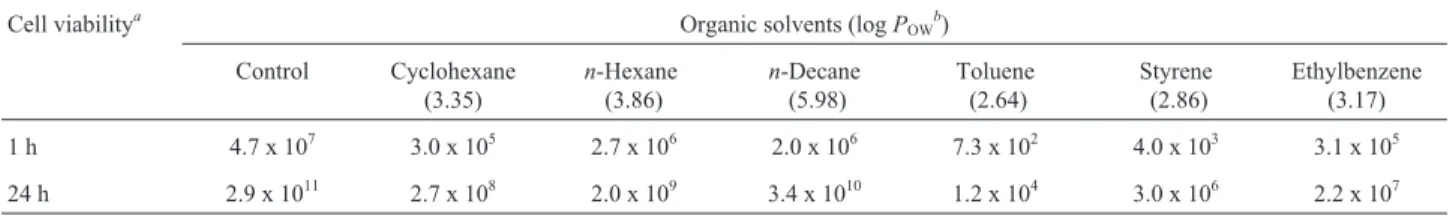
al-Table 1- Viability ofR. erythropolisIBBPo1cells after 1% organic solvents exposure.
Cell viabilitya Organic solvents (logPOWb)
Control Cyclohexane (3.35)
n-Hexane (3.86)
n-Decane (5.98)
Toluene (2.64)
Styrene (2.86)
Ethylbenzene (3.17)
1 h 4.7 x 10⁷ 3.0 x 10⁵ 2.7 x 10⁶ 2.0 x 10⁶ 7.3 x 10² 4.0 x 10³ 3.1 x 10⁵
24 h 2.9 x 10¹¹ 2.7 x 10⁸ 2.0 x 10⁹ 3.4 x 10¹⁰ 1.2 x 10⁴ 3.0 x 10⁶ 2.2 x 10⁷
kanes (cyclohexane,n-hexane,n-decane) with logPOW
be-tween 3.35 and 5.98, as compared with the survival rates of cells (102-107cfu mL-1) exposed to 1% aromatics (toluene, styrene, ethylbenzene) with logPOWbetween 2.64 and 3.17.
The survival rates drastically reduced from 107and 1011to 102and 104, respectively, whenR. erythropolisIBBPo1cells were exposed to 1% toluene. This is in agreement with a previous study which found that organic solvents with lower log POWvalue (e.g., benzene, toluene) bound more abundantly to bacterial cells thus being more toxic for them (Sikkemaet al., 1995; Torreset al., 2011).
Cell morphology
Different adaptation responses of R. erythropolis
IBBPo1 cells were observed by SEM (Figure 1a-1h) and TEM (Figure 1i-1p) studies, 1 and 24 h respectively after 1% alkanes (cyclohexane, n-hexane, n-decane) and aromatics (toluene, styrene, ethylbenzene) exposure. R. erythropolis IBBPo1 cells exposed to 1% alkanes and aromatics were free within the water phase as those in the control cells (Figure 1a), embedded in a polymeric layer (Figure 1b), closely grouped around polymeric structures of bacterial origin (Figure 1c, 1d), on the surface of biofilms (Figure 1e, 1f) or linked together as clusters (Fig-ure 1g, 1h). All these struct(Fig-ures were not observed inR. erythropolisIBBPo1control cells. Similar results were
viously obtained by Rocha et al. (2011) for a Gram-negative bacterium Pseudomonas aeruginosa ATCC 55925 grown on 0.5% heating oil or pure alkanes (i.e., C7-C18n-alkanes, C19 branched alkane).
According to the literature (Urai et al., 2007; Gutiérrezet al., 2009; Rocha et al., 2011; Torres et al., 2011), the structures depicted only when the cells are ex-posed to hydrocarbons (e.g., biofilms, cells clusters) play a significant role in toxic compounds tolerance and they pro-tect the cells from different environmental stresses. Fur-thermore, the polymers produced by some bacterial strains play an important role in sequestering molecules of solvent (i.e., benzene) within its immediate environment, thereby reducing solvent contact with its cell membrane and con-ferring it some degree of tolerance (Gutiérrezet al., 2009).
InR. erythropolisIBBPo1cells exposed to 1% alkanes (cyclohexane,n-hexane,n-decane), the cell membrane was intact (Figure 1j-1l) just like in the case of the control cells (Figure 1i), and no accumulation of solvents was seen in the cytoplasm. In cells exposed to 1% aromatics (toluene, sty-rene, ethylbenzene), the accumulation of solvent in the cy-toplasm of cells with a disturbed cell membrane and an increase in the cell size were observed (Figure 1m-1p). The observed cytoplasmatic electron-transparent inclusions were similar to those reported previously for other bacterial strains, and their formation is a general cell adaptation re-sponse to hydrocarbon growth (Rochaet al., 2011). When the large solvent inclusions occupied all the cytoplasm and the cell wall integrity was altered, the death of R. erythropolisIBBPo1cells was also observed (Figure 1p).
Membrane permeability
In general, solvents exert their toxic effect by altering the cell membrane permeability (Gaur and Khare, 2009), which leads to the inactivation and denaturation of mem-brane-embedded proteins, and the promotion of leakages of ions and intracellular macromolecules, such as nucleic ac-ids (Isken and de Bont, 1998). The exposure for 1 and 24 h ofR. erythropolisIBBPo1cells to 1% alkanes (cyclohexane, n-hexane,n-decane) and aromatics (toluene, styrene, ethyl-benzene) had different effects on membrane permeability (Table 2).
The release of nucleic acids was higher after aromatics (0.509-0.834) and alkanes (0.277-0.370) expo-sure, compared with the controls (0.156, 0.234). Similar re-sults were earlier obtained by Gaur and Khare (2009) for a Gram-negative bacteriumPseudomonas aeruginosaPseA grown in the presence of cyclohexane and tetradecane. These two organic solvents affect membrane integrity and structure dramatically, thereby altering the permeability and incurring toxicity. The higher release of nucleic acid into the growth medium whenR. erythropolisIBBPo1cells were grown in minimal medium in the presence of 1% aromatics (toluene, styrene, ethylbenzene) supports the re-sults of TEM studies, which showed changes in the cyto-plasmic membrane integrity of cells exposed to these toxic solvents.
Membrane lipids
Changes in the composition of phospholipids, glyco-lipids and mycolic acids ofR. erythropolishave been sug-gested to depend on the availability and structure of the carbon source (de Carvalhoet al., 2009). The TLC analysis revealed the existence of some differences between phos-pholipids and glycolipids extracted from R. erythropolis
IBBPo1control cells and those extracted from cells exposed 1 and 24 h to 1% alkanes (cyclohexane,n-hexane,n -dec-ane) and aromatics (toluene, styrene, ethylbenzene) (Figu-re 2a-2d).
The phospholipids (Figure 2a, 2b) found in R. erythropolisIBBPo1control cells were identified based on theirRf(retardation factor) values as phosphatidylinositol (PI withRf0.45), cardiolipin (CL withRf0.75) and fatty
ac-ids (FA withRf0.79). The phosphatidylethanolamine (PE)
was not detected in these extracts. Although the major lipid components of coryneform and nocardioform bacteria are cardiolipin and phosphatidylethanolamine (Kolomytseva
et al., 2005), only cardiolipin was detected in the R. erythropolisIBBPo1cells extracts. The phospholipids found inR. erythropolisIBBPo1cells 1 and 24 h after 1% alkanes and aromatics exposure were cardiolipin (with Rf
0.70-0.75) and fatty acids (with Rf 0.76-0.80).
Phosphati-dylinositol was detected only in barely detectable quanti-ties in all extracts, exceptn-decane. An elevated level of cardiolipin was detected in extracts of R. erythropolis
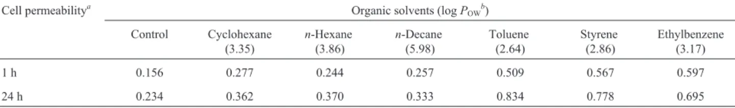
Table 2- Nucleic acid release byR. erythropolisIBBPo1cells after 1% organic solvents exposure.
Cell permeabilitya Organic solvents (logPOWb)
Control Cyclohexane (3.35)
n-Hexane (3.86)
n-Decane (5.98)
Toluene (2.64)
Styrene (2.86)
Ethylbenzene (3.17)
1 h 0.156 0.277 0.244 0.257 0.509 0.567 0.597
24 h 0.234 0.362 0.370 0.333 0.834 0.778 0.695
IBBPo1cells 1 and 24 h after 1% alkanes and aromatics ex-posure (except styrene, ethylbenzene), as compared with those of the respective controls; no such changes were ob-served in cells exposed to styrene and ethylbenzene. It is known that phospholipids, as the major component of bac-terial cells membranes, are responsible for their structural organization and selective permeability. Therefore, the in-crease in the content of cardiolipin and fatty acids in the membranes of cells cultured in the presence of the toxic compounds is the result of adaptation of the bacterial cells and has a defensive nature (Kolomytseva et al., 2005; Martínkováet al., 2009).
The glycolipids (Figure 2c, 2d) found, based on their
Rf values, in R. erythropolis IBBPo1 control cells were trehalolipids (THL1withRf0.30, THL2withRf0.61) and
fatty acids (FA withRf0.72). The glycolipids found inR. erythropolisIBBPo1cells 1 and 24 h after 1% alkanes and aromatics exposure were trehalolipids (THL1withRf
0.26-0.29, THL2withRf0.57-0.63) and fatty acids (FA withRf
0.70-0.74). THL1was not detected in extracts of cells ex-posed 1 h to toluene and for 24 h to cyclohexane, toluene and styrene.Rfvalues of THL1and THL2were similar to data given in the literature, and these value correspond to trehalose monomycolate and trehalose dimycolate, which are regular extractable components of the rhodococcal cell envelope (Niescheret al., 2006).
An elevated level of THL2was detected in extracts of R. erythropolis IBBPo1 control cells, as compared with those of the cells exposed for 1 and 24 h to 1% alkanes and aromatics. This is not surprising because the trehalolipids could be released in high quantities into the growth medium when the cells are exposed to 1% organic solvents, as com-pared with the control cells. We observed previously thatR. erythropolisIBBPo1is a good biosurfactant producer (emul-sification index E24 = 100%), compared with other Rhodococcusstrains (Stancu, 2014). The biosurfactants re-leased into the growth medium modify the cell surface hydrophobicity and/or promote emulsification and/or solu-bilization of organic solvents thus accelerating their bio-degradation (Philpet al., 2002; Geshevaet al., 2010). Such solvent-tolerant bacteria provide the key for the use of oth-erwise toxic solvents in whole-cell two-phase biotrans-formations, by overcoming the toxic effects of substrates and products (Heipieperet al., 2007).
Carotenoid pigments
Carotenoids are hydrophobic molecules typically as-sociated with cytoplasmic membrane and/or noncovalently bound to specific proteins. These pigments form an integral part of the complex membrane structure of some bacteria and influence membrane fluidity, by increasing its rigidity and mechanical strength (Godinho and Bhosle, 2008). It has been suggested that the presence of carotenoids may
change the effectiveness of the membrane as a barrier to water, oxygen, and other molecules. Some bacteria may be accumulating carotenoids as part of their responses to vari-ous environmental stresses, and thus aiding their survival in this habitat (Godinho and Bhosle, 2008). Therefore, we fur-ther investigated the effect of the 1% alkanes (cyclohexane,
n-hexane,n-decane) and aromatics (toluene, styrene, ethyl-benzene) to the carotenoids synthesis in R. erythropolis
IBBPo1cells. The UV/visible absorption scanning spectra of the pigment extract ofR. erythropolisIBBPo1cells showed absorption maxima at 340 nm. Carotenoid pigments ex-tracted fromR. erythropolisIBBPo1cells 1 and 24 h after 1% alkanes and aromatics exposure showed the same ab-sorption maxima. The TLC plate developed with chloro-form-methanol mixture showed the separation of the carotenoid pigments into 6 fluorescent spots (Figure 2e). The carotenoids synthesized by differentR. erythropolis
strains were previously characterized to be 4-keto-g -caro-tene as the major carotenoid and sometimesg-carotene as the minor carotenoids (Taoet al., 2006). However, other carotenoids (e.g., phytoene, lycopene, chlorobactene, b -carotene) were also described inR. erythropolis(Taoet al., 2006). The carotenoids (Figure 2e, 2f) found, based on their
Rf values, in R. erythropolis IBBPo1 control cells were phytoene (Pe withRf0.04), lycopene (Ly ory,y-carotene
with Rf 0.10), 4-keto-g-carotene (KgC with Rf 0.23),
chlorobactene (CB orF,y-carotene withRf0.29),g
-caro-tene (gC with Rf0.53),b-carotene (bC withRf0.73). The
carotenoids found inR. erythropolisIBBPo1cells 1 and 24 h after 1% alkanes and aromatics exposure were phytoene (withRf0.04-0.06), lycopene (withRf0.07-0.10),
4-keto-g-carotene (with Rf 0.20-0.24), chlorobactene (with Rf
0.26-0.30),g-carotene (withRf0.51-0.53),b-carotene (with Rf0.71-0.73). An elevated level of lycopene (which is an
important intermediate in the biosynthesis of g-carotene andb-carotene) was detected in extracts ofR. erythropolis
IBBPo1cells 1 and 24 h after 1% alkanes exposure (espe-cially in the case ofn-decane), as compared with those of the control cells; no such changes were observed in cells exposed to 1% aromatics (except ethylbenzene).
16S rRNA gene expression
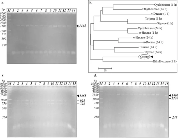
Because rhodococci commonly exhibit considerable genomic instabilities that can be specifically selected (Larkinet al., 2006), we further investigated the expression of 16S rRNA gene afterR. erythropolisIBBPo1cells expo-sure to organic solvents. Genomic DNA extracted from R.
PCR product of 16S rRNA gene (1465 bp fragment) fromR. erythropolisIBBPo1 cells was digested byEcoRI andXbaI restriction endonucleases (Figure 3c, 3d). The
EcoRI recognition site exists in the sequence of 16S rRNA gene fromR. erythropolisIBBPo1, whileXbaI recognition site did not exist in this bacterium. When 16S rRNA gene fromR. erythropolisIBBPo1cells (control) was digested by EcoRI, two distinct bands (925+725 bp) were observed (Figure 3c). The same RFLP pattern was obtained forR. erythropolisIBBPo1cells exposed 1 and 24 h to 1% alkanes (cyclohexane,n-hexane,n-decane) and aromatics (toluene, styrene, ethylbenzene).
In the 16S rRNA phylogenetic tree obtained using the neighbor-joining method (Figure 3b), control (KF059972.1) formed a tight cluster with only 16S rRNA gene sequence of the cells exposed 1 h to ethylbenzene. Moreover, considerable modifications in 16S rRNA gene sequence of R. erythropolis IBBPo1 cells were observed 24 h after ethylbenzene exposure, 1 and 24 h after 1% al-kanes (cyclohexane, n-hexane, n-decane) and aromatics
(toluene, styrene) exposure, as compared with the control (Figure 3b). It is not surprising to observe DNA sequence modification after organic solvent exposure, because these toxic compounds are metabolically activated in cells to yield highly reactive bay region dihydrodiol epoxide deriv-atives which can effectively attack DNA, leading to disrup-tion of normal cellular funcdisrup-tions (Weiet al., 1984).
Conclusions
Solvent-tolerant bacteria belonging to the genus
Rhodococcusare increasingly recognized as very good can-didates for the biodegradation of toxic compounds, because of their ability to degrade a wide range of organic com-pounds, hydrophobic cell surfaces, biosurfactant produc-tion, and ubiquity and robustness in the environment (Pini
et al., 2007; Martinkovaet al., 2009; Torreset al., 2011). Organic solvents with log POW values below 5 are
considered extremely toxic to bacteria. However we have previously shown thatR. erythropolisIBBPo1 cells had a
good tolerance (40-100%) to both alkanes (cyclohexane, n-hexane,n-decane) and aromatics (toluene, styrene, ethyl-benzene) with logPOWvalues between 2.64 and 5.98. Addi-tionally,R. erythropolisIBBPo1was found to utilize these toxic organic solvents as the sole carbon source. Modifica-tions in the cell viability, cell morphology, membrane per-meability, lipid profile, carotenoid pigments profile and 16S rRNA gene were revealed inR. erythropolisIBBPo1 cells grown 1 and 24 h on minimal medium in the presence of 1% alkanes (cyclohexane, n-hexane, n-decane) and aromatics (toluene, styrene, ethylbenzene). The acquired results showed higher survival rates whenR. erythropolis
IBBPo1 cells were exposed to 1% alkanes (cyclohexane, n-hexane,n-decane) with logPOWbetween 3.35 and 5.98,
compared with those of the cells exposed to 1% aromatics (toluene, styrene, ethylbenzene) with logPOWbetween 2.64
and 3.17. Due to its environmental origin and its metabolic potential,R. erythropolisIBBPo1is an excellent candidate for bioremediation of soils contaminated with crude oils and other toxic compounds. Bioremediation of soil contam-inated with 5% (w/v) Poeni crude oil was studied for a pe-riod of 30 days, under laboratory condition (data not shown). The amount of crude oil degraded by R. erythropolisIBBPo1after 15 and 30 days incubation were 34% and 85%, respectively. Moreover, the carotenoids pro-duced by this nonpathogenic Gram-positive bacterium can have a variety of other potential applications (medicine, cosmetics, food industry).
Acknowledgments
This study was funded by project no. RO1567-IBB05/2014 from the Institute of Biology Bucharest of Ro-manian Academy. The author is grateful to Ana Dinu and Alexandru Brînzan for technical support.
References
Altschul SF, Gish W, Miller Wet al.(1990) Basic local alignment search tool. J Mol Biol 215:403-410.
Das P, Mukherjee S, Sen R (2009) Substrate dependent produc-tion of extracellular biosurfactant by a marine bacterium. Biores Technol 100:1015-1019.
de Carvalho CCCR, Wick LY, Heipieper HJ (2009) Cell wall ad-aptations of planktonic and biofilm Rhodococcus erythropoliscells to growth on C5 to C16n-alkane hydro-carbons. Appl Microbiol Biotechnol 82:311-320.
Gaur R, Khare SK (2009) Cellular response mechanisms in Pseu-domonas aeruginosa PseA during growth in organic sol-vents. Lett Appl Microbiol 49:372-377.
Gesheva V, Stackebrandt E, Vasileva-Tonkova E (2010) Biosur-factant production by halotolerant Rhodococcus fascians
from Casey Station, Wilkes Land, Antarctica. Curr Microbiol 61:112-117.
Godinho A, Bhosale S (2008) Carotenes produced by alkaliphilic orange-pigmented strain ofMicrobacterium arborescens
-AGSB isolated from coastal sand dunes. Indian J Mar Sci 37:307-312.
Gutiérrez T, Learmonth R, Couperwhite I (2009) Analysis of ben-zene-induced effects on Rhodococcus sp. 33 reveals that constitutive processes play a major role in conferring toler-ance. Sci World J 9:209-223.
Hall TA (1999) BioEdit: a user-friendly biological sequence alignment editor and analysis program for Windows 95/98/NT. Nucl Acids Symp Ser 41:95-98.
Heipieper HJ, Neumann G, Cornelissen S et al. (2007) Sol-vent-tolerant bacteria for biotransformations in two-phase fermentation systems. Appl Microbiol Biotechnol 74:961-973.
Isken S, de Bont JAM (1998) Bacteria tolerant to organic solvents. Extremophiles 2:229-238.
Kolomytseva MP, Solyanikova IP, Golovlev ELet al.(2005) Het-erogeneity of Rhodococcus opacus 1CP as a response to stress induced by chlorophenols. Appl Biochem Microbiol 41:474-479.
Larkin MJ, Kulakov LA, Allen CCR (2006) Biodegradation by members of the genusRhodococcus:biochemistry, physiol-ogy, and genetic adaptation. Adv Appl Microbiol 59:1-29. Marchesi JR, Sato T, Weightman AJet al.(1998) Design and
evaluation of useful bacterium-specific PCR primers that amplify genes coding for bacterial 16S rRNA. Appl Environ Microbiol 64:795-799.
Martínková L, Uhnáková B, Pátek Met al.(2009) Biodegradation potential of the genus Rhodococcus. Environ Int 35:162-177.
Niescher S, Wray V, Lang Set al.(2006) Identification and struc-tural characterisation of novel trehalose dinocardio-mycolates from n-alkane-grownRhodococcus opacus1CP. Appl Microbiol Biotechnol 70:605-611.
Philp JC, Kuyukina MS, Ivshina IBet al.(2002) Alkanotrophic
Rhodococcus ruber as a biosurfactant producer. Appl
Microbiol Biotechnol 59:318-324.
Pini F, Grossi C, Nereo Set al.(2007) Molecular and physiologi-cal characterisation of psychrotrophic hydrocarbon-degra-ding bacteria isolated from Terra Nova Bay (Antarctica). Eur J Soil Biol 43:368-379.
Rocha CA, Pedregosa AM, Laborda F (2011) Biosurfactant-mediated biodegradation of straight and methyl-branched alkanes byPseudomonas aeruginosaATCC 55925. AMB Express 1:1-10.
Sambrook J, Fritsch EF, Maniatis T (1989) Molecular Cloning, A Laboratory Manual. 2nded. Cold Spring Harbor Laboratory Press, Cold Spring Harbor, New York.
Segura A, Hurtado A, Rivera Bet al.(2008) Isolation of new tolu-ene-tolerant marine strains of bacteria and characterization of their solvent-tolerance properties. J Appl Microbiol 104:1408-1416.
Sikkema J, de Bont JAM, Poolman B (1995) Mechanisms of membrane toxicity of hydrocarbons. Microbiol Rev 59:201-222.
Stancu MM, Grifoll M (2011) Multidrug resistance in hydrocar-bon-tolerant Gram-positive and Gram-negative bacteria. J Gen Appl Microbiol 57:1-18.
Stancu MM (2014) Physiological cellular responses and adapta-tions ofRhodococcus erythropolisIBBPo1to toxic organic
Tamura K, Peterson D, Peterson Net al.(2011) MEGA5: Molecu-lar Evolutionary Genetics Analysis using Maximum Likeli-hood, Evolutionary Distance, and Maximum Parsimony Methods. Molec Biol Evol 28:2731-2739.
Tao L, Wagner LW, Rouvière PEet al.(2006) Metabolic engi-neering for synthesis of aryl carotenoids inRhodococcus. Appl Microbiol Biotechnol 70:222-228.
Torres S, Pandey A, Castro GR (2011) Organic solvent adaptation of Gram positive bacteria: applications and biotechnological potentials. Biotechnol Adv 29:442-452.
Urai M, Yoshizaki H, Anzai Het al.(2007) Structural analysis of an acidic, fatty acid ester-bonded extracellular polysaccha-ride produced by a pristine assimilating marine bacterium,
Rhodococcus erythropolis PR4. Carbohydr Res 342:933-942.
Wei SJC, Desai SM, Harvey RGet al.(1984) Use of short DNA oligonucleotides for determination of DNA sequence
modi-fications induced by benzo[a]pyrene diol epoxide. Proc Nati Acad Sci USA 81:5936-5940.
Whyte LG, Greer CW, Inniss WE (1996) Assessment of the biodegradation potential of psychrotrophic microorganisms. Can J Microbiol 42:99-106.
Yoo M, Kim D, Choi KYet al.(2012) Draft genome sequence and comparative analysis of the superb aromatic-hydrocarbon degrader Rhodococcus sp. strain DK17. J Bacteriol 194:4440.
Zhang Y, Qin F, Qiao Jet al.(2012) Draft genome sequence of Rhodococcus sp. strain P14, a biodegrader of high-mole-cular-weight polycyclic aromatic hydrocarbons. J Bacteriol 194:3546.
Associate Editor: Valeria Maia de Oliveira