Braz. Dent. J. vol.14 número1
Texto
Imagem
Documentos relacionados
amostras foram caracterizadas por HPLC-DAD-ESI/MS em termos de compostos fenólicos e testou-se a sua citotoxicidade em diferentes linhas celulares tumorais humanas (MCF-7,
– Bloco de Natação ter contribuído para “publicitar” as respetivas piscinas, pelo facto destas serem um pouco resguardadas e não estarem à vista da população; também o facto
9 – Porcentagem de frutos nas classes de diâmetro das cultivares Royal Gala, Fuji Suprema e Pink Lady®, sobre os porta-enxertos M-9 e combinação Maruba/M9. Vacaria, RS, safra
Com o intuito de avaliar a atividade detoxificadora sobre a patulina testaram-se 21 isolados de leveduras com atividade antagônica contra Penicillium spp n ° 18 isolado de maçã.. Para
Certamente que apenas colocar os conteúdos da Educação Física Adaptada como parte constituinte do conhecimento a ser desenvolvido nas aulas ou nos currículos dos cursos de
The effect caused by the TLC purified yellow pigment on the growth and proliferation of Human cervical cancer cell line (HeLa) and Human liver cancer cell line
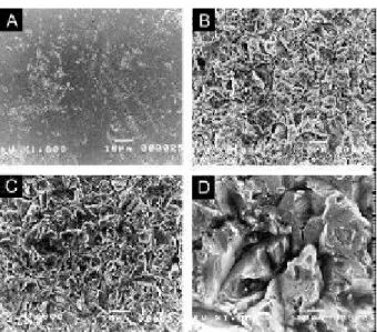
The aim of this study was to investigate the effect of acute systemic sepsis on osteoclast activity, bone mechanical, bone composition and surface roughness of cortical tibia..
The aim of this study was to investigate the effect of acute systemic sepsis on osteoclast activity, bone mechanical, bone composition and surface roughness of cortical tibia..
