In vitro
activity of Amazon plant extracts against
Enterococcus faecalis
Adriana Lígia de Castilho
1, Juliana Paola Correa da Silva
1,
Cintia Helena Coury Saraceni
1, Ingrit Elida Collantes Díaz
2,
Mateus Luís Barradas Paciencia
2, Antonio Drauzio Varella
2, Ivana Barbosa Suffredini
1,21Programa de Graduação em Odontologia, Universidade Paulista, São Paulo, SP, Brazil. 2Laboratório de Extração, Núcleo de Pesquisas em Biodiversidade, São Paulo, SP, Brazil.
Submitted: November 2, 2012; Approved: March 14, 2014.
Abstract
Previous studies analyzing 2,200 plant extracts indicated anti-enterococcal activity in 25 extracts ob-tained from Brazilian forests’ plants. In the present study, these extracts were subjected to micro-dilution broth assay (MDBA) and disk diffusion assay (DDA) using planktonic Enterococcus faecalisATCC®29212TMand were submitted to phytochemical analysis in TLC and HPLC. Three extracts obtained fromIpomoea alba(MIC < 40mg/mL),Diclinanona calycina(MIC£40mg/mL) andMoronobea coccinea(40 < MIC < 80mg/mL; MBC = 80mg/mL) showed significant bactericidal activity in the MDBA and four extracts obtained from I. alba (14.04 ± 0.55 mm diameter) S. globulifera(14.43±0.33 mm and 12.18±0.28 mm diameter) andConnarus rubervar.ruber(13.13
±0.18 mm diameter) were active in DDA. Residues H2O obtained fromPsidium densicomum(mean
of 16.78 mm diameter) and fromStryphnodendron pulcherrimum(mean of 15.97 mm diameter) have shown an improved antibacterial activity after fractionation if compared to that obtained from the re-spective crude extracts. Antioxidant activity was observed in some residues of the active extracts. TLC analysis showed that phenolic compounds are likely to be found in active extracts. Three mole-cules were isolated from S. globulifera and were identified by 13C NMR lupeol, a-amyrin and 3b-hydroxyglutin-5-ene. The present chemical and biological findings suggest that these extracts are a potential source of new anti-Enterococcuscompounds to be introduced in endodontic therapy.
Key words: Enterococcus faecalis, endodontitis, antibacterial activity, Amazon rain forest, bio-prospection.
Introduction
Enterococcus faecalisis one of the main nosocomial pathogens (Horneret al., 2005) which may be resistant to a wide range of antimicrobial agents, causing several dis-eases. These bacteria inhabit the gastrointestinal and geni-tourinary tracts and have been related to infectious endocarditis and surgical wound infection (Pasticciet al., 2008). In the oral cavity,E. faecalisis likely to be found in caries lesions, in periodontal diseases (Soutoet al., 2006), and mainly in endodontic infections (Hancocket al., 2001, Duggan and Sedgley, 2007), in which pulp may become in-fected via dentinal tubules, through carious lesions, and via periodontal disease. Studies have shown that remainingE.
faecaliscontributes to the failure of endodontic therapy. The suppression and control ofE. faecalisin dental proce-dures is paramount to diminish invasion of bacteria in dentinal tubules (Love, 2001), as well as to avoid strains to get resistance to several antibiotics (Aslangulet al., 2005) and irrigating solutions, to avoid bacteria adaptation to en-vironments where nutrients are scarce, and to abolish the establishment of any relationship with other bacteria, as in biofilms, environment and virulence factors (Kayaoglu and Orstavik, 2004).
Chemical and mechanical site preparation in the treat-ment of caries lesions and periodontal and endodontic in-fections aim to reduce infection and provide a favorable Brazilian Journal of Microbiology 45, 3, 769-779 (2014) Copyright © 2014, Sociedade Brasileira de Microbiologia
ISSN 1678-4405 www.sbmicrobiologia.org.br
Send correspondence to I.B. Suffredini. Centre for Research in Biodiversity, Extraction Laboratory, Paulista University; Av. Paulista 900, 1 andar, Bela Vista, 01310-100 São Paulo, SP, Brazil. E-mail: [email protected].
environment for an effective treatment. Chemical prepara-tion in endodontics is synonym of sodium hypochloride (SH). Currently, there is a limited number of irrigating sub-stances with antimicrobial properties that can be used in periodontal and endodontic treatment, as well as in the treatment of carious lesions. Most of the chemical sub-stances show adverse factors, such as toxicity, non-physio-logical pH, unpleasant odor and taste, and inability to degrade organic matter and smear layer. One of the most widely indicated substance is SH diluted to 1% (Estrelaet al., 2008).
The use of SH as irrigation solution is part of the pro-tocols established to prepare root canals to endodontic treatments, considering that removal of any microorganism is fundamental to avoid any further relapse. One of the main microorganisms found in recurrent failure in endodontic treatment isE. faecalis, which is a bacterium that exhibits increased virulence in pathological conditions of endo-dontic disease. For that reason, this bacterium is one of the main target for drug action in the treatment of this oral pathological condition (Pinheiroet al., 2004; Pinheiro et al., 2003). Considering the undesirable effects related to SH, it has become increasingly necessary to find new tools to assist in the fight againstE. faecalis.
Systematic studies with plant and animal extracts (Alvianoet al., 2008) have shown that it is possible to find compounds with pharmaceutical potential, such as some antibacterial chemotherapeutic agents (penicillin, erythro-mycin, tetracyclin). Universidade Paulista (UNIP), Brazil, introduced in 1996 a large-scale bioprospecting program targeting the identification of antibacterial compounds in plant extracts native to Brazilian forests. Throughout the program, over 2,000 plant extracts were tested against sev-eral microorganisms, such asStaphylococcus aureus, Esch-erichia coli,Pseudomonas aeruginosa,Candida albicans, Streptococcus mutans, S. sanguinis, and Enterococcus faecalis(Suffrediniet al., 2006a; Suffrediniet al., 2006b; Suffrediniet al., 2004; Suffrediniet al., 2002). Based on these results, the present study aimed the study of the anti-bacterial activity of 25 plant extracts and their residues against traditional antibacterial assays and to search for chemical groups that may occur in the active extracts and residues, emphasizing an organic extract obtained from Symphonia globulifera, which was available in higher amounts to develop initial chemical studies.
Material and Methods
Plant collection and extraction preparation
For plant collection and extraction, our team obtained a collecting permit issued by the Brazilian Institute of Envi-ronment and Renewable Natural Resources - IBAMA and a permit to access genetic resources issued by the Genetic Heritage Management Council - CGen of the Brazilian Ministry of the Environment. Collected plants were
taxo-nomically identified and voucher specimens were depos-ited at UNIP Herbarium, São Paulo, Brazil. Organic and aqueous extracts were obtained by a 24-h maceration with dichloromethane: methanol (1:1), followed by a 24-h mac-eration with distilled water (Millipore®, Bedford, MA, USA) (Youneset al., 2007; Youneset al., 2000).
Test compounds and controls
Twenty-five previously selected extracts, here de-signed as EB 55, EB 321, EB 352, EB 429, EB 841, EB 1247, EB 1257, EB 1259, EB 1298, EB 1373, EB 1389, EB 1395, EB 1461, EB 1493, EB 1497, EB 1525, EB 1543, EB 1549, EB 1637, EB 1743, EB 1765, EB 1905, EB 1923, EB 1991 and EB 1999, which taxonomical and collection data are listed in table 1, were diluted to a series of dilutions that led to final test concentrations in the microdilution broth as-say of 12,500, 10,000, 7,500, 5,000, 2,500, 1,300, 600, 310, 160, 80, and 40mg/mL,, being 20 times more diluted than the initial extracts dilutions, according to the established protocol). Dimethylsulfoxide and water were used as sol-vent in the dilution procedure. Sodium hypochlorite 1% (SH) was used as positive control, and was obtained from a formulated SH 10% solution (Fórmula & Ação - Com-pounding Pharmacy). A 1% SH commercial solution (Asfer®, São Paulo, SP, Brazil) was also used as positive
control. The same extracts were tested in disk diffusion as-say at a concentration of 200 mg/mL, diluted in dime-thylsulfoxide 50% in water. All organic extracts were diluted in dimethylsulfoxide 50% (DMSO50).
Enteroroccus faecalis
The biosafety level 2 bacteria used in all assays were obtained from ATCC. Bacteria was acquired lyophilized, in Loops®(Oxoid Ltd, London, England), and was seeded
in Müeller-Hinton agar (Oxoid Ltd, London, England), put in an incubator for 24 h, at 36 °C. this plate was called “mother-plate”, and was left to be used within 30 days, if kept under refrigeration. 24-h fresh colonies were acquired before each assay, so, bacteria were at the same 4thpassage
during every experiment performed in the present work.
Disk diffusion assay and the determination of the growth inhibition zone diameter
Disk diffusion assay was done as usually described for plant extracts analysis (Soutoet al., 2006), in strict ster-ile conditions. A 0.5 McFarland saline suspension was pre-pared from a fresh colony ofEnterococcus faecalisATCC®
29212TM. Assay was performed in sterile Müeller-Hinton
inhibi-Amazon
plants
against
E.
faecalis
771
Table 1- List of extracts showing activity againstEnterococcus faecalisATCC®29212TMand the corresponding minimal inhibitory concentrations obtained from microdilution broth assay.
Extract number* Herbarium reference Collect date Organ Family Gender Species 1 x 10⁵cfu MIC (
mg/mL)
55 PSC 396 05/12/1997 Leaves Fabaceae
Caesalpinioideae
Macrolobium multijugum 5,000 < MIC < 12,500
321 PSC 414 06/12/1997 Leaves and flowers Myrtaceae Psidium densicomum 5,000 < MIC < 12,500
352 IBS 9 25/06/1998 Stem Fabaceae Mimosoideae Stryphnodendron pulcherrimum 2,500 < MIC < 5,000
429 PSC 144 30/05/1997 Stem Proteaceae Roupala sp. 7,500 < MIC < 12,500
841 AAO 3458 26/08/1999 Aerial organs Myrsinaceae Rapanea parvifolia 200 < MIC < 300
1247 IBS 53 30/09/2001 Aerial organs Myrtaceae Psidium densicomum 5,000 < MIC < 12,500
1257 AAO 3717 10/08/2001 Aerial organs Clusiaceae Symphonia globulifera 200 < MIC < 300
1259 IBS 56 30/09/2001 Aerial organs Piperaceae Piper sp. 7,500 < MIC < 12,500
1298 AAO 3721 11/08/2001 Stem Lauraceae Ocotea myriantha 300 < MIC < 600
1373 IBS 110 06/12/2001 Leaves Clusiaceae Garcinia cf. macrophylla > 12,500
1389 IBS 142 08/12/2001 Stem Clusiaceae Moronobea coccinea 1,300 < MIC < 2,500
1395 IBS 143 08/12/2001 Leaves Fabaceae
Caesalpinioideae
Hymenaea parvifolia 80 < MIC < 200
1461 AAO 4015 12/05/2002 Aerial organs Sapindaceae Paullinia cf. alata 7,500 < MIC < 12,500
1493 AAO 4031 13/05/2002 Aerial organs Convolvulaceae Ipomoea alba < 40
1497 IBS 164 13/06/2002 Aerial organs Moraceae Sorocea duckei 200 < MIC < 300
1525 IBS 63 01/11/2001 Stem Connaraceae Connarus ruber var. ruber 600 < MIC < 1,300
1543 AAO 3711 10/08/2001 Aerial organs Clusiaceae Tovomita longifolia > 12,500
1549 AAO 4027 12/05/2002 Aerial organs Combretaceae Combretum rotundifolium 200
1637 AAO 4120 20/07/2002 Aerial organs Annonaceae Diclinanona calycina £40
1743 IBS 204 12/07/2003 Aerial organs Connaraceae Connarus cf. ruber var. sprucei 1,300 < MIC < 2,500
1765 IBS 142 08/12/2001 Flores Clusiaceae Moronobea coccinea 40 < MIC < 80
1905 AAO 4148 21/07/2002 Stem Clusiaceae Symphonia globulifera 2,500 < MIC < 5,000
1923 AAO 4029 12/05/2002 Aerial organs Fabaceae Faboideae Vatairea guianensis 2,500 < MIC < 5,000
1991 AAO 4148 21/07/2002 Aerial organs Clusiaceae Symphonia globulifera 80 < MIC < 200
1999 AAO 4199 22/08/2002 Aerial organs Myristicaceae Iryanthera laevis > 12,500
tion zones were measured horizontally and vertically with a caliper rule.
Statistical analysis for disk diffusion assays
One-way ANOVA and Tukey’s post-test analysis was applied in the evaluation of growth inhibition zone di-ameters resulted from the antibacterial activity of plant ex-tracts and their residues, SH1%F = formulated sodium hypochlorite 1%; SH1%C = commercial sodium hypo-chlorite 1% (standard drugs used as positive control), against Enterococcus faecalis ATCC® 29212TM. Results
were significant if p < 0.05.
Microdilution broth assay and determination of minimal inhibitory concentration and minimal bactericidal concentration
The extracts were tested by the microdilution broth assay (MDBA), in completely sterile conditions, adapted to high-throughput conditions (Suffrediniet al., 2006b), in a biosafety level 2 Laboratory Unit, using sterilized Müeller-Hinton broth medium (Oxoid Ltd, London, Eng-land), in 96-well microplates, with inocula adjusted to a 0.5 McFarland standard, or 1 x 10⁸colony-forming unit per mL (cfu/mL), obtained from fresh colonies of Enterococcus faecalisATCC®29212TM(Suffrediniet al., 2006a; Suffredini et al., 2006b; Suffredini et al., 2004) grown on sterilized Müeller-Hinton agar medium (Oxoid Ltd, London, England). Concentrations of bacterial suspen-sion, such as 1 x 10², 1 x 10³, 1 x 10⁴, 1 x 10⁵, 1 x 10⁶, and 1 x 10⁷cfu/mL, were prepared in sterilized Müeller -Hinton broth medium (Oxoid Ltd, London, England) starting from the 1 x 10⁸colony-forming unit per mL (cfu/mL) suspen-sion. Thus, a 190-mL aliquot of the inoculum was dispensed into the wells, and 10mL aliquots of extract, residue or stan-dard compound solutions were then added to the inoculum. Microplates were incubated at 37 °C for 24 h. Inhibition of bacterial growth was assessed visually, and bacterial sus-pensions from all test wells were subcultured in sterile agar medium in order to evaluate bactericidal activity.
Following a procedure similar to that described above in sterile MDBA, minimal inhibitory concentration (MIC) and minimal bactericidal concentration (MBC) were ob-tained for the 25 plant extracts (PE), against E. faecalis. Solvent dimethylsulfoxide were tested as negative control.
Liquid-liquid partition and biological assay of residues
Each one of the 25 extracts was partitioned with three solvents of different polarities resulting in non polar resi-dues (chloroform residue, or Residue CHCl3, or EB-CHCl3
where EB refers to the extract number), intermediate-pola-rity residues (butanol residue, or Residue BuOH, or EB-BuOH where EB refers to the extract number), and polar residues (aqueous residue, or Residue H2O, or EB-H2O
where EB refers to the extract number). The solvents were
completely evaporated, followed by lyophilization, resulting in 75 residues. Organic solvents were completely removed under vacuum. All residues were re-suspended in 50% dimethylsulfoxide (DMSO50) or in distilled water (both tested as negative controls), at the concentration of 200 mg/mL, to be tested, as follows.
For preparation of 200 mg/mL solutions, aqueous res-idues were diluted with Milli-Q distilled water (Millipore®,
Bedford, MA, USA) and chloroform and butanol residues were diluted in DMSO50, in order to be assayed by the MDBA, according to the technique previously described, in bacterial suspensions containing 1 x 10⁶cfu/mL.
Thin-layer chromatography
Thin-layer chromatography (TLC) was performed with extracts and residues. Silica gel GF chromatography sheets (Merck®, Whitehouse Station, New Jersey, USA)
were used and the following mobile phase were prepared: System 1 - hexane and ethyl acetate (4:1) applied on chro-matographic sheet 1; System 2 - chloroform and ethyl ace-tate (1:1) applied on chromatographic sheet 2; and System 3 - chloroform, ethyl acetate and methanol (2:2:1); System 4 - ethyl acetate, methanol and water (100:13.5:10); System 5 - ethyl acetate, glacial acetic acid, formic acid and water (100:11:11:26). Revelation of spots was done with potas-sium hydroxide (A); Kedde Reagent (B), Dragendorff Re-agent (C), NP ReRe-agent (D), 25% sulfuric acid solution (E) andb-caroten (F). Ultraviolet light at 254 and/or 366 nm (uv254 or uv356) was used to develop some of the chroma-tograms (Suffrediniet al., 2004).
Evaluation of antioxidant potential
Evaluation of the antioxidant potential of extracts and residues was performed using the b-carotene technique (Pratt and Miller, 1984), in System 3 described above. A mixture of two solutions composed of (a) 9-mgb-carotene dissolved in 30 mL of chloroform and (b) two drops of linoleic acid dissolved in 60 mL of ethanol was used to re-veal antioxidant substances in chromatogram. The chroma-tograms were exposed to natural light for at least 2 h in order to catalyze the antioxidant reaction between the natu-ral product and the development system.
Fractionation of EB 1257
Extract EB1257, obtained from the aerial organs ofS. globulifera, was used in the chemical assays.
Column chromatography fractionation
In order to isolate major compounds, 16.7668 g of ex-tract 1257 were submitted to liquid-liquid partition using the technique first described, which resulted in Residue CHCl3 (7212.6 mg, or 43.02% yield) Residue BuOH
(3815.3 mg, or 22.76% yield) and Residue H2O
(5738.9 mg, or 34.23% yield). Residue CHCl3was
sta-tionary phase, and 100% hexane, 100% dichloromethane and 100% methanol, as mobile phase, and resulted in hex-ane fraction (3031.2 mg, or 42.03% yield), dichlorome-thane fraction (1230.9, or 17.07% yield mg) and methanol fraction (2950.5 mg, or 40.91% yield).
Chromatographic techniques used in the isolation of major compounds - thin layer chromatography and normal phase open column chromatography
The hexane fraction obtained from Residue CHCl3,
which was obtained from EB1257, was submitted to frac-tionation in column chromatography using silica gel 60-200mm as stationary phase and a gradient with increas-ing polarity composed by mixtures of hexane, ethyl acetate and methanol as mobile phase, which resulted in 28 frac-tions. The fractions were compared by TLC and were grouped according to their similarity, after analysis with sulfuric acid 25% and heating, resulting in fractions 10-11, 12, 13, 14-15, 16, 17,.., 28. Fraction 13, named UNIP147 was sent to1H NMR and e13C NMR, in CDCl
3.
Fraction 13 (or UNIP147) was obtained as a crystal cream sample (30 mg). 1H NMR was obtained but it showed to be complex. So, a13C NMR was obtained in or-der to support molecules’ identification. Three molecules were identified and their spectra were compared to that in the literature.
Results
Taxonomic information of the 25 plants included in the present study is provided in Table 1 and extracts are re-ferred to in the text by their extract number.
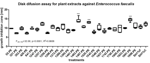
Figure 1 represents the comparison among means ob-tained from inhibition growth zone diameters of the ex-tracts and positive / negative controls, in the disk diffusion assay against 0.5 McFarland inoculum ofE. faecalis.
One-way ANOVA followed by Tukey’s post-test indicated that the means were significant different (F(26, 135) = 32.06;
p < 0.0001;R2 = 0.8606, 27 groups). Results generated from both DMSO solutions (media = 0.00 mm growth inhi-bition zone diameters) were not included in statistical anal-ysis due to the lack of homocedasticity if those groups were included. The following treatments showed to be as effi-cient as SH1%F (p > 0.05): 55.PE, 352.PE, 841.PE, 1247.PE, 1257.PE, 1298.PE, 1373.PE, 1389.PE, 1497.PE, 1549.PE, 1743.PE, 1765.PE, 1905.PE, 1923.PE, 1999.PE. The following treatments showed to be significantly more efficient than SH1%F (p < 0.05): 1493.PE, 1525.PE and 1991.PE. Considering SH1%C, the following treatments showed equivalent antibacterial activity (p > 0.05): 1257.PE, 1298.PE, 1389.PE, 1497.PE, 1525.PE and 1999.PE.Lastly, the following treatments were signifi-cantly more efficient than SH1%C (p < 0.05): 1493.PE 1991.PE.
Figure 2 represents the comparison among means ob-tained from inhibition growth zone diameters of the ex-tracts, residues and standard drugs, in the disk diffusion assay against 0.5 McFarland inoculum ofE. faecalisusing one-way ANOVA followed by Tukey’s post-test. Results generated from both DMSO solutions (mean = 0.00 mm growth inhibition zone diameters) were not included in sta-tistical analysis due to the lack of homocedasticity if those groups were included. The following treatments showed to be as efficient as SH1%F (p > 0.05): 55.BuOH, 321.BuOH, 352.BuOH, 1257.CHCl3, 1257.Aq, 1389.Aq, 1525.Aq and
1991.CHCl3. The following treatments were more effective
than SH1%F (p < 0.05): 321.Aq, 352.Aq, 841.BuOH, 1257.BuOH, 1259.BuOH, 1298.BuOH, 1389.BuOH, 1493.CHCl3, 1493.BuOH, 1525.BuOH and 1991.BuOH.
In relation to SH1% C, the following treatments were statis-tically equivalent antibacterial activity (p > 0.05):
Amazon plants againstE. faecalis 773
Figure 1- Two-way ANOVA and Bonferroni’s post-test analysis related to growth inhibition zone diameters obtained from the antibacterial activity in disk diffusion assay of plant extracts and their chloroform, buthanol and aqueous residues (concentration of 200 mg/mL), formulated and commercial so-dium hypochloride 1% (used as positive controls) and dimethylsulfoxide 50% and pure (used as negative control), againstEnterococcus faecalisATCC®
29212TM. SH1%F = formulated sodium hypochlorite 1%; SH1%C = commercial sodium hypochlorite 1%. Residues are nominated as XX.CHCL3,
55.BuOH, 352.BuOH, 841.BuOH, 1257.BuOH, 1259.BuOH, 1298.BuOH, 1389.BuOH, 1525.BuOH and 1991.CHCl3. Finally, the following treatments were more
effective than SH1%C (p < 0.05): 321.Aq, 352.Aq, 1493.CHCl3, 1493.BuOH and 1991.BuOH.
Table 2 shows the antioxidant activity of residues. Antioxidant compounds are prone to be found in residues BuOH obtained from extracts EB 55, EB 841, EB 1257, EB 1373, EB 1389, EB 1525, EB 1905, EB 1991 and EB 1999. Also, antioxidant compounds are prone to be found in resi-dues CHCl3obtained from extract EB 1637 and in residues
H2O obtained from extracts EB 1525 and EB 1905.
TLC analysis of EB 321, EB 352, EB 1257, EB 1493, EB 1525, EB 1765, EB 1991 and EB 321 indicated that phenolic compounds might be present, once a weak blue fluorescence could be seen after NP and uv356 develop-ment. Triterpenes or essential oils (due to sulfuric acid de-velopment) may be present in EB 321. EB 352 may also have phenolic compounds (due to the yellow spots that could be seen after NP and uv356 development) and triter-penes or essential oils. EB 1257 may have coumarins or anthrone-like compounds, once the development with KOH showed a brown spot under visible light, and may have phenolic compounds related to orange spots observed after NP and uv 356 development. Triterpenes may also be present in this extract. EB1493 may have triterpenes as ma-jor compounds, once only the sulfuric acid solution re-vealed the the chromatogram. EB 1525 may have couma-rins and triterpenes. EB 1765 may have antrone-like compounds (development under KOH and visible light), a
brown spot was observed after development with Dragen-dorff’s Reagent and NP followed by uv356 observation showed two orange and one yellow-green spots, suggesting the presence of phenolic compounds. Lastly, EB 1991 showed a yellow spot after development with KOH, sug-gesting anthrone-like compounds, 3 orange and 1 yel-low-green spot, after development with NP and uv356.
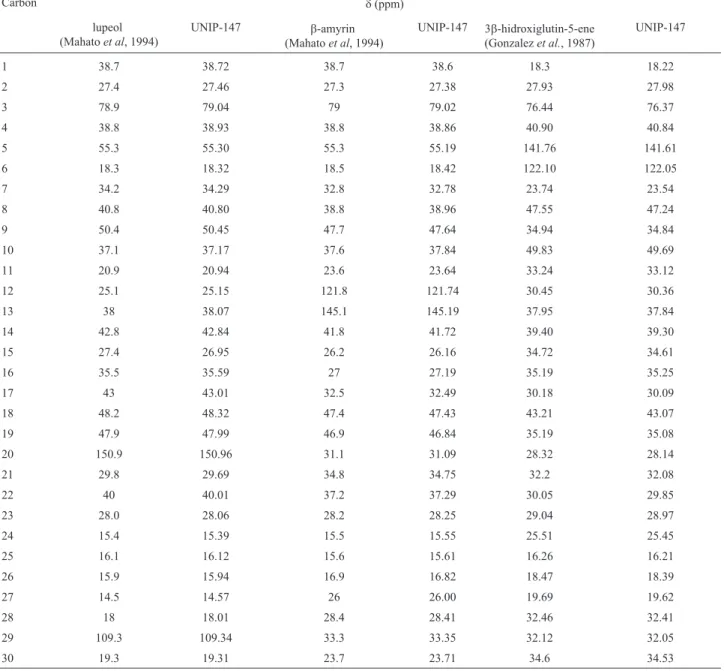
EB 1257 was fractionated and compounds lupeol,
b-amyrin and 3b-hydroxyglutin-5-ene were isolated (Fig-ure 3) and their struct(Fig-ure was identified by 13C NMR
(125 MHz, CDCl3) analysis and comparison with literature
(Table 3).
Discussion
E. faecalisis an important pathogen related to dis-eases that affect the oral cavity. In endodontic infections, especially in recurrent infection, its presence is commonly associated with treatment failure (Kayaogluet al., 2011; Cowan, 1999). SH is a compound of choice in Dentistry, and is widely used in different concentrations, in Endodontics. Nonetheless, the compound has undesirable effects, which are often disregarded when these substances are elected because of their antimicrobial therapeutic bene-fit. Therefore, substances that have similar therapeutic effi-cacy and fewer side effects should be tracked and used in dental therapy. Natural products, such as propolis (Kaya-ogluet al., 2011), are being investigated for their antibacte-rial activity.
Two experimental models were selected to evaluate the potential antimicrobial activity of plant extracts against
Figure 2- One-way ANOVA and Tukey’s post-test analysis related to growth inhibition zone diameters obtained from the antibacterial activity in disk diffusion assay of plant extracts, residues (concentration of 200 mg/mL), formulated and commercial sodium hypochloride 1% (used as positive con-trols), againstEnterococcus faecalisATCC®29212TM. SH1%F = formulated sodium hypochlorite 1%; SH1%C = commercial sodium hypochlorite 1%.
E. faecalis: DDA and MDBA, understanding that both to-gether might contribute to the prospection of new lead com-pounds in a complementary way. It is not the scope of the present manuscript to determine which assay is more suit-able to be used - and discussions on the subject have widely been made before (Cowan, 1999). Briefly, natural products are complex mixture of substances that have different phys-ical-chemical characteristics. Such differences may inter-fere with the characteristics of each DDA or MDBA assay due to the way chemicals can diffuse (in DDA) or dilute (in MDBA) in each type of medium. The use of both experi-ments seems to be complementary so far.
Based on analyses done with extracts in MDBA (Ta-ble 1) - which compared data from various concentrations of treatments tested against bacterial concentration of 1.0 x 10e5 CFU/mL, it was possible to identify three promising extracts: EB 1493, EB 1637 and EB 1765. EB 1493 was
ob-tained from aerial organs of Ipomoea alba L. (Convolvulaceae).I. albawas barely studied in terms of its chemical constituents or pharmacological activity. The use of its latex was reported (Hosleret al., 1999). Sweet potato is one of the species belonging toIpomoeagenus. Clavine alkaloids occur in I. muricata (Maruia and Srivastava, 2009), as well as ipobscurines that are indol alkaloids from the macrolactam-type (Jenett-Siemset al., 2003) and alka-loids occur in some toxic species, as inI. carnea (Hara-guchi et al., 2003). Other compounds as anthocyanins (Steed and Truong, 2008) and polyphenolic compounds (Kurataet al., 2007) also occur inIpomoeaspecies.
Extract 1637, obtained from Diclinanona calycina (Annonaceae), has shown a significant activity in the assay. Previous study made withD. calycina, tested against di-verse microorganisms in an agar-diffusion model, has shown its activity againstStreptococcus oralisand
Myco-Amazon plants againstE. faecalis 775
Figure 3- Molecular structure of UNIP-147 isolated compounds (A) lupeol, (B)b-amyrin and (C) 3b-hydroxyglutin-5-ene.
Table 2- Results corresponding to the antioxidant activity of residues, which represent reaction tob-caroten.
Extract number CHCl3 BuOH H2O Extract number CHCl3 BuOH H2O
55 - X - 1493 - -
-321 - - - 1497 - -
-352 - - - 1525 - X X
429 - - - 1543 - -
-841 - X - 1549 - -
-1247 - - - 1637 X -
-1257 - X - 1743 - -
-1259 - - - 1765 - -
-1298 - - - 1905 - X X
1373 - X - 1923 - -
-1389 - X - 1991 - X
-1395 - - - 1999 - X
-1461 - -
bacterium smegmatis(Carneiroet al., 2008). Annonaceae species were chemically studied before. Alkaloids (Pérez and Cassels, 2010) and acetogenins (McLaughlin, 2008) are the main classes of active compounds occurring in the family.
Extract EB 1765 was obtained from the flowers of Moronobea coccinea, a species belonging to Clusiaceae. Polycyclic polyprenylated acylphoroglucinols and oxi-dized derivatives (Martiet al., 2010) were isolated fromM. coccinea, as well as xanthone derivatives and flavonoids (Mkoungaet al., 2009).
DDA showed a different group of active extracts: EB 1257, EB 1493, EB 1525 and EB 1991. Extracts 1257 and 1991 were obtained from aerial organs ofS. globulifera,
Clusiaceae in different periods of time. Chemistry of Ipomoea(EB 1493) species was previously discussed. Ex-tract 1525 was obtained from the stem ofConnarus ruber var.ruber(Connaraceae). It was observed that there is a lack of chemical data related to the genusConnarus. Ex-tracts EB 1257 and EB 1991 were obtained from the same species, S. globulifera, Clusiaceae, but plants were col-lected in different periods of time. Reports relate biological activity maybe related to the presence of polycyclic poly-prenylated acylphoroglucinols and oxidized derivatives (Martiet al., 2010). Xanthones are also present in the spe-cies (Panaet al., 2010). Chemical studies were performed with EB 1257 due to extract availability. So, isolation and identification of major compounds were performed and
re-Table 3-13C NMR (125 MHz, CDCl
3) data for sample UNIP-147.
Carbon d(ppm)
lupeol (Mahatoet al, 1994)
UNIP-147 b-amyrin
(Mahatoet al, 1994)
UNIP-147 3b-hidroxiglutin-5-ene (Gonzalezet al., 1987)
UNIP-147
1 38.7 38.72 38.7 38.6 18.3 18.22
2 27.4 27.46 27.3 27.38 27.93 27.98
3 78.9 79.04 79 79.02 76.44 76.37
4 38.8 38.93 38.8 38.86 40.90 40.84
5 55.3 55.30 55.3 55.19 141.76 141.61
6 18.3 18.32 18.5 18.42 122.10 122.05
7 34.2 34.29 32.8 32.78 23.74 23.54
8 40.8 40.80 38.8 38.96 47.55 47.24
9 50.4 50.45 47.7 47.64 34.94 34.84
10 37.1 37.17 37.6 37.84 49.83 49.69
11 20.9 20.94 23.6 23.64 33.24 33.12
12 25.1 25.15 121.8 121.74 30.45 30.36
13 38 38.07 145.1 145.19 37.95 37.84
14 42.8 42.84 41.8 41.72 39.40 39.30
15 27.4 26.95 26.2 26.16 34.72 34.61
16 35.5 35.59 27 27.19 35.19 35.25
17 43 43.01 32.5 32.49 30.18 30.09
18 48.2 48.32 47.4 47.43 43.21 43.07
19 47.9 47.99 46.9 46.84 35.19 35.08
20 150.9 150.96 31.1 31.09 28.32 28.14
21 29.8 29.69 34.8 34.75 32.2 32.08
22 40 40.01 37.2 37.29 30.05 29.85
23 28.0 28.06 28.2 28.25 29.04 28.97
24 15.4 15.39 15.5 15.55 25.51 25.45
25 16.1 16.12 15.6 15.61 16.26 16.21
26 15.9 15.94 16.9 16.82 18.47 18.39
27 14.5 14.57 26 26.00 19.69 19.62
28 18 18.01 28.4 28.41 32.46 32.41
29 109.3 109.34 33.3 33.35 32.12 32.05
sulted in lupeol,a-amyrin and 3b-hydroxyglutin-5-ene as isolated compounds, widely known for their biological and pharmacological activities.
All 25 extracts were submitted to an initial fraction-ation, resulting in three residues to each extracts: CHCl3,
BuOH and H2O. It was observed that after extracts 321 and
352 were fractionated, a significant improvement of the an-tibacterial activity could be observed in their H2O residue.
For that reason, both extracts were also chosen to be studied in a near future. Extract 321, obtained from the leaves of Psidium densicomum(Myrtaceae), belongs to a group of plants (guajava family) well known to contain terpenes, triterpenoids and flavonoids (Hoet al., 2012; Gutierrezet al., 2008). Extract 352 was obtained from aerial parts of Stryphnodendron pulcherrimum(Fabaceae), species taxo-nomically related to a medicinal plant known in Brazil as barbatimao, that was already studied against oral patho-gens (Pereiraet al., 2011) to contain tannins as the main chemical class of compounds (Santoset al., 2002).
As fractionation of the 25 extracts was done exactly in the same way to obtain residues, added to the fact that all residues were tested at the same time, in both biological as-says, it is possible to suggest a comparative analysis based on the results obtained from the biological assays of resi-dues. Antibacterial results obtained from the residues when compared to their original extracts may take different courses: (a) biological activity has been missing during fractionation, and synergism was lost; (b) biological ity is concentrated in one fraction and its antibacterial activ-ity may be improved or not, in relation to the original extract; (c) biological activity appears in two or more frac-tions and the antibacterial activity may have improved or not, in relation to the original extract. Based on that pre-mises, a large number of extracts can be considered for fur-ther analyses, which is profitable but time-consuming. For that reason, it is primordial to establish a method according to which the extracts and their residues are going to be com-pared among each other, and the more adequate way is to rank MIC’s and MBC’s values of treatments when MDBA is used, and to compare growth inhibition zones of treat-ments by analysis of variance ANOVA, as applied in the present work when DDA is used. If that idea is applied, it is possible to create a ranking establishing which extract and/or residue is best to be prioritized in the studies.
As the current study aims the identification of a new compound to be used in root canal treatment, it is important to compare the efficacy of extracts treatments to SH, which is one of the compounds of choice, for this application. SH was prepared at the concentration of 1% to be used as stan-dard drug, despite its wide range of concentrations found in literature, ranging from 1 to 5.25% that are available in the marketing. Studies reporting the efficacy of SH in biofilms obtained fromE. faecalisand/orS. mutanswere done and this compound showed better efficacy when used against biofilms obtained from planktonic cultures (Jianget al.,
2011). Although a direct comparison between SH and plant extracts is not feasible, since SH is an isolated compound and plant extracts are highly complex mixtures of com-pounds of various origins, the results obtained for SH pro-vide an important reference for a comparative, albeit indirect, analysis of the antibacterial activity of extracts. Moreover, it is highly expected that when active substances are isolated from the extracts, they may become more ac-tive than the original extract, which is going to be further done.
Antioxidant activity was also evaluated, and resulted that 55.BuOH, 841.BuOH, 1257.BuOH, 1373.BuOH, 1389.BuOH, 1525.BuOH, 1525.H2O, 1637.CHCl3,
1905.BuOH, 1905.H2O, 1991.BuOH, 1999.BuOH showed
activity in theb-caroten TLC assay. Further evaluation of isolated compounds shall be done in the near future. Chem-ical data found in the literature is consistent with results we obtained in the TLC analysis of the main chemical classes as phenolic compounds, triterpenes and alkaloids, and will support future isolation procedures to be done with all seven active extracts.
Results related to the efficacy of the extracts against planktonic cultures do not assure that the extracts are going to be as effective against biofilms, and for that reason, fur-ther in vitro studies regarding the activity of extracts against biofilms are being developed today as well as the evaluation of the extracts in toxicological assays, in order to gather information to supportin vivostudies (Barrellaet al., 2012) with the extracts.
Conclusions
Nature is a potential source of new drugs to be used in Dentistry. In the present work, seven plant extracts ob-tained from species native to the Amazon rain forest showed significant activity againstE. faecalisand showed that antioxidant compounds may be present as well, indi-cating that possibly phenolic compounds may be involved in antibacterial activity. Also, statistical design here pro-posed also contributes to optimize the decision-making of which extract shall be prioritized for chemical studies so as to isolate active compounds that might be introduced in endodontic therapy.
Acknowledgments
Authors thank FAPESP for their financial support
References
Alviano WS, Alviano DS, Diniz CG, Antoniolli AR, Alviano CS, Farias LM, Carvalho MA, Souza MM, Bolognese AM (2008)In vitroantioxidant potential of medicinal plant ex-tracts and their activities against oral bacteria based on Bra-zilian folk medicine. Arch Oral Biol 53:545-552.
Aslangul E, Ruimy R, Chau F, Garry L, Andremont A, Fantin B (2005) Relationship between the level of acquired resistance
to gentamicin and synergism with amoxicillin in
Enterococcus faecalis. Antimicrob. Agents Chemother 49:4144-4148.
Barrella GE, Suffredini IB, Ribeiro FV, Cirano FR, Pimentel SP (2012) Evaluation of the effect of an organic extract ob-tained fromIpomoea albaL. on experimental periodontitis in rats. Braz Oral Res 26:158-164.
Carneiro AL, Teiceira MF, Oliveira VM, Fernandes OC, Cauper GS, Pohlit AM (2008) Screening of Amazonian plants from Adolpho Ducke forest reserve, Manaus, state of Amazonas, Brazil, for antimicrobial activity. Mem Inst Oswaldo Cruz 103:31-38.
Cowan MM (1999) Plant products as antimicrobial agents. Clin Microbiol Rev 12:564-582.
Duggan JM, Sedgley CM (2007) Biofilm formation of oral and endodonticEnterococcus faecalis.J Endod 33:815-818. Estrela C, Silva JA, de Alencar AH, Leles CR, Decúrcio DL
(2008) Efficacy of sodium hypochlorite and chlorhexidine againstEnterococcus faecalis- A systematic review. J Appl Oral Sci 16:364-368.
Gutierrez RM, Mitchell S, Solis RV (2008)Psidium guajava: a re-view of its traditional uses, phytochemistry and pharmacol-ogy. J Ethnopharmacol 117:1-27.
Hancock HH 3rd, Sigurdsson A, Trope M, Moiseiwitsch J (2001) Bacteria isolated after unsuccessful endodontic treatment in a North American population. Oral Surg Oral Med Oral Pathol Oral Radiol and Endod 91:579-586.
Haraguchi M, Gorniak SL, Ikeda K, Minami Y, Kato A, Watson AA, Nash RJ, Molyneux RJ, Asano N (2003). Alkaloidal components in the poisonous plant, Ipomoea carnea
(Convolvulaceae). J Agric Food Chem 51:4995-5000. Ho R, Violette A, Cressend D, Raharivelomanana P, Corrupt PA,
Hostettmann K (2012) Antioxidant potential and radical-scavenging effects of flavonoids from the leaves ofPsidium cattleianum grown in French Polynesia. Nat Prod Res 26:274-277.
Hosler D, Burkett SL, Tarkanian MJ (1999) Prehistoric polymers: rubber processing in ancient Mesoamerica. Science 284:1988-1991.
Horner R, Liscano MG, Maraschin MM, Salla A, Meneghetti B, Dal Forno NLF, Righi RA (2005) Suscetibilidade antimi-crobiana entre amostras deEnterococcusisoladas no Hospi-tal Universitário de Santa Maria. J Bras Patol Med Lab 41391-41395.
Jenett-Siems K, Weigl R, Kaloga M, Schulz J, Eich E (2003) Ipobscurines C and D: macrolactam-type indole alkaloids from the seeds of Ipomoea obscura. Phytochemistry 62:1257-1263.
Jiang LM, Hoogenkamp MA, van der Sluis LWM, Wesselink PR, Crielaard W, Deng DM (2011) Resazurin metabolism assay for root canal disinfectant evaluation on dual-species bio-films. J Endod 37:31-35.
Kayaoglu G, Orstavik D (2004) Virulence factors of
Enterococcus faecalis: relationship to endodontic disease. Crit Rev Oral Biol Med 15:308-320.
Kayaoglu G, Ömürlü H, Akca G, Gürel M, Gençay Ö, Sorkun K, Salih B (2011) Antibacterial activity of propolisvs. conven-tional endodontic disinfectants against Enterococcus faecalisin infected dentinal tubules J Endod 37:376-381.
Kristich AJ, Li YH, Cvitkovitch DG, Dunny GM (2004) Esp-independent biofilm formation byEnterococcus faecalis. J Bacteriol 186:154-163.
Kurata R, Adachi M, Yamakawa O, Yoshimoto M (2007) Growth suppression of human cancer cells by polyphenolics from sweetpotato (Ipomoea batatas L.) leaves. J Agric Food Chem 55:185-190.
Love MR (2001)Enterococcus faecalis- a mechanism for its role in endodontic failure. Int Endod J 34:399-405.
Marti G, Eparvier V, Moretti C, Prado S, Grellier P, Hue N, Thoison O, Delpech B, Guéritte F, Litaudon M (2010) Anti-plasmodial benzophenone derivatives from the root barks of
Symphonia globulifera (Clusiaceae). Phytochemistry 71:964-974.
Maurya A, Srivastava SK (2009) Large-scale separation of clavine alkaloids from Ipomoea muricata by pH-zone-refining centrifugal partition chromatography. J Chromatogr B Analyt Technol Biomed Life Sci 877:1732-1736.
McLaughlin JL (2008) Paw paw and cancer: annonaceous aceto-genins from discovery to commercial products. J Nat Prod 71:1311-1321.
Mkounga P, Fomum ZT, Meyer M, Bodo B, Nkengfack AE (2009) Globulixanthone F, a new polyoxygenated xanthone with an isoprenoid group and two antimicrobial biflavonoids from the stem bark of Symphonia globulifera. Nat Prod Commun 4:803-808.
Pana E, Cao S, Brodie PJ, Miller JS, Rakotodrajaona R, Rato-voson F, Birkinshaw C, Andriantsiferana R, Rasamison VE, Kingston DG (2010) An antiproliferative xanthone of
Symphonia pauciflora from Madagascar rainforest. Nat Prod Commun 5:751-754.
Pasticci MB, Mencacci A, Moretti A, Palladino N, Maria Lapa-lorcia L, Bistoni F, Baldelli F (2008)In vitroantimicrobial activity of ampicillin-ceftriaxone and ampicillin-ertapenem combinations against clinical isolates of Enterococcus faecalis with levels of aminoglycoside resistance. Open Microbiol J 2:79-84.
Pereira EM, Gomes RT, Freire NR, Aguiar EG, Brandão MG, Santos VR (2011)In vitroantimicrobial activity of Brazilian medicinal plant extracts against pathogenic microorganisms of interest in Dentistry. Planta Med 77:401-404.
Pérez EG, Cassels BK (2010) Alkaloids from the genusDuguetia. Alkaloids Chem Biol 68:83-156.
Pinheiro ET, Gomes BP, Ferraz CC, Sousa EL, Teixeira FB, Souza-Filho FJ (2003) Microorganisms from canals of root-filled teeth with periapical lesions. Int Endod J 36:1-11. Pinheiro ET, Gomes BPFA, Drucker DB, Zaia AA, Ferraz CC,
Souza-Filho FJ (2004) Antimicrobial susceptibility of
Enterococcus faecalis isolated from canals of root filled teeth with periapical lesions. Int Endod J 37:756-763. Pratt DE, Miller EE (1984) A flavonoid antioxidant in Spanish
peanuts (Arachia hypogoea). JAOCS 61:1064-1067. Santos SC, Costa WF, Ribeiro JP, Guimarães DO, Ferri PH,
Ferreira HD, Seraphin JC (2002) Tannin composition of barbatimao species. Fitoterapia 73:292-299.
Steed LE, Truong VD (2008) Anthocyanin content, antioxidant activity, and selected physical properties of flowable pur-ple-fleshed sweetpotato purees. J Food Sci 73:S215-S21.
Suffredini IB, Varella AD, Oliveira AA, Younes RN (2002)In vi-troanti-HIV and antitumor evaluation of Amazonian plants belonging to the Apocynaceae family. Phytomedicine 9:175.
Suffredini IB, Sader HS, Gonçalves AG, Reis AO, Gales AC, Varella AD, Younes RN (2004) Screening of antibacterial active extracts obtained from plants native to Brazilian Am-azon rain forest and Atlantic forest. Braz J Med Biol Res 37:379-384.
Suffredini IB, Paciencia MLB, Nepomuceno DC, Younes RN, Varella AD (2006) Antibacterial and cytotoxic activity of
Brazilian plant extracts Clusiaceae. Mem Inst Oswaldo Cruz 101:287-290.
Suffredini IB, Paciencia MLB, Varella AD, Younes RN (2006) Antibacterial activity of Brazilian Amazon plant extracts. Braz J Infect Dis 10:400-402.
Wagner H, Bladt S (1996) Plant Drug Analysis. A thin layer chro-matography atlas. 2 ed. Springer, Berlin.
Younes RN, Varella AD, Suffredini IB (2007) Discovery of new antitumoral and antibacterial drugs from Brazilian plant ex-tracts using high throughput screening. Clinics 62:763-768. Younes RN, Varella AD, Suffredini IB (2000) Extração e rastrea-mento de novas drogas em plantas brasileiras. Acta Oncol Bras 20:15-19.
All the content of the journal, except where otherwise noted, is licensed under a Creative Commons License CC BY-NC.