OCCURRENCE OF KILLER CANDIDA GLABRATA CLINICAL ISOLATES
Arroyo-Helguera O1, *, De Las Penas Alejandro2, Castaño Irene2
1
Instituto de Salud Pública, Universidad Veracruzana, Av. Luís Castelazo Ayala S/N, Industrial Animas, 91190 Xalapa, Veracruz,
México; 2División de Biología Molecular, Instituto Potosino de Investigación Científica y Tecnológica, Camino a la Presa San José 2055, Lomas 4ª sección, 78216 San Luis Potosí, México.
Submitted: January 14, 2011; Returned to authors for corrections: August 18, 2011; Approved: June 07, 2012.
ABSTRACT
In this work we characterized the occurrence of killer activity in 64 Candida glabrata clinical isolates under
different conditions. We found that only 6.25 % of the clinical isolates tested were positive for killer activity
against a Saccharomyces cerevisiae W303 sensitive strain. Sensitivity of killer activity to different values of
pH and temperatures was analyzed. We found that the killer activity presented by all isolates was resistant
to every pH and temperature tested, although optimal activity was found at a range of pH values from 4 to 7
and at 37°C. We did not observe extrachromosomal genetic elements associated with killer activity in any of
the positive C. glabrata isolates. The killer effect was due to a decrease in viability and DNA fragmentation
in sensitive yeast.
Key words: Candida glabrata; killer activity; yeast; Saccharomyces
INTRODUCTION
C. glabrata has emerged as an important pathogen in
humans and now is the second most common Candida species
isolated from bloodstream infections, after Candida albicans
(17, 18). This emergence has been attributed to a low
susceptibility to azole compounds and to the high rate at which
C. glabrata develops resistance to antifungals, requiring the
use of alternative antifungal therapy (5, 6, 15, 19, 22). Some
yeast strains of Candida and other genera secrete into the
extracellular medium proteins or glycoproteins also known as
killer toxins with toxic effects on sensitive yeasts (1). It has
been reported that the capacity to produce killer proteins can
confer advantage over sensitive strains when competing for
nutrients available in their host or in the environment (25).
Several studies propose such killer proteins as potential novel
antimycotic biocontrol agents for fungal pathogens and for
treatment of human fungal infections (20, 26). In C. glabrata
Muller et al. (16) reports three killer yeast from 182 human
clinical isolates, but none of this killer yeast or killer toxin
properties were characterized.
Killer proteins were described first in Saccharomyces
cerevisiae and soon after in many other yeast such as Candida,
Cryptococcus, Debaryomyces, Kluyveromyces, etc. The genetic
elements that encode for a killer phenotype are double stranded
RNA molecules (dsRNA) encapsulated in virus like particles,
linear double stranded DNA plasmids (dsDNA) or nuclear
genes (23) and these genetic elements that encode for killer
Arroyo-Helguera, O. et al. Killer C. glabrata
proteins have been isolated from several yeast genera, and
killer phenotypes are classified into at least 11 groups (27, 30).
The mechanisms by which killer toxins kill sensitive yeast are:
disruption of the membrane function through ion channel
formation (9), blockage DNA synthesis, and arrest in the G1
phase of the cell cycle and by caspase-mediated apoptosis (21).
In this work, we analyzed the occurrence of killer
phenotype in a collection of 64 clinical isolates of C. glabrata
and we partially characterized the killer toxin, including pH
and temperature sensitivity of the killer activity. We also
determined the presence of extra chromosomal genetic
elements and killer effect in yeast viability.
MATERIALS AND METHODS
Strains
Laboratory strains and human clinical isolates used in this
work are described in Table 1.
Table 1. Strains used in this study.
Strain Parent Genotype Source or Reference
Saccharomyces cerevisiae W303 MATa ura3-1 leu2-3,112 his3-11,15 trp1 ade2-1 ade3::hisG
Lab collection (IPICYT) Saccharomyces cerevisiae
(Containing a 4.5 Kb dsRNAs)
S288C αMKT+ (28)
Candida glabrata BG14 BG1, clinical isolate ura3∆::Tn903 G418R killer -
(2)
Candida glabrata CBS138 Clinical isolate killer + Reference strain (ATCC2001)
clinical isolates 1 through 64 MC1 through MC64
Clinical isolates (Lavaniegos-Sobrino et al. 2009)
Culture media
All yeast were grown in standard yeast media YEPD broth
containing 10 g/L yeast extract, 20 g/L peptone, 2 % glucose
were supplied by Fisher. YPD-MB agar (YPD containing
0.01% methylene blue and 2.0% agar) was used for killer
phenotype determination. YPD with 2.0% agar was used for
viability assay.
Toxin crude extracts preparation and determination of killer activity
Killer cells were inoculated from stock culture and grown
in 250 mL Erlenmeyer flasks containing 100 mL of YEPD
medium at 30°C and 200 r.p.m. of shaking. Samples of 10 mL
of C. glabrata culture samples were centrifuged at 3500 rpm
for 10 min at 4°C. The supernatant was filtered through sterile
0.22 µm pore size polivinyliden fluoride membrane
(Millipore). A volume of ethanol was added to the cell-free
supernatant to achieve a final concentration of 70% v/v,
incubated 4°C for 1 h and centrifuged at 16,000 g for 40 min.
The pellet was dried and resuspended in 1 mL of
citrate/phosphate buffer (1 mM sodium citrate/phosphate pH
7). Killer extracts were maintained at -20°C. To evaluate killer
activity, 100 µl of killer extracts were impregnated on filters (8
mm diameter) placed on YPD-MB agar plates; each inoculated
with 1 x 10⁶ cells of the sensitive strain S. cerevisiae W303. The plates were incubated at 30°C for 72 h. The killer strains
were identified by the presence of a death halo (precipitate of
methylene blue) of the sensitive cells surrounding the filter
containing the killer extract. The diameter of the inhibition
zone around each filter was measured and the area was
calculated.
Effect of the pH and temperature on killer activity
In order to analyze the effect of pH on killer toxin activity,
killer yeast were grown in YEPD medium adjusted to different
killer extracts and were obtained maintained at -20°C. Killer
extracts from killer yeast grown at pH 7.0 were adjusted to
different pH values ranging from 4 to 9 and then 100 µL aliquots
of the samples were added to determine killer activity on
YPD-MB agar, previously plated with the sensitive yeast suspension.
Three plates of each pH were incubated at 25, 28, 30, 37°C for
72h.
Extraction of total nucleic acids and DNA fragmentation assay
Cells were collected by centrifugation at 3500 rpm for 10
min, resuspended in lysis buffer (50 mM Tris, 10 mM EDTA, 150
mM NaCl, 1% Triton and 1% SDS) plus 500 µL of
phenol:chloroform:isoamylic alcohol (25:24:21) followed by
incubation at 44 ºC for 30 min. The aqueous phase was recovered
and washed twice with 1 volume of cold ethanol and the pellet was
resuspended in 10 mM Tris. For DNA fragmentation assays, total
DNA was extracted from S. cerevisiae (W303) treated cells as
described previously with modifications (3). Briefly, cells were
collected by centrifugation at 3500 rpm for 10 min, resuspended in
lysis buffer and 0.5 mm zirconia beads (Biospec Products). DNA
was extracted as described above. The pellet was resuspended in
10 mM Tris with 1 mg/ml RNase and incubated at 37°C for 0.5 h.
Ethanol (95% v/v) and 3 M sodium acetate solution (pH 5.2) was
added and the samples were stored at 20°C overnight. The DNA
samples were analyzed by electrophoresis on a 1% agarose gel (at
60 V/30 mA). 1 kb DNA ladder (Invitrogen) was used as marker.
The gel was visualized on a gel-doc system (Biorad) after staining
with ethidium bromide.
Curing of nucleic acids
The killer yeasts were plated on YPD-agar plates at a density
of 107 cells/plate and was subjected to ultraviolet light irradiation
(254 nm) at a dose of 20,000 μJ/cm2 for 10 s (lethality about
80-98%) with a UV cross-linker (Stratalinker UV). The UV-irradiated
plates were incubated at 30°C for 4 days. The presence or absence
of plasmid was examined by electrophoresis of nucleic acids. As
positive control for plasmid curing, we used a strain that contains a
4.5 kb double-stranded RNAs (dsRNAs) from (S. cerevisiae strain
S288C) (28).
Cell viability assay
To examine killer toxin activity in liquid media, yeast strains
were inoculated in YPD medium and incubated with shaking at
30°C in the presence or absence of the killer extracts for 72 hours,
serial dilutions of each culture were made and aliquots were plated
onto YPD agar plates. The colony forming units (CFU) of the
yeast was evaluated after 3 days of incubation at 30°C, the results
were expressed as % CFU of treated yeast cells compared with
untreated control cells.
Statistical analysis
Cell viability results are presented as mean ± S.D. of minimal
three independent measurements. In addition, Tukey test was
employed for further determination of the significance of
differences between control and treated cells.
RESULTS
Killer activity of some Candida glabrata clinical isolates
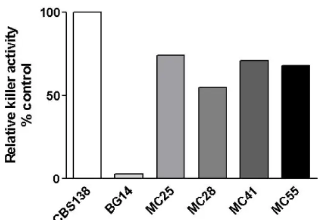
We screened 64 clinical isolates from C. glabrata for ability
to kill sensitive S. cerevisiae strain W303. We found that only four
(6.25%) of all the clinical isolates showed killer activity and by
comparison, C. glabrata CBS-138 (ATCC2001) a previously
identified killer yeast (16) produced more killer activity than any
of the four clinical isolates identified in this study. Only the killer
activity from clinical isolates MC28 was lower than the other
isolates (Fig. 1).
Figure 1. Killer activity of four C. glabrata clinical isolates.
Arroyo-Helguera, O. et al. Killer C. glabrata
Effect of pH and temperature on killer activity
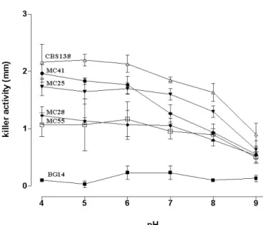
As shown in Fig. 2, killer yeast extracts were adjusted to
pH values of 4-9 in order to analyze the effect of pH on killer
protein stability. We found that killer activity was almost
constant over the pH range tested and little difference was
observed when C. glabrata culture media used for the
cultivation of the clinical isolates had their pH adjusted at 4 to
7, but at pH values from 8 to 9 pH killer activity was reduced
considerably. Fig. 3 shows the effect of changes in pH on the
secretion of killer toxin by C. glabrata strains grown at
different pH values. Secretion of killer toxin by all producing
C. glabrata clinical isolates was constant between pH values
from 4 to 7, while secretion was markedly decreased when C.
glabrata clinical isolates were grown at pH values from 8 to 9.
We then determined whether killer activity was affected
by the temperature of incubation during the growth assay. As
shown in Fig. 4, culture extracts from all C. glabrata clinical
isolates and from the positive control CBS138, showed
stronger killer activity when S. cerevisiae sensitive yeast was
grown at 37°C and 28°C or 30°C.
Figure 2. Effect of pH on stability of killer activity. Culture
filtrates were adjusted to the appropriate pH (4-9) with 0.1 M
citrate phosphate buffer and kept at 4°C. Determination of
killer activity was assayed. n= 5.
Figure 3. Effect of the pH in the C. glabrata growth media on
the production of killer activity. YPD aliquots were adjusted to
the appropriate pH values (4-9) with 0.1 M citrate phosphate
buffer. Killer activity was assayed. n=4.
Figure 4. Temperature effect on killer activity. The killer
activity was assayed using culture filtrates at pH 7.0 and
incubated at 25, 28, 30 and 37 °C for 72 h. n=4.
Plasmid isolation and curing
removal extrachromosomal genetic elements as dsRNA and
dsDNA that encoded killer phenotype (27, 28). This killer
plasmids loss by UV is by DNA damage and formation of
pyrimidine dimmers that disturbs DNA replication, RNA
synthesis and plasmid replication (7). In order to test whether
C. glabrata killer yeast contain extrachromosomal genetic
elements, total nucleic acids were purified and analyzed by
agarose gel electrophoresis. None of the killer clinical isolates
of C. glabrata or CBS138 reference strain presented any
extrachromosomal band of nucleic acids. Only in our positive
control S. cerevisiae S288C strain, an extrachromosomal band
of about 4.5 kb was found as described (Fig. 5 A) (28). In order
to cure possible extrachromosomal nucleic acids not identified
by agarose gel electrophoresis, we irradiated with UV all four
killer C. glabrata clinical isolates at 20,000 μJ/cm2 (87-98 %
of killing). Surviving colonies from all four clinical isolates
were tested for killer activity, and all of them, as well as
surviving colonies from CBS138 strain retained the killer
activity after the exposition to UV (data no shown). These data
strongly suggest that the gene is localized in the chromosome.
Figure 5. Detection of total nucleic acids isolated from C.
glabrata clinical isolates by agarose gel electrophoresis. BG14
(killer negative), CBS138 (killer yeast), S. cerevisiae (contain a
4.5 kb dsRNA plasmid used as positive control). gDNA,
genomic DNA. dsRNA, double stranded RNA. n=3.
Killer toxin from C. glabrata induces DNA fragmentation and loss of viability.
Incubation of sensitive yeast S. cerevisiae with killer toxin
extracts results in gradual loss of viability after 24, 48 and 72 h
treatment. The relative CFU decrease is time dependent in the
clinical isolates, but the loss of viability is more pronounced
with killer toxin from CBS138 (Fig. 6). To determine if the
loss of viability occurs through induction of DNA
fragmentation in the sensitive yeast strain of S. cerevisiae (in
an apoptotic-like pathway), DNA was extracted from sensitive
yeast S. cerevisiae treated with killer toxin for 72 h and
resolved in an agarose gel. Figure 7 shows DNA fragmentation
(visible as DNA ladders) of varying sizes only in cells treated
with killer culture extracts but not in control cells treated with
used media from non-killer C. glabrata clinical isolates (Fig. 7
compare lanes 5-9 with lanes 1-3).
Figure 6. Sensitivity of S. cerevisiae to killer extract from C.
glabrata clinical isolates. A, % survival (CFU %) of S.
cerevisiae in liquid media incubated with killer extracts, log
CFU (%) is the relative number of colony forming units; time
0= 5 x 10 6 cells. CFU was measured at intervals by diluting
Arroyo-Helguera, O. et al. Killer C. glabrata
Figure 7. Survival of S. cerevisiae and DNA fragmentation induced by killer toxins from C. glabrata clinical isolates.
Agarose electrophoresis with total DNA from S. cerevisiae
treated with killer extracts for 72 h at 30°C. n=3.
DISCUSSION
The present study describes the killer phenotype from a
collection of C. glabrata clinical isolates, our data showed low
occurrence (6.25%) of killer yeast from a collection of 64
clinical isolates. This low prevalence of killer pathogenic
strains has been previously reported. Kandel and Stern (10),
screening 234 strains from Cryptococcus, Torulopsis, Candida,
and Trichosporon and found killing activity only in 5%
Candida and 7% in Cryptococcus, therefore it appears that
occurrence of pathogenic killer yeast is limited.
The majority of killer yeast did not produce killer toxin at
pH values higher than pH 5.6 and the optimal pH for killer
activity oscillates between 4 to 4.6 (20). We found that
CBS138 and our C. glabrata clinical isolates produce killer
toxin over a wide range of pH and the optimal killer activity
was at pH 4 to 8 at 37°C, in agreement with Yokomori et al.
(29) where it is shown that SW-55 killer toxin from genus
Candida had killer activity over a broad pH range and
temperature. These properties of C. glabrata killer proteins
could have potential antimycotic activity against fungal
pathogens, although further studies are necessary. In agreement
with previous studies where it was shown that the killer protein
from Candida sp SW-55 is encoded in the chromosome (29),
we found that none of the clinical isolates of C. glabrata with
killer activity have extrachromosomal molecules, suggesting
that the genes responsible for this activity are encoded in the
genome (23). Furthermore, the fact that killer phenotype was
not cured with UV radiation strengthens this notion (Data no
shown).
In yeast, apoptotic cell death can be induced by different
exogenous and intrinsic stresses like H2O2, UV irradiation, acetic acid, cell aging, and high pheromone concentration (4, 8,
11-13, 24). Based on the data presented here, we suggest that
killer yeast toxins from C. glabrata clinical isolates and
CBS138 decrease viability of S. cerevisiae sensitive strain,
probably through an apoptotic cell death as suggested by DNA
fragmentation observed in sensitive yeast. This process has
been reported previously with killer toxin k1 and k28 where
apoptosis is mediated through yeast caspase Yca1p and the
generation of ROS (21). More studies are necessary to analyze
the apoptotic cell mechanism induced by C. glabrata killer
toxins.
ACKNOWLEDGEMENTS
This study had financial support from Instituto Potosino
de Investigación Científica y Tecnológica (IPICYT) and
CONACyT grants No. CB-2005-48304 to ICN and
CONACyT-Salud No. 13927 to APN and PROMEP-SEP grant
No. PROMEP/103.5/10/5006 and from Instituto de Salud
Pública of Universidad Veracruzana grants POA 2010-2011 to
Murillo for his contribution to this study and Irene Xochihua
Rosas for proofreading.
REFERENCES
1. Bendova, O. (1986). The killer phenomenon in yeasts. Folia Microbiol.,
31, 422-33.
2. Cormack, B.P.; Falkow, S. (1999). Efficient homologous and illegitimate recombination in the opportunistic yeast pathogen Candida glabrata. Genetics., 151, 979-987.
3. Coyle, B.; Kinsella, P.; McCann, M.; Devereux, M.; O'Connor, R.; Clynes, M.; Kavanagh, K. (2004). Induction of apoptosis in yeast and mammalian cells by exposure to 1,10-phenanthroline metal complexes.
Toxicol In Vitro., 18, 63-70.
4. Del Carratore, R.; Della, C.C.; Simili, M.E.; Taccini, M. Scavuzzo, and S. Sbrana. (2002). Cell cycle and morphological alterations as indicative of apoptosis promoted by UV irradiation in S. cerevisiae. Mutat Res.,
513, 183-91.
5. Fidel, P.L.; Vazquez, J.A.; Sobel, J.D. (1999). Candida glabrata: review of epidemiology, pathogenesis, and clinical disease with comparison to
C. albicans. Clin Microbiol Rev., 12, 80-96.
6. González, G.M.; Elizondo, M.; Ayala, J. (2008). Trends in species distribution and susceptibility of bloodstream isolates of Candida collected in Monterrey, Mexico, to seven antifungal agents: results of a 3-year (2004 to 2007) surveillance study. J Clin Microbiol., 46, 2902-2905.
7. Gunge, N.; Takahashi, S.; Fukuda, K.; Ohnishi, T.; Meinhardt, F. (1994). UV hypersensitivity of yeast linear plasmids. Curr Genet., 26:369-373. 8. Herker, E.; Jungwirth, H.; Lehmann, K.A.; Maldener, C.; Frohlich, K.U.;
Wissing, S.; Buttner, S.; Fehr, M.; Sigrist, S.; Madeo, F. (2004). Chronological aging leads to apoptosis in yeast. J Cell Biol., 164, 501-7. 9. Kagan, B.L. (1983). Mode of action of yeast killer toxins: channel
formation in lipid bilayer membranes. Nature., 302, 709-11.
10. Kandel, J.S.; Stern, T.A. (1979). Killer phenomenon in pathogenic yeast.
Antimicrob Agents Chemother., 15, 568-71.
11. Laun, P.; Pichova, A.; Madeo, F.; Fuchs, J.; Ellinger, A.; Kohlwein, S.; Dawes, I.; Frohlich, K.U.; Breitenbach, M. (2001). Aged mother cells of
Saccharomyces cerevisiae show markers of oxidative stress and apoptosis. Mol Microbiol., 39, 1166-73.
12. Lavaniegos-Sobrino, M.T.; Ramírez-Zavaleta, C.Y.; Ponce de León, A.; Sifuentes-Osornio, J.; Bobadilla-Del Valle, M.; Rangel-Cordero, A.; De Las Peñas, A.; Castaño, I. (2009). Genotyping of the MTL loci and susceptibility to two antifungal agents of Candida glabrata clinical isolates. Mem Inst Oswaldo Cruz., 104, 775-82.
13. Ludovico, P.; Sousa, M.J.; Silva, M.T.; Leao, C.; Corte-Real, M. (2001).
Saccharomyces cerevisiae commits to a programmed cell death process
in response to acetic acid. Microbiology., 147, 2409-15.
14. Madeo, F.; Frohlich, E.; Ligr, M.; Grey, M.; Sigrist, S.J.; Wolf, D.H.; Frohlich, K.U. (1999). Oxygen stress: a regulator of apoptosis in yeast. J Cell Biol., 145, 757-67.
15. Messer, S.A.; Diekema, D.J.; Boyken, L.; Tendolkar, S.; Hollis, R.J.; Pfaller, M.A. (2006). Activities of micafungin against 315 invasive clinical isolates of fluconazole-resistant Candida spp. J Clin Microbiol.,
44, 324-6.
16. Muller, H.; Hennequin, C.; Gallaud, J.; Dujon, B.; Fairhead, C. (2008). The asexual yeast Candida glabrata maintains distinct a and alpha haploid mating types. Eukaryot Cell., 7, 848-58.
17. Nguyen, M.H.; Yu, V.L.; Morris, A.J. (1996). Candida infection of the arteriovenous fistula used for hemodialysis. Am J Kidney Dis., 27, 596-8. 18. Pertowski, C.A.; Baron, R.C.; Lasker, B.A.; Werner, S.B.; Jarvis, W.R. (1995). Nosocomial outbreak of Candida albicans sternal wound infections following cardiac surgery traced to a scrub nurse. J Infect Dis.,
172, 817-22.
19. Pfaller, M.A.; Boyken, L.; Hollis, R.J.; Kroeger, J.; Messer, S.A.; Tendolkar, S.; Diekema, D.J. (2008). In vitro susceptibility of invasive isolates of Candida spp. to anidulafungin, caspofungin, and micafungin: six years of global surveillance. J Clin Microbiol., 46, 150-6.
20. Polonelli, L.; Conti, S.; Gerloni, M.; Magliani, W.; Chezzi, C.; Morace, G. (1991). Interfaces of the yeast killer phenomenon. Crit Rev Microbiol., 18, 47-87.
21. Reiter, J.; Herker, E.; Madeo, F.; Schmitt, M.J. (2005). Viral killer toxins induce caspase-mediated apoptosis in yeast. J Cell Biol., 168, 353-8. 22. Richter, S.S.; Galask, R.P.; Messer, S.A.; Hollis, R.J.; Diekema, D.J.;
Pfaller, M.A. (2005). Antifungal susceptibilities of Candida species causing vulvovaginitis and epidemiology of recurrent cases. J Clin Microbiol., 43, 2155-62.
23. Schmitt, M.J.; Breinig, F. (2002). The viral killer system in yeast: from molecular biology to application. FEMS Microbiol Rev., 26, 257-76. 24. Severin, F.F.; Hyman, A.A. (2002). Pheromone induces programmed
cell death in S. cerevisiae. Curr Biol., 12, R233-5.
25. Starmer, W.T.; Ganter, P.F.; Aberdeen, V.; Lachance, M.A.; Phaff, H.J. (1987). The ecological role of killer yeasts in natural communities of yeasts. Can J Microbiol., 33, 783-96.
26. Walker, G.M.; McLeod, A.H.; Hodgson, V.J. (1995). Interactions between killer yeasts and pathogenic fungi. FEMS Microbiol Lett., 127, 213-22.
27. Wickner, R.B. (1979). The killer double-stranded RNA plasmids of yeast. Plasmid., 2, 303-22.
28. Wickner, R.B. (1983). Killer systems in Saccharomyces cerevisiae: three distinct modes of exclusion of M2 double-stranded RNA by three species of double-stranded RNA, M1, L-A-E, and L-A-HN. Mol Cell Biol., 3, 654-61.
Arroyo-Helguera, O. et al. Killer C. glabrata
Candida killer yeast with a novel killer property. Agric Biol Chem., 52, 2797–2801.
30. Young, T.W.; Yagiu, M. (1978). A comparison of the killer character in
different yeasts and its classification. Antonie Van Leeuwenhoek., 44, 59-77.