483 483 483 483 483 Mem Inst Oswaldo Cruz, Rio de Janeiro, Vol. 100(5): 483-490, August 2005
Value of morphotyping for the characterization of
Candida albicans
clinical isolates
Giovanni M Giammanco/+, M aria M anuel Lopes* , Roney S Coimbra* * /+ +, Sarina Pignato* * * , Patrick AD Grimont* * , Francine Grimont* * , Graciete Freitas* ,
Giuseppe Giammanco* * *
Dipartimento di Igiene e Microbiologia, Università di Palermo, via del Vespro 133, I-90127 Palermo, Italia *Departamento de Microbiologia, Faculdade de Farmácia, Universidade de Lisboa, Lisboa, Portugal **Unité Biodiversité des Bactéries Pathogènes
Emergentes, Unité INSERM 389, Institut Pasteur, Paris, France ***Dipartimento “G.F. Ingrassia” Igiene e Sanità Pubblica, Università di Catania, Catania, Italia
Until recently, morphotyping, a method evaluating fringe and surface characteristics of streak colonies grown on malt agar, has been recommended as a simple and unexpensive typing method for Candida albicans isolates. The discriminatory power and reproducibility of Hunter’s modified scheme of Phongpaichit’s morphotyping has been evaluated on 28 C. albicans isolates recovered from the oral cavity of asymptomatic human immunodeficiency virus-positive subjects, and compared to two molecular typing methods: randomly amplified polymorphic DNA (RAPD) fingerprinting, and contour clamped homogeneous electric field (CHEF) electrophoretic karyotyping. Morpho-logical features of streak colonies allowed to distinguish 11 different morphotypes while RAPD fingerprinting yielded 25 different patterns and CHEF electrophoresis recognized 9 karyotypes. The discriminatory power calcu-lated with the formula of Hunter and Gaston was 0.780 for morphotyping, 0.984 for RAPD fingerprinting, and 0.630 for karyotyping. Reproducibility was tested using 43 serial isolates from 15 subjects (2 to 6 isolates per subject) and by repeating the test after one year storage of the isolates. While genetic methods generally recognized a single type for all serial isolates from each of the subjects studied, morphotyping detected strain variations in five subjects in the absence of genetic confirmation. Poor reproducibility was demonstrated repeating morphotyping after one year storage of the isolates since differences in at least one character were detected in 92.9% of the strains.
Key words: Candida albicans - karyotyping - morphotyping - randomly amplified polymorphic DNA fingerprinting
Although different species of Candida, such as C. glabrata, C. tropicalis, C. krusei, and C. dubliniensis,
are at present recognized as increasing opportunistic pathogens specially in human immunodeficiency virus (HIV) infected individuals and AIDS patients, C. albicans
still remains the most common yeast isolated in humans (Anaissie 1992, Bart-Delabesse et al. 1993, Coleman et al. 1998, Sullivan et al. 2004). The different species of Can-dida can be identified by commercially available biochemi-cal tests, but the characterization of isolates is necessary for epidemiological investigation and to ascertain if new candidiasis episodes are caused by relapse or due to rein-fection. With this aim, a variety of molecular typing tech-niques has been used to distinguish Candida isolates, including multilocus enzyme electrophoresis, electro-phoretic karyotyping, total genomic DNA restriction analy-sis, Southern blot hybridization with repetitive DNA probes, and randomly amplified polymorphic DNA analy-sis-RAPD (Magee & Magee 1987, Bostock et al. 1993, Schonian et al. 1993, Clemons et al. 1997, Pujol et al. 1997, Taylor et al. 1999, Meyer et al. 2001).
+Corresponding author. E-mail: [email protected] ++CNPq-Brazil fellowship
Received 4 January 2005 Accepted 27 April 2005
Electrophoretic karyotyping (EK) is a well-established method for typing Candida spp. (Monod et al. 1990). In particular, pulsed-field gel electrophoresis (PFGE) by con-tour clamped homogeneous electric field (CHEF) of C. albicans allows to subdivide this species in several karyo-types (Magee & Magee 1987, Soll 2000) and to distin-guish it from phenotypically close species like C. du-bliniensis (Sullivan & Coleman 1998, Giammanco et al. 2000). EK has been used to characterize strains of C. albicans responsible for recurrent oropharingeal can-didiasis (Whelan et al. 1990, Schmid et al. 1992, Bart-Delabesse et al. 1993, Pfaller et al. 1994, Lupetti et al. 1995) and in epidemiological investigations to identify the sources of hospital acquired C. albicans infections (McNeil et al. 1999). Strain substitution or stable C. albicans colonization can be demonstrated by this method (Sangeorzan et al. 1994, Barton et al. 1995).
ex-484 484 484 484
484 M orphotyping characterization of C. albicans • Giovanni M Giammanco et al.
pensive and technically demanding, and are not available in many routine clinical laboratories.
Conversely, phenotyping methods like auxonotyping, enzimotyping, or resistotyping do not need specialized equipment and are easy to perform. However, they all suf-fer from poor discriminatory power (Otero et al. 1995). Among phenotyping methods, Phongpaichit’s mor-photyping (Phongpaichit et al. 1987), a method evaluat-ing frevaluat-inge and surface characteristics of streak colonies grown on malt agar, has been shown in the past to have good discriminatory capacity (Otero et al. 1995). Hunter et al. (1989) modified the reading system in order to achieve better reproducibility and were able to relate strains of proven virulence with a distinct morphotype. This test is based on the evaluation of only four simple growth pa-rameters such as fringe distribution, width, texture, and streak surface topography of linear colonies. However, concern has been expressed about the reproducibility of such a scheme, as C. albicans has been shown to un-dergo phenotypic switching (Slutsky et al. 1985). High-frequency morphological variation not related to genomic DNA fingerprinting results has been demonstrated in re-current isolates of C. albicans (O’Connell et al. 1995) and in conjunction with antifungal types alteration (Maffei et al. 1997).
Until recently, morphotyping has been recommended as a simple typing method to carry out in routine labora-tories where other methods are impractical because of contraints of resources (Khan et al. 2003, Liguori et al. 2004), but its discriminatory power and reproducibility as a strain marker needs to be compared to molecular typing methods.
In the present study, a collection of unrelated clinical strains of C. albicans isolated from the oral cavity of 28 HIV-positive subjects (one strain per subject) was used in order to verify whether colony morphotyping accord-ing to Hunter’s system (Hunter et al. 1989) could be a valuable typing method. Results have been compared with karyotyping and RAPD fingerprinting. The discrimina-tory power of the three typing methods was assessed calculating the numerical Index of Discrimination (ID) pro-posed by Hunter and Gaston (1988). Reproducibility was tested using 43 serial oropharyngeal isolates obtained from 15 asymptomatic HIV-positive subjects (2 to 6 iso-lates per subject) who had not undergone therapy with antimycotic drugs and were likely to harbour resident strains. To assess the stability of their morphological char-acters, 14 serial isolates were re-tested for morphotyping after one year storage in agar slants.
MATERIALS AND METHODS
Yeast strains -A collection of 28 clinical isolates of C. albicans was used for studying the discriminatory power of the typing methods. The collection consisted of con-secutive clinical isolates (one per patient) obtained from oral swabs collected between January 1996 and Decem-ber 1996 during one year of monitoring of HIV-positive subjects and submitted to the laboratory of the Istituto di Igiene e Medicina Preventiva of the University of Catania (Catania, Italy). Fifteen of the subjects under study were submitted to monthly oral sampling during the
monitor-ing and a total of 43 serial isolates (two to six isolates per subject) were obtained. Serial isolates were used to as-sess the reproducibility of the typing. During all the moni-toring period the subjects were asymptomatic for can-didiasis and other opportunistic infections and were not treated with any antifungal drugs. Identification of iso-lates to species level was performed by the API ID 32 C system (BioMérieux, Marcy-l’Etoile, France) and germ-tube formation in human serum. Strains were stored in Sabouraud agar slants for one year at room temperature before being re-tested for morphotyping.
RAPD fingerprinting -The DNA extraction was per-formed in accordance with Hoffman’s protocol (1993). The single repeat sequence (GACA)4 was used as primer in the PCR experiments as described by Schonian et al. (1993). Briefly, 200 µM of each deoxinucleotide (Amersham Pharmacia Biotech, Uppsala, Sweden), 16 pmol primer, 25 ng template DNA, 2.5 U Taq DNA polymerase (Perkin Elmer Corp., Applied Biosystems, New Jersey, US), 10 mM Tris/HCl (pH 8.3), 50 mM KCl, 1.5 mM MgCl2 , and 3 mM Mg-acetate were added to a 50 µl reaction volume. The cycle parameters were 33 cycles of 20 s at 94°C, 1 min at 43°C, 20 s at 72°C, followed by a final extension cycle of 6 min at 72°C, performed on a Hybaid thermocycler (Teddington Middlesex, UK). The RAPD products were separated by electrophoresis through a 1.2% agarose gel in 0.5 x TBE (89 mM Tris-borate, 2 mM EDTA) buffer at 2 volts/cm for 16 h. The gels were stained with ethidium bromide and photographed. Lambda phage DNA digested with BstE II was used for the DNA fragments size determi-nation. The Taxotron software (Taxolab Software, Institut Pasteur, Paris, France) compared the RAPD fingerprints and a distance coefficient was calculated using the comple-ment of the Dice index. A dendrogram was drawn on the basis of the distance matrix generated by the unweighted pair group method of averages (UPGMA) algorithm.
EK -C. albicans DNA for EK was prepared according to Monod et al. (1990). PFGE was performed in a CHEF-DR III system (Bio-Rad Laboratories, Hercules, CA), us-ing 0.8% Pulsed-Field-Certified Agarose (Bio-Rad Labo-ratories) in TAE buffer (40 mM Tris-acetate; 1 mM EDTA; pH 8.2). Two program blocks were used. For block one, switch time was 120 s and run time 24 h. For block two, switch time was 240 s and run time 36 h. Angle was fixed at 106°, gradient at 3.5 volts/cm, and temperature at 13°C. Intact chromosomes from Saccharomyces cerevisiae
strain YPH80 were used as molecular weight markers. Gels were stained in ethidium bromide. Gel images were elec-tronically captured and karyotypes were analyzed by the Taxotron software (Taxolab Software, Institut Pasteur, Paris, France) as already described for RAPD fingerprints.
to-485 485 485 485 485 Mem Inst Oswaldo Cruz, Rio de Janeiro, Vol. 100(5), August 2005
pography. The characters used to produce the mor-photyping code are resumed in Table I. Mormor-photyping was repeated using the same protocol after one year stor-age of the strains in Sabouraud agar tubes.
Evaluation of the discriminatory power - In order to assess the discriminatory power of RAPD, karyotyping and morphotyping for the typing of C. albicans isolates, the numerical index of discrimination (ID) proposed by Hunter and Gaston (1988) has been calculated. This Index is based on the Simpson’s index of diversity and is the result of the following equation:
s
ID = 1- [1 / N (N - 1)] Σ nj (nj - 1) j = 1
where N is the total number of examined strains, s is the total number of types described, and nj is the number of strains belonging to the jth type.
RESULTS
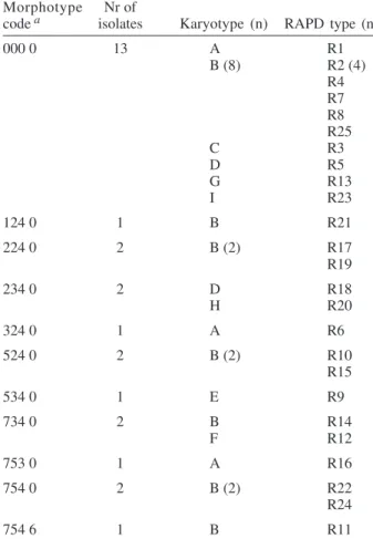
Overall, the C. albicans isolates were indistinguish-able by classical biochemical methods since they showed the same substrate assimilation profile corresponding to the API ID 32C code 734 734 00 15. Morphotyping, karyo-typing and RAPD fingerprinting analysis results are re-sumed in Table II.
Analysis of the 28 isolates using RAPD fingerprint-ing with primer (GACA)4 originated 25 different amplifica-tion patterns (from R1 to R25) showing nine to 17 frag-ments of various intensity and ranging between 0.96 and 5.58 kbp in size (Figs 1, 2). Software analysis demonstrated that almost all the patients presented a specific RAPD profile. Only R2 pattern was shared by four patients.
TABLE I
Four digit morphotyping coding according to Hunter et al. (1989)
Feature Code Description
Fringe
Distribution 0 Absent
1 Discontinuous: < 20% of margin 2 Discontinuous: 20-50% of margin 3 Discontinuous: 50-90% of margin
5 Continuous at periphery only or strands conspicuously fan shaped 7 Continuous; filamentous outgrowths parallel
Width 0 Absent
2 ≤ 2 mm
3 3-5 mm
5 ≥ 6 mm
Texture 0 Absent
1 Very coarse
2 Coarse
3 Intermediate
4 Fine
Streak surface topography 0 Smooth
1 Nodular
2 Pitted
4 Crateriform
5 Crateriform plus wrinkles or folds 6 Wrinkles or folds
8 Hairy
EKs were determined by software analysis of the elec-trophoretic patterns revealing five to seven bands of dif-ferent size. On the basis of the number and migration dis-tances of DNA bands, nine different karyotypes (A to I) were recognized among the 28 isolates (Fig. 3). Type B karyotype was the most frequent (17 out of 28 isolates, 60.7%), followed by type A (three isolates), type D (two isolates), and types C, E, F, G, H, and I (one isolate each). Eleven different morphotypes could be identified us-ing the abbreviated code proposed by Hunter et al. (1989). Morphotype code 000 0 was the most common (13 out of 28 isolates, 46.4 %), followed by 224 0, 234 0, 524 0, 734 0, and 754 0 (two isolates each). Five other morphotypes (codes: 124 0, 324 0, 534 0, 753 0, and 754 6) were repre-sented by one isolate each.
The discriminatory power of RAPD fingerprinting with primer (GACA)4 calculated with the formula of Hunter and Gaston (1988) was 0.984. The ID was lower for karyo-typing (0.630) and for morphokaryo-typing (0.780), with the larger typing cluster constituting 60.7% and 46.4% of the iso-lates, respectively (Table III). In both cases IDs were lower than the minimal index of 0.90 preconized as a desirable limit for results to be interpreted with confidence.
486 486 486 486
486 M orphotyping characterization of C. albicans • Giovanni M Giammanco et al.
DISCUSSION
The availability of a number of unrelated clinical iso-lates recovered during several months from the oral cav-ity of HIV-seropositive subjects gave us the opportuncav-ity to evaluate the usefulness of colony morphotyping on malt agar according to Hunter’s scheme as an epidemio-logical typing method for C. albicans, by comparing this phenotypic typing method with DNA-based typing meth-ods such as RAPD fingerprinting and karyotyping for the ability to discriminate among isolates.
Morphotyping is extremely simple and cheap to per-form, requiring only malt agar plates. Moreover, special morphological markers, like discontinuous fringes, have been found to be associated with virulent strains and an increased risk of death in the case of deep infection (Hunter et al. 1989). Unfortunately, reproducibility of typ-ing schemes based on colony morphology has already been reported to be affected by phenotypic variation (Slutsky et al. 1985), but a better in vitro reproducibility has been claimed for Hunter’s scheme (Hunter et al. 1989, Otero et al. 1995). Hunter’s scheme clusters Phongpaichit’s morphotypes in a lower number, simplifying plates read-ing.
In our hands, the discriminatory capacity of this method, though lower than the 0.90 ID limit for results to be interpreted with confidence, was good compared to karyotyping. In fact, EK revealed a moderate variety of patterns among the isolates from different subjects. On the contrary, RAPD fingerprinting with the primer (GACA)4 showed a specific RAPD profile for each of the patients with the exception of a cluster of four isolates which produced identical patterns. Although we had no evidence of these isolates being epidemiologically linked, they were indistinguishable by all of the three typing methods used, since they also shared the same morpho-type and karyomorpho-type.
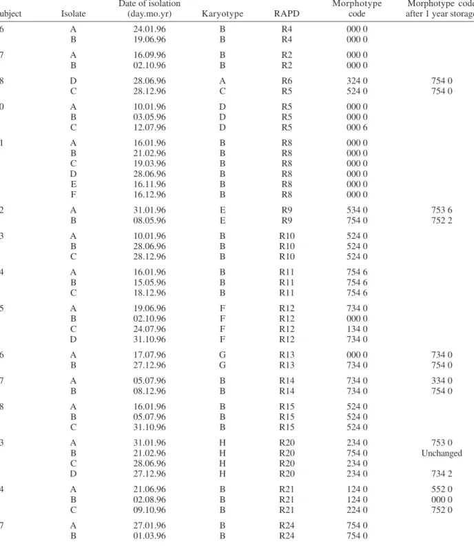
Serial isolates belonging to the same subject which were likely to be the same strain have been used to assess reproducibility of typing results.
Reproducibility of the two genotyping methods was excellent, since for all but one patient they both assigned to a single type all of the serial isolates. On the contrary, in 6 out of 15 subjects with multiple isolates morphotypes variation not in agreement with genotyping were detected. In subject nr 15 four genotypically identical serial isolates yielded three different morphotype codes, with the last isolate recovering the parental code. Poor reproducibility of morphotyping had already been signalled when recur-rent isolates of C. albicans had been studied (O’Connell
TABLE II
Morphotypes, karyotypes, and randomly amplified polymorphic DNA (RAPD) fingerprinting typing for 28
clinical isolates of Candida albicans
Morphotype Nr of
code a isolates Karyotype (n) RAPD type (n)
000 0 13 A R1
B (8) R2 (4) R4 R7 R8 R25
C R3
D R5
G R13
I R23
124 0 1 B R21
224 0 2 B (2) R17
R19
234 0 2 D R18
H R20
324 0 1 A R6
524 0 2 B (2) R10
R15
534 0 1 E R9
734 0 2 B R14
F R12
753 0 1 A R16
754 0 2 B (2) R22
R24
754 6 1 B R11
a: based on fringe distribution, width, texture, and streak surface topography (Hunter et al. 1989).
TABLE III
Index of discrimination (ID) a of typing methods used to discriminate among 28 Candida albicans strains
Typing method Nr of types Size (% of strains in largest cluster) ID
RAPD fingerprinting 25 14.3 0.984
Morphotyping 11 46.4 0.780
Karyotyping 9 60.7 0.630
a: according to Hunter and Gaston (1988).
487 487 487 487 487 Mem Inst Oswaldo Cruz, Rio de Janeiro, Vol. 100(5), August 2005
Fig. 1: dendrogram showing the genetic distance of the 25 (R1-R25) randomly amplified polymorphic DNA patterns obtained in this study with the primer (GACA)4. The distance matrix was generated by using the complement of Dice similarity coefficient and the unweighted pair group method of averages (UPGMA) algorithm.
Fig. 2: randomly amplified polymorphic DNA patterns obtained with the primer (GACA)4 for some of the Candida albicans isolates. Lanes L: molecular weight marker (Lambda phage DNA -BstEII digested); 1: R5 pattern; 2: R7 pattern; 3: R9 pattern; 4: R1 pattern; 5: R2 pattern; 6: R8 pattern.
et al. 1995, Maffei et al. 1997) and phenotypic switching had been ascribed to selective pressure due to antifungal therapy (Maffei et al. 1997, Takasuka et al. 1998). In our study, phenotypic switching affecting morphological char-acters of colonies seems to have occurred even if none of our patients had been submitted to antifungal therapy.
A further reproducibility test performed after one year of storage in Sabouraud agar tubes revealed that all but one of fourteen serial isolates from six subjects showed at least one change in their morphotyping code. Differences in environmental conditions such as pH have already been demonstrated to affect the expression of several genes in
C. albicans (De Bernardis et al. 1998). Therefore, growth conditions and time of harvesting, as well as modifica-tions in the oral cavity environment, can compromise the reproducibility of biotyping methods such as mor-photyping.
488 488 488 488
488 M orphotyping characterization of C. albicans • Giovanni M Giammanco et al.
TABLE IV
Randomly amplified polymorphic DNA (RAPD) fingerprinting patterns, karyotypes and morphotypes, of Candida albicans serial isolates
Date of isolation Morphotype Morphotype code
Subject Isolate (day.mo.yr) Karyotype RAPD code after 1 year storage
6 A 24.01.96 B R4 000 0
B 19.06.96 B R4 000 0
7 A 16.09.96 B R2 000 0
B 02.10.96 B R2 000 0
8 D 28.06.96 A R6 324 0 754 0
C 28.12.96 C R5 524 0 754 0
10 A 10.01.96 D R5 000 0
B 03.05.96 D R5 000 0
C 12.07.96 D R5 000 6
11 A 16.01.96 B R8 000 0
B 21.02.96 B R8 000 0
C 19.03.96 B R8 000 0
D 28.06.96 B R8 000 0
E 16.11.96 B R8 000 0
F 16.12.96 B R8 000 0
12 A 31.01.96 E R9 534 0 753 6
B 08.05.96 E R9 754 0 752 2
13 A 10.01.96 B R10 524 0
B 28.06.96 B R10 524 0
C 28.12.96 B R10 524 0
14 A 16.01.96 B R11 754 6
B 15.05.96 B R11 754 6
C 18.12.96 B R11 754 6
15 A 19.06.96 F R12 734 0
B 02.10.96 F R12 000 0
C 24.07.96 F R12 134 0
D 31.10.96 F R12 734 0
16 A 17.07.96 G R13 000 0 734 0
B 27.12.96 G R13 734 0 754 0
17 A 05.07.96 B R14 734 0 334 0
B 08.12.96 B R14 734 0 754 0
18 A 16.01.96 B R15 524 0
B 05.07.96 B R15 524 0
C 31.10.96 B R15 524 0
23 A 31.01.96 H R20 234 0 753 0
B 21.02.96 H R20 754 0 Unchanged
C 28.06.96 H R20 234 0
D 27.12.96 H R20 234 0 734 2
24 A 21.06.96 B R21 124 0 552 0
B 02.08.96 B R21 124 0 000 0
C 09.10.96 B R21 224 0 752 0
27 A 27.01.96 B R24 754 0
B 01.03.96 B R24 754 0
in routine epidemiological investigations (Khan et al. 2003). In accordance with previous reports (Bostock et al. 1993, Schonian et al. 1993, Holmberg & Feroze 1996, Clemons et al. 1997, Pujol et al. 1997),RAPD analysis con-firmed to be highly discriminatory and suitable for large-scale epidemiological studies of C. albicans as well as for inquiries in epidemic outbreaks. In laboratories not
equipped for molecular methods,isolates should be sent to reference laboratories for epidemiological studies.
489 489 489 489 489 Mem Inst Oswaldo Cruz, Rio de Janeiro, Vol. 100(5), August 2005
REFERENCES
Anaissie E 1992. Opportunistic mycoses in the immunocom-promised host: experience at a cancer center and review.
Clin Infect Dis14 (Suppl. 1): S43-S53.
Bart-Delabesse E, Boiron P, Carlotti A, Dupont B 1993. Can-dida albicans genotyping in studies with patients with AIDS developing resistance to fluconazole. J Clin Microbiol 31: 2933-2937.
Barton RC, van Belkum A, Scherer S 1995. Stability of karyo-type in serial isolates of Candida albicans from neutro-penic patients. J Clin Microbiol33: 794-796.
Bostock A, Khattak MN, Matthews R, Burnie J 1993. Comparation of PCR fingerprinting, by random amplifica-tion of polymorphic DNA, with other molecular typing methods for Candida albicans. J Gen Microbiol139: 2179-2184.
Clemons KV, Feroze F, Holmberg K, Stevens DA 1997. Com-parative analysis of genetic variability among Candida albicans isolates from different geographic locales by three genotypic methods. J Clin Microbiol35: 1332-1336.
Coleman DC, Rinaldi MG, Haynes KA, Rex JH, Summerbell RC, Anaissie EJ, Li A, Sullivan DJ 1998. Importance of
Candida species other than Candida albicans as opportu-nistic pathogens. Med Mycol36 (Suppl. 1): 156-165.
De Bernardis F, Muhlschlegel FA, Cassone A, Fonzi WA 1998. The pH of host niche controls gene expression in and viru-lence of Candida albicans. Infect Immun66: 3317-3325.
Giammanco GM, Pignato S, Salvo S, Giammanco G 2000. Car-bohydrate assimilation profiles of the first Italian Candida dubliniensis clinical isolates recovered from an HIV-infected individual. Res Microbiol151: 889-891.
Gyanchandani A, Khan ZK, Farooqui N, Goswani M, Ranade SA 1998. RAPD analysis of Candida albicans strains re-coveredfrom different immunocompromised patients (ICP) reveals an apparently non-random infectivity of the strains.
Biochem Mol Int44: 19-27.
Hoffman CS 1993. Preparation of yeast DNA, RNA and pro-teins. In FM Ausubel, R Brent, RE Kingston, DD Moore,
JG Seidman, JA Smith, K Struhl (eds), Current Protocols in Molecular Biology, John Wiley & Sons, Inc., Hoboken, NJ, Vol. 2. Section 13.
Holmberg K, Feroze F 1996. Evaluation of an optimized sys-tem for random amplified polymorphic DNA (RAPD)-analysis for genotypic mapping of Candidaalbicans strains.
J Clin Lab Anal10: 59-69.
Hunter PR, Fraser CAM, Mackenzie DWR 1989. Morphotype markers of virulence in human candidal infections. J Med Microbiol28: 85-91.
Hunter PR, Gaston MA 1988. Numerical index of the discrimi-natory ability of typing systems: an application of Simpson’s index of diversity. J Clin Microbiol26: 2465-2466.
Khan ZU, Chandy R, Metwali KE 2003. Candida albicans
strain carriage in patients and nursing staff of an intensive care unit: a study of morphotypes and resistotypes. My-coses46: 479-486.
Liguori G, Marinelli A, Galdiero E, Arnese A, Di Onofrio V, Lucariello A, Marinelli P 2004. Candida spp. morphotype differentiation on Sabouraud-Triphenyltetrazolium-Agar (STTZ-Agar) under three different experimental conditions.
New Microbiol27: 193-197.
Lupetti A, Guzzi G, Paladini A, Swart K, Campa M, Senesi S 1995. Molecular typing of Candida albicans in oral can-didiasis: kariotype epidemiology with human immunodefi-ciency virus-seropositive patients in comparison with that with healthy carriers. J Clin Microbiol33: 1238-1242.
Maffei CM, Paula CR, Mazzocato TS, Franceschini S 1997. Phenotype and genotype of Candida albicans strains iso-lated from pregnant women with recurrent vaginitis.
Mycopathologia137: 87-94.
Magee BB, Magee PT 1987. Electrophoretic karyotypes and chromosome numbers in Candida species. J Gen Microbiol 133: 425-430.
McNeil MM, Lasker BA, Lott TJ, Jarvis WR 1999. Postsurgi-cal Candida albicans infections associated with an extrin-sically contaminated intravenous anesthetic agent. J Clin Microbiol37: 1398-1403.
490 490 490 490
490 M orphotyping characterization of C. albicans • Giovanni M Giammanco et al.
Meunier JR, Grimont PAD 1993. Factors affecting reproduc-ibility of random amplified polymorphic DNA fingerprint-ing. Res Microbiol144: 373-379.
Meyer W, Maszewska K, Sorrell TC 2001. PCR fingerprinting: a convenient molecular tool to distinguish between Can-dida dubliniensis and Candida albicans. Med Mycol39: 185-193.
Monod M, Porchet S, Baudraz-Rosselet F, Frenk E 1990. The identification of pathogenic yeast strains by electrophoretic analysis of their chromosomes. J Med Microbiol29: 123-129.
O’Connell B, Coleman DC, Bennett D, Sullivan D, McCann SR, Keane CT 1995. An epidemiological study of Candida
species infection in cancer patients using genetic finger-printing and morphotyping. J Hosp Infect31: 211-217.
Otero L, Vazquez F, Palacio V, Vazquez S, Carreno F, Mendez FJ 1995. Comparison of seven phenotyping methods for
Candida albicans. Eur J Epidemiol11: 221-224.
Pfaller MA, Rhine-Chalberg J, Redding SW, Smith J, Farinacci G, Fothergill AW, Rinaldi MG 1994. Variations in fluconazole susceptibility and electrophoretic karyotype among oral isolates of Candida albicans from patients with AIDS and oral candidiasis. J Clin Microbiol32: 59-64.
Phongpaichit S, Mackenzie DWR, Fraser C 1987. Strain differ-entiation of Candida albicans by morphotyping. Epid In-fect99: 421-428.
Pujol C, Joly S, Lockhart SR, Noel S, Tibayrenc M, Soll DR 1997. Parity among the randomly amplified polymorphic DNA method, multilocus enzyme electrophoresis, and Southern blot hybridization with the moderately repetitive DNA probe Ca3 for fingerprinting Candida albicans. J Clin Microbiol35: 2348-2358.
Robert F, Lebreton F, Bougnoux ME, Paugam A, Wassermann D, Schlotterer M, Tourte-Schaefer C, Dupouy-Camet J 1995. Use of random amplified polymorphic DNA as a typing method for Candida albicans in epidemiological surveillance of a burn unit. J Clin Microbiol33: 2366-2371.
Sangeorzan JA, Bradley SF, He X, Zarins LT, Ridenour GL, Tiballi RN, Kauffman CA 1994. Epidemiology of oral
candidiasis in HIV-infected patients: colonization, infec-tion, treatment, and emergence of fluconazole resistance.
Am J Med 97: 339-346.
Schmid J, Odds FC, Wiselka MJ, Nicholson KG, Soll DR 1992. Genetic similarity and maintenance of Candida albicans
strains from a group of AIDS patients, demonstrated by DNA fingerprinting. J Clin Microbiol30: 935-941.
Schonian G, Meusel O, Tietz HJ, Meyer W, Graser Y, Tausch I, Presber W, Mitchell TG 1993. Identification of clinical strains of Candida albicans by DNA fingerprinting with the polymerase chain reaction. Mycoses36: 171-179.
Slutsky B, Buffo J, Soll DR 1985. High-frequency switching of colony morphology in Candida albicans. Science 230: 666-669.
Soll DR 2000. The ins and outs of DNA fingerprinting the infectious fungi. Clin Microbiol Rev13: 332-370.
Sullivan D, Coleman D 1998. Candida dubliniensis: character-istics and identification. J Clin Microbiol36: 329-334.
Sullivan D, Moran GP, Pinjon E, Al-Mosaid A, Stokes C, Vaughan C, Coleman DC 2004. Comparison of the epidemi-ology, drug resistance mechanisms, and virulence of Can-dida dubliniensis and Candida albicans. FEMS Yeast Res4: 369-376.
Takasuka T, Baily GG, Birch M, Anderson MJ, Law D, Den-ning DW 1998. Variation in morphotype, karyotype and DNA type of fluconazole resistant Candida albicans from an AIDS patient. J Infect36: 57-62.
Taylor JW, Geiser DM, Burt A, Koufopanou V 1999. The evolutionary biology and population genetics underlying fungal strain typing. Clin Microbiol Rev12: 126-146.
Tyler KD, Wang G, Tyler SD, Johnson WM 1997. Factors affecting reliability and reproducibility of amplification-based DNA fingerprinting of representative bacterial patho-gens. J Clin Microbiol35: 339-346.