GLIOSARCOMA
Report of four cases with immunohistochemical findings
Tiago Noguchi Machuca
1, Daniel Monte-Serrat Prevedello
2,
Leonora Zozula Blind Pope
3, Salo Semelman Haratz
1,
João Cândido Araújo
4, Luiz Fernando Bleggi Torres
5ABSTRACT - Gliosarcoma (GSa) is a rare primary central nervous system neoplasm (CNS) characterized by biphasic histological pattern with both glial and sarcomatous components. Our objective is to describe the clinical, morphological and immunohistochemical features of four cases of GSa and to discuss its pathogene-tic mechanisms. The male:female ratio was 3:1. The mean age was 39 years, ranging from 19 to 48. Hea-dache was the commonest clinical symptom. All patients underwent craniotomy with microsurgery and total resection of the tumor. Diagnosis was suspected due to microscopic architecture and confirmed by detection of reticulin fibers through histochemical techniques. Immunohistochemical analysis was positive for p53 in both glial and sarcomatous cells in all four cases. EGFR was focally positive in glial cells in one case. Our findings support monoclonal origin of GSa involving the TP53 tumor-suppressor gene. However, alternative pathways cannot be ruled out.
KEY WORDS: brain neoplasms, gliosarcoma, immunohistochemistry.
Gliossarcoma: relato de quatro casos com achados imuno-histoquímicos
RESUMO - Gliossarcoma (GSa) é uma neoplasia primária rara do sistema nervoso central, caracterizada por padrão histológico bifásico incluindo componentes tanto glial como sarcomatoso. São discutidos os aspec-tos clínicos, morfológicos e imunohistoquímicos de quatro casos de GSa e seus mecanismos patogêneticos. A relação masculino/feminino foi 3:1. A média de idade foi 39 anos, variando de 19 a 48. Cefaléia foi a ma-nifestação predominante. Todos os pacientes foram submetidos a craniotomia com microcirurgia e ressecção total do tumor. O diagnostico foi suspeitado devido à arquitetura microscópica e foi confirmada por pre-sença de fibras de reticulina através de técnicas de histoquímica. A análise imuno-histoquímica foi positiva para p53 tanto em células gliais como em células sarcomatosas nos quatro casos. EGFR foi localmente posi-tivo em células gliais em apenas um caso. Esses achados apoiam uma origem monoclonal do GSa relaciona-da com alteração no Tp53, gene supressor de tumor. No entanto, outras vias alternativas na gênese des-ses tumores não podem ser afastadas.
PALAVRAS-CHAVE: neoplasias cerebrais, gliossarcoma, imuno-histoquímica.
genetic studies failed to support this theory, sugges-ting a monoclonal origin for both histological com-ponents1.
GSa corresponds to less than 2% of all glioblasto-mas1, with a peak of incidence from the fourth to the sixth decades of life, mean age 53 years. Male: female ratio is 1.8:1 and the clinical presentation, natural history and radiologic profile are similar to those of primary glioblastoma1,3.
We studied four cases of gliosarcoma aiming to
Department of Neurosurgery and Pathology, Hospital Nossa Senhora das Graças, Curitiba, Paraná, Brazil (HNSG); and Department of Pathology, Universidade Federal do Paraná, Curitiba, Paraná, Brazil (UFPR): 1Medical student, UFPR; 2Resident in Neurosurgery,
HNSG; 3Resident in Pathology, Hospital de Clínicas, UFPR; 4Adjunct Professor of Neurosurgery, UFPR and Coordinator of the Program
of Medical Residency of the Department of Neurosurgery, HNSG; 5Professor of Pathology, Universidade Federal do Paraná; Chairman
of the Laboratory of Anatomical Pathology Service, HNSG. Tiago Noguchi Machuca is supported by a grant from UFPR/National Treasure. Salo Semelman Haratz is supported by a grant from PIBIC/CNPq.
Received 24 November 2003, received in final form 27 February 2004. Accepted 29 March 2004.
Dr. Luiz Fernando Bleggi Torres - Department of Pathology - Hospital de Clínicas, Universidade Federal do Paraná - Rua General Carneiro 181 - 80060-900 Curitiba PR - Brasil. E-mail: [email protected]
describe its clinical and pathological features. Im-munohistochemical analyses were performed to confirm diagnosis, assess histogenetic origin and evaluate the proliferation index in all cases.
METHOD
The cases were selected from the files of the “CNS Tumor Bank”, which is part of the section of Neuropa-thology of the Department of PaNeuropa-thology, Federal Uni-versity of Parana, in Curitiba, from 1990 to 2002. This tumor bank collects around 95% of brain tumors diagno-sed in the city of Curitiba, a 1.5 million inhabitants city in Southern region of Brazil. Clinical data were obtained by reviewing the medical records of all patients.
Histologic sections from all four cases were reviewed and original available paraffin-embedded blocks were recut and stained with hematoxylin/eosin and reticulin. The sarcomatous component was classified and both com-ponents were quantified. Mitotic figures per high pow-er field wpow-ere counted and type and extension of necro-sis assessed.
Immunohistochemical study using the avidin-biotin method was performed with antibodies to GFAP, vimen-tin, CD34, p53, EGFR and Ki-67. Positive and negative con-trols were obtained for each antibody. GFAP and vimentin were classified as focal positivity, diffuse positivity and negative. CD34 and EGFR were classified as negative or positive. Ki-67 and p53 were considered positive only when nuclear staining was present and were graded as + when less than 1% of nuclei were stained; ++ when between 1% and 5% were stained, and +++ when more than 5% of stained nuclei were observed.
RESULTS
The CNS Tumor Bank of Curitiba has 3873 biop-sy cases, with 92.4% of these corresponding to CNS primary tumors. From the CNS primary tumors, we obtained 5 GSa (0.13%). Due to lack of appropri-ate mappropri-aterial from one of the cases, we were able to histologically review and perform immunohisto-chemical analysis in only four cases.
Clinical findings
Case 1. A 43-years old male had experienced headaches over the 40 days before seeking med-ical assistance and a progressive loss of strength and sensation in the left lower limb. On admission he was confused, with left side hemiplegia. Both CT and MRI showed an irregular enhancing lesion in the right parietal and temporal lobes with a significant mass due to surrounding edema, causing a midli-ne shift to the left. The patient was placed on cor-ticosteroids and submitted to microsurgery
thro-ugh a right temporo-parietal craniotomy. The tumor, which was grayish and hard, was macroscopically totally removed without significant blood loss. The patient improved the motor deficit mainly in the left upper limb and was discharged on the 5th postope-rative day. He died 3 months after the procedure.
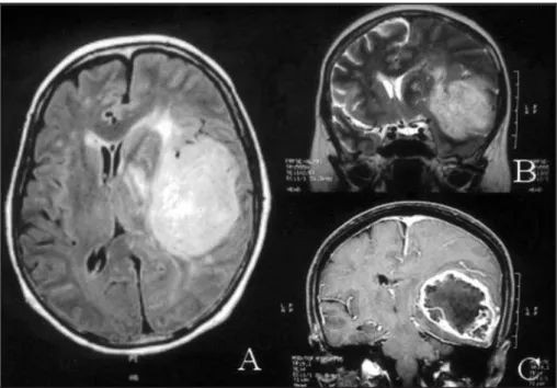
Case 2.A 44-years old male presented with the clinical picture resembling uncinate fits one month before admission, which consisted in the anomalous perception of the smell of paints, followed by fain-ting spells and nonspecific headaches. He was tak-en to the hospital due to rectak-ent mtak-ental confusion, memory deficits and expressive aphasia, which were evident on clinical examination, besides right hemiparesis. Both CT and MRI showed an irregu-lar infiltrating enhancing mass lesion in the left fron-to-temporal lobes with slight surrounding edema (Figure 1). The patient was submitted to tempo-ral lobectomy including the mesial structures. He received adjuvant radiotherapy during the follow-ing month. He persisted aphasic, but not confused during the next three months, without other clin-ical signs, when the symptoms recurred. He died 6 months after the surgical procedure.
Case 3.A 48-years old male had complained of pain in the left temporal area for 4 years, which he presumed was an earache. The patient was pre-viously blind of the left eye and 2 months before admission, headaches worsened and he developed a left palpebral ptosis. He was initially seen by an ophthalmologist, who diagnosed glaucoma. Further work-up, including CT and MRI showed an irreg-ular infiltrating enhancing mass lesion in the left temporo-parietal lobes with surrounding edema and compression of the left cerebral peduncle. On admission he was blind of the left eye with asso-ciated left palpebral ptosis. Left facial numbness, right hemiparesis and right hyperreflexia were also present. The patient was placed on corticosteroids and submitted to left temporo-parietal cranioto-my. The tumor, which was grayish, soft and bloody, was macroscopically totally removed. The patient received radiotherapy during the following month and died 5 months after the surgical procedure.
scan and MRI showed an irregular enhancing mass lesion in the right parieto-occipital lobes involving midline structures, invading right lateral ventricle and infiltrating corpus callosum. The patient was placed on corticosteroids and reoperated through the same approach. The tumor, which was macro-scopically totally removed, was grayish, soft, and bloody. The lesion, interpreted initially as a malig-nant meningioma, had the diagnosis of gliosarco-ma established trough immunohistochemical analy-sis. She died 4 months after the second procedure. The main clinical findings are summarized in Table 1.
Histological and immunohistochemical findings
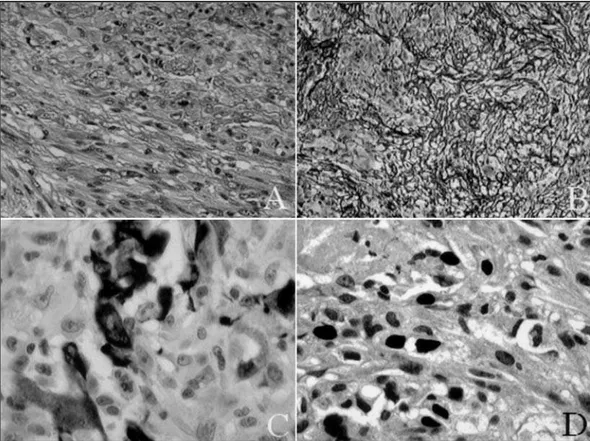
All four cases displayed sarcomatous and glial components. The glial component was remarkably similar to a glioblastoma, with nuclear pleomor-phism, high mitotic index, marked vascular
prolif-eration and foci of necrosis, either focal or geogra-phical (Fig 2A).
The sarcomatous component consisted of groups of spindle cells arranged in a fashion that resembled fibrosarcoma in all cases (Fig 2A). In order to evaluate the mesenchymal component, histochemical study to reticulin fibers was performed and demonstrat-ed a rich network of fibrils in all cases (Fig 2B).
The glial component comprised greater than 50% of the neoplasm in only one case. In the re-maining three cases, the sarcomatous component comprised 70%, 80% and 90% of the sampled neoplasm, respectively. Mitotic figures per 5 high-power fields in the glial neoplasm ranged between 8 and 2 (mean 6.25). In the sarcomatous areas, the number of mitotic figures per 5 high-power fields ranged between 6 and 2 (mean 3).
Immunohistochemical findings are illustrated in Table 2, Figures 2C and 2D.
Fig 1. MR images of case 2 displaying a bulging lesion with marked peripheral edema and midline deviation localized on the fronto-parietal region (A, axial view; B, coronal view). Peripheral enhancement of the lesion was observed after contrast medium injection (C).
Table 1. Clinical findings of gliosarcomas.
Case Age Sex Presenting symptom Site of lesion Follow-up
1 43 male headache R temporo-parietal † in 3 months
2 44 male olfactory allucinations L fronto-temporal † in 6 months
3 48 male headache L temporo-parietal † in 5 months
4 21 female headache R parieto-occipital † in 4 months
DISCUSSION
GSa is a rare morphological variant of glioblasto-ma, predominant in males, with age ranging from the fourth to the sixth decades, with a slight predi-lection for the temporal lobes1,3,4-6. In our present report, GSa accounted for 1.06% of all glioblasto-mas, with a mean age of 39. However, the occurren-ce of a GSa in a 22-year old patient is uncommon. We failed to observe an anatomical site of
predilec-tion. In spite of some reports of metastatic spread7, none of the patients reported herein evolved with systemic dissemination.
On clinical basis, primary glioblastomas and glio-sarcomas are indistinguishable, both presenting with short clinical course, low median survival and similar peak of incidence1,3,6,8-11; hence the inclusion of GSa in the same clinical trials of glioblastomas3. The mean duration of symptoms in our patients
Table 2. Immunohistochemical findings of gliosarcomas.
Case GFAP Vimentin CD34 p53 EGFR Ki-67
1 G – D G – F G – N G +++ G – N G +++
S – N S – D S – N S ++ S – N S +
2 G – D G – F G – N G +++ G – N G +
S – N S – D S – N S +++ S – N S +
3 G – D G – F G – N G +++ G – N G ++
S – N S – D S – N S +++ S – N S ++
4 G – D G – F G – N G + G – P G ++
S – N S – D S – N S ++ S – N S ++
G, glial component; S, sarcomatous component; D, diffuse positivity; F, focal positivity; N, negative; P, positive; +, staining of less than 1% of nuclei; ++, staining of 1 to 5% of nuclei; +++, staining of more than 5% of nuclei; GFAP, glial-fibrillary-acidic protein; EGFR, epidermal growth factor.
(17 months) was somewhat longer due to a parti-cular case presenting with symptoms for 48 months.
The treatment does not differ from that perfor-med on glioblastomas, consisting of surgical resec-tion and, depending on clinical status, radiother-apy and/or chemotherradiother-apy3,6. Patients who receive radiotherapy have a longer overall median survival (mean 10.6 months) when compared to patients submitted to surgery only (mean 6.2 months), ac-cording to Perry et al.6. However, one should notice that patients eligible for radiation therapy pres-ent with higher performance status, younger age and more favorable prognostic factors. In our series only two patients were eligible for adjuvant ther-apy and the mean overall survival was 4.5 months, a rate consistent with the marked positivity for pro-liferation index marker Ki-67 observed on immuno-histochemical evaluation.
The histogenesis of GSa has been subject of debate. Feigin et al., who defined the sarcomatous component as a proliferation of glioblastoma blo-od vessels, first assessed it2. However, immunohisto-chemical studies have failed to detect endothelial markers in the sarcomatous component6,12,13. In the present study, only blood vessels stained positi-vely for CD34.
The monoclonal theory was first proposed by Biernat et al., who demonstrated identical p53 mutations in both tumor areas14. Subsequently, Boerman et al., using comparative genomic hybridi-zation, fluorescence in situ hybridihybridi-zation, cytoge-netic analysis and microsatellite analysis, described genetic alterations shared by both tumor compo-nents15. Reis et al., assessing the genetic profile of 19 GSa patients, have found identical PTEN muta-tion, p53nuclear accumulation, p16 deletion and CDK4 amplification in both tumor areas11. Recently, Actor et al. have reported that 57% off all chromo-somal imbalances detected by comparative genom-ic hybridization of eight GSa were shared by both components. These authors have also detected identical p53 mutations in both glial and sarcoma-tous areas in 13 of 38 patients16. In our study, p53 nuclear accumulation was detected by immunohis-tochemistry in all 4 cases studied and the degree of staining was similar in glial and sarcomatous regions.
The frequency of p53mutations observed in GSa is considered to be intermediate between primary and secondary glioblastomas, with the former pre-senting approximately 11% and the latter, 67%17. We attribute the finding of p53positivity in both components in all of our cases to the considerably
small cohort. However, its role in the tumorigen-esis of GSa cannot be ruled out.
Actor et al. have described EGFR overexpression in 8% of 38 tumors, with staining of both compo-nents in only one case16. Previous studies have re-ported EGFR overexpression and amplification as absent11. In our study, we detected EGFR immunos-taining in only one case, which showed positivity in the glial area and negativity in the sarcomatous one.
In conclusion, GSa in our series present clinical and some genetic features similar to primary glio-blastomas. An extremely low rate of EGFR overex-pression remains as the most striking difference bet-ween these two entities. Furthermore, the TP53 tu-mor-supressor gene mutation seems to play a pathogenetic role in GSa and the authors believe there is strong evidence concerning its monoclon-al origin. However, different molecular and genet-ic events may partgenet-icipate in the pathogenesis of GSa and deserve further investigation.
REFERENCES
1. Ohgaki H, Biernat W, Reis R, Hegi M, Kleihues P. Gliosarcoma. In Klei-hues P, Cavenee WK (eds). Pathology and genetics of tumors of the nerv-ous system. Lyon: IARC Press 2000:42-44.
2. Feigin IM, Allen LB, Lipkin L,Gross SW. The endothelial hyperplasia of the cerebral blood vessels and its sarcomatous transformation. Cancer 1958;11:264-277.
3. Meis JM, Martz KL, Nelson JS. Mixed glioblastoma multiforme and sar-coma: a clinicopathologic study of 26 Radiation Therapy Oncology Group cases. Cancer 1991;67:2342-2349.
4. Jack CR Jr, Bhansali DT, Chason JL, et al. Angiographic features of gliosarcoma. AJNR 1987;8:117-122.
5. Morantz RA, Feigin I, Ransohoff J III. Clinical and pathological study of 24 cases of gliosarcoma. J Neurosurg 1976;45:398-408.
6. Perry JR, Ang LC, Bilbao JM, Muller PJ. Clinicopathologic features of prima-ry and postradiation cerebral gliosarcoma. Cancer 1995;75:2910-2918. 7. Witwer BP, Salamat MS, Resnick DK. Gliosarcoma metastatic to the
cer-vical spinal cord: case report and review of the literature. Surg Neurol 2000;54:373-379.
8. Burger PC, Scheithauer BW. Gliosarcoma. In Burger PC, Scheithauer BW (eds). Tumors of the central nervous system. Washington DC: Armed Forces Institute of Pathology, 1994:68-70.
9. Kaschten B, Flandroy P, Reznik M, Hainaut H, Stevenaert A. Radiation-induced gliosarcoma: case report and review of the literature. J Neurosurg 1995;83:154-162.
10. Lutterbach J, Guttenberger R, Pagenstecher A. Gliosarcoma: a clinical study. Radiotherapy Oncol 2001;61:57-64.
11. Reis RM, Konu-Lebleblicioglu D, Lopes JM, Kleihues P, Ohgaki H. Genetic profile of gliosarcomas. Am J Pathol 2000;156:425-432. 12. Grant JW, Steart PV, Aguzzi A, Jones DB, Gallagher PJ. Gliosarcoma:
an immunohistochemical study. Acta Neuropathol 1989;79:305-309. 13. Sreenan JJ, Prayson RA. Gliosarcoma: a study of 13 tumors, including
p53 and CD34 immunohistochemistry. Arch Pathol Lab Med 1997; 121:129-133.
14. Biernat W, Aguzzi A, Sure U, Grant JW, Kleihues P, Hegi ME. Identical mutations of the p53 tumor supressor gene in the gliomatous and the sarcomatous components suggests a common origin from glial cells. J Neuropathol Exp Neurol 1995;54:651-656.
15. Boerman RH, Anderl K, Herath J, et al. The glial and mesenchymal ele-ments of gliosarcomas share similar genetic alterations. J Neuropathol Exp Neurol 1996;55:973-981.
16. Actor B, Cobbers JMJL, Buschges R, et al. Comprehensive analysis of genomic alterations in gliosarcoma and its two tissue components. Genes Chromosomes Cancer 2002;34:416-427.