DOI: 10.5216/cab.v14i2.18930
uPAR EXPRESSION IN CANINE NORMAL PROSTATE AND WITH
PROLIFERATIVE DISORDERS
MARIANA RODRIGUES FALEIRO1,DENISE CAROLINE TOLEDO1,MARCELA MARCONDES PINTO RODRIGUES2,RENEE LAUFER AMORIM3,LUIZ AUGUSTO BATISTA BRITO4,ADILSON DONIZETI
DAMASCENO4,VERIDIANA MARIA BRIANEZI DIGNANI DE MOURA4
1Pós-Graduanda da Escola de Veterinária e Zootecnia da Universidade Federal de Goiás, Goiânia, GO, Brasil.
2Pós-Doutoranda da Universidade Estadual Paulista, Botucatu, SP, Brasil. 3Professora Doutora da Universidade Estadual Paulista, Botucatu, SP, Brasil.
4Professores Doutores da Escola de Veterinária e Zootecnia da Universidade Federal de Goiás, Goiânia, GO, Brasil.
ABSTRACT Prostatic lesions such as prostatic intraepithelial neoplasia
(PIN) and proliferative inflammatory atrophy (PIA) are studied in human and canine species due to their malignance potential. The plasminogen activator (PA) system has been suggested to play a central role in cell adhesion, angiogenesis, inflammation, and tumor invasion. The urokinase-type plasminogen activator receptor (uPAR) is a component of the PA, with a range of expression in tumor and stromal cells. In this study, uPAR expression in both canine normal prostates and with proliferative disorders (benign prostatic hyperplasia-BPH, proliferative inflammatory atrophy-PIA, prostatic intraepithelial neoplasia-PIN, and carcinoma-PC) was evaluated by immunohistochemistry in a tissue microarray
(TMA) slide to establish the role of this enzyme in extracellular matrix (ECM) remodeling and in the processes of tissue invasion. A total of 298 cores and 355 diagnoses were obtained, with 36 (10.1%) normal prostates, 46 (13.0%) with BPH, 128 (36.1%) with PIA, 74 (20.8%) with PIN and 71 (20.0%) with PC. There is variation in the expression of uPAR in canine prostate according to the lesion, with lower expression in normal tissue and with BPH, and higher expression in tissue with PIA, PIN and PC. The high expression of uPAR in inflammatory and neoplastic microenvironment indicates increased proteolytic activity in canine prostates with PIA, PIN, and PC.
KEYWORDS:CD87; PIA; PIN; prostatic carcinoma; TMA.
EXPRESSÃO DE uPAR NA PRÓSTATA CANINA NORMAL E COM LESÕES PROLIFERATIVAS
RESUMO Lesões prostáticas como a neoplasia intraepitelial prostática (PIN) e a atrofia inflamatória proliferativa (PIA) são estudadas na espécie humana e canina devido ao seu potencial de malignidade. O sistema de ativador de plasminogênio (PA) tem sido sugerido como um importante mecanismo na adesão celular, angiogênese, inflamação e invasão tumoral. O receptor ativador de plasminogênio do tipo uroquinase (uPAR) é um componente do PA, expresso em células tumorais e
(10,1%) próstatas normais, 46 (13,0%) com HPB, 128 (36,1%) com PIA, 74 (20,8%) com PIN e 71 (20,0%) com CP. Há variação na expressão de uPAR na próstata canina de acordo com a lesão, com menor expressão nas glândulas normais e com HPB, e maior naquelas com
lesões displásicas e (PIA e PIN) e neoplásicas (CP). A superexpressão de uPAR nos microambientes inflamatório e neoplásico indica aumento da atividade proteolítica em próstatas caninas com PIA, PIN e CP.
PALAVRAS-CHAVE: carcinoma prostático; CD87; PIA; PIN; TMA.
INTRODUCTION
Researchers have studied the canine prostate due to its similarities with the human prostate regarding the natural occurrence of diseases and the hormonal influence in their development, for instance, benign prostatic hyperplasia (BPH) and prostatic carcinoma (PC) (LEROY & NORTHRUP, 2009).
Some dysplastic lesions that affect man´s prostate are considered premalignant, as the prostatic intraepithelial neoplasia (PIN), because they show morphological similarities to cancer or because they involve potentially carcinogenic factors (DE MARZO et al., 2006). The proliferative inflammatory atrophy (PIA) is another affection that has been constantly investigated due to the controversy regarding its premalignant potential (WATERS & BOSTWICK, 1997; WANG et al., 2009).
In dogs, PIN has been considered a premalignant lesion and it can be observed in cases of prostatic carcinoma (WATERS & BOSTWICK 1997; WATERS et al., 1997; MADEWELL et al., 2004; MATSUZAKI et al., 2010). RODRIGUES et al. (2010) and TOLEDO et al. (2010) mentioned PIA in canine prostates.
The evolution process of PIA and PIN is followed by tumor invasion through the extracellular matrix (ECM). This condition comprises the interaction phases between neoplastic cells and ECM, with hydrolytic destruction by proteolytic enzymes and migration of neoplastic cells through the altered extracellular environment (DEL MAESTRO et al., 1990; AMBIRU et al., 1997).
The plasminogen activator (PA) system is among the proteolytic enzymes involved in tumor invasion. This system is composed of a serine protease urokinase-type plasminogen activator (uPA) and its receptor (uPAR), a serine protease tissue-type PA (tPA), plasminogen and its multiple receptors, besides three inhibitors (plasminogen activator inhibitors PAI-1, PAI-2, and protease nexin 1) (WANG, 2001; BOCK & WANG, 2004).
The coordinated expression of this system has been suggested to play a central role in cell
adhesion, migration, and invasion (PEI et al., 1999; BOCK & WANG, 2004), as well as the degradation of basement membrane and ECM, and the development of cancer metastasis (COHEN et al., 1991; DANO et al., 1994; VASSALLI, 1994; KOBLINSKI et al., 2000).
Components of the PA system and in particular uPAR are suited for routine analysis because of the high levels of antigen found not only within cancer tissue but also within serum, making it readily accessible for measurement (GAO et al. 2001; PLOUGAR et al., 2002; BOCK & WANG, 2004; SEHGAL et al., 2006). The association among high levels of uPAR, higher histological grades, and advanced stages of prostate cancer (STEWART et al., 2004) makes any high uPAR expression in cancer not only possible but also valuable as it is an attractive therapeutic target (MAZAR, 2001; BOCK & WANG, 2004; SEHGAL et al., 2006).
The assessment of uPA and uPAR in prostate cancer was initially performed in primary tumors of this organ in humans and in models of the prostatic disease like rats and mice (GILARDONI et al., 2003; PULUKURI et al., 2007). BAILEY et al. (2006) were the first ones to report the constitutive expression of these proteins only in normal prostates of dogs, without reports on ill tissues.
The expression of uPAR in both normal canine prostatic tissue and with proliferative disorders (BPH, PIN, PIA and PC) was verified in order to evaluate the role of this enzyme in ECM remodeling and tissue invasion processes.
MATERIAL AND METHODS
239 uPar expression in canine normal prostate and with proliferative disorders
The prostate tissue microarray (TMA) was carried out according to criteria described by KONONEN et al. (1998) and BUBENDORF et al. (2001). From the previous defined areas, core biopsies were taken from 298 prostatic paraffin-embedded samples using Tissue Microarrayer (Beencher Instruments®, Silver Spring, USA). Tissue
cores with a dimension of 1.0 mm from each specimen were punched and arrayed on a recipient paraffin block. Three-µm-sections were obtained from the recipient block and mounted on silanized glass slides for HE and immunohistochemical tests.
Immunohistochemistry was performed in one TMA slide, which was deparaffinized, rehydratated and washed in distilled water. For anti-uPAR mouse monoclonal antibody, clone R4 (Dako M7294), we used a 1:25 dilution and antigen retrieval in water bath at 96ºC for 40min, with pre-heated TRIS-EDTA buffer and pH9.0. Endogenous peroxidase activity was blocked and incubation with primary antibody was carried out in a wet chamber, at 4ºC, for 18h. Advance HRP signal amplification system (Dako K 4068) was used and the reaction
was visualized by DAB (Diaminobenzidine, Dako, K3468-1). Sections were counterstained with Mayer's hematoxylin, washed, dehydrated, cleared, mounted, and examined by light microscopy.
Dog small intestine was used like positive tissue control for uPAR. The primary antibody was replaced by TRIS buffer, pH7.4, on canine prostate for the negative antibody control.
The intensity of cytoplasmic reactivity of the antibody in epithelial and periacinar stromal cells was scored in 0 = negative, 1 = discrete, 2 = moderate and 3 = intense. Regarding the number of both epithelial and periacinar stromal stained cells the scores were 0 = negative; 1 = 1 - 25%; 2 = 26 - 50%; 3 = 51 - 75% and 4 = 76 -100%.
Kruskal-Wallis and Mann Whitney tests as well as descriptive data were used to compare the scores of percentage of positive cells and the intensity. Data were analyzed using Excel 2007 and SPSS (Statistical Package for Social Science, version 16.0) software. All values were considered at 5% of significance level.
Table 1 - Histomorphological criteria for the prostatic canine tissue classification.
Diagnoses Architectural features Cytoplasm Nucleus Chromatin Nucleolus membrane Basement inflammatory Periacinar infiltrate
Normal prostate
One epithelial layer with cuboidal or columnar cells
and fine fibrovascular
stroma
Abundant Not change Uniform None Intact None
BPH hyperplasia or Epithelial hypertrophy
Variable Not change Uniform None Intact None
PIA discrete
Atrophic acini, more than one epithelial layer
Reduced and hypochromatic
Discrete changes
in size and form Condensed
One or more
evident Intact Sparse
PIA moderate Atrophic acini, more than one epithelial layer
Reduced and
hypochromatic Discrete changes in size and form Condensed One or more evident Intact mononuclear cells Aggregates of
PIA intense
Atrophic acini, more than one epithelial layer
Reduced and hypochromatic
Discrete changes
in size and form Condensed
One or more evident Intact
Mononuclear cells forming follicles
PIN
Hypercellular epithelium, stacked, laminated
and irregular epithelial cells
Reduced and hypochromatic
Variable in size
and form Coarse
Large and evident
May be
ruptured None
Carcinoma
Cellular proliferation, variation in acini and cell size and
form
Variable Variable in size and form Coarse Large and evident Ruptured None
RESULTS
From the 298 TMA cores we obtained 355 different diagnoses and since in 19.1% of the cores presented multiple diagnoses (for example: PIN foci and a PC area). From 355 diagnosis, 36 (10.1%) were normal tissues, 46 (13.0 %) BPH, 128 (36.1 %) PIA, 74 (20.8 %) PIN, and 71 (20.0 %) PC. Concerning PIA (n=128), we observed that 71 (55.5 %) were discrete (PIA-D), 39 (30.5 %) moderate (PIA-M) and 18 (14.0 %) intense (PIA-I).
Immunohistochemistry staining for uPAR was cytoplasmic (Figure 1). In acinar and periacinar compartments there was difference in uPAR protein expression according to the diagnosis, regarding the number and intensity of positive cells (p<0.05).
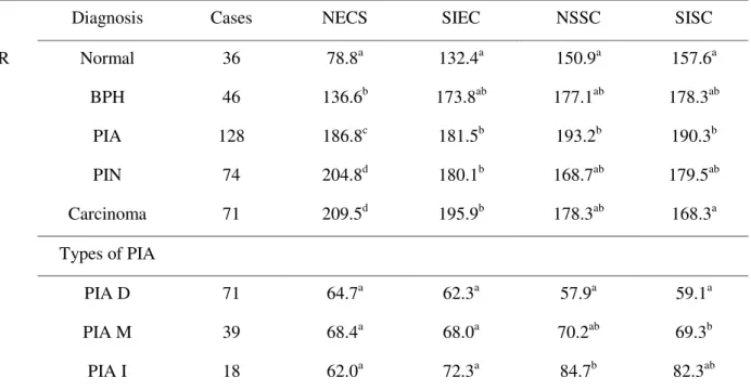
The number of epithelial cells stained
(NECS) for uPAR was statistically different between all diagnoses, except between PIN and carcinoma. For staining intensity of epithelial cells (SIEC), there was a significant difference between normal prostates and those with PIA, PIN, and carcinoma. Regarding the number of stained stromal cells (NSSC) there was difference between normal glands and those with PIA. Moreover, for the uPAR staining intensity of stromal cells (SISC), there was difference between normal prostates and those with PIA, as well as between those with PIA and carcinoma (Table 2).
Regarding the types of PIA, there was difference between PIA-D and PIA-I in relation to the number of stained stromal cells and between PIA-D and PIA-M in staining intensity of stromal cells for uPAR (Table 2).
241 uPar expression in canine normal prostate and with proliferative disorders
Table 2 - Comparison between diagnoses in relation to the number of stained cells and staining intensity of uPAR in epithelial and stromal cells
Diagnosis Cases NECS SIEC NSSC SISC
uPAR Normal 36 78.8a 132.4a 150.9a 157.6a
BPH 46 136.6b 173.8ab 177.1ab 178.3ab
PIA 128 186.8c 181.5b 193.2b 190.3b
PIN 74 204.8d 180.1b 168.7ab 179.5ab
Carcinoma 71 209.5d 195.9b 178.3ab 168.3a
Types of PIA
PIA D 71 64.7a 62.3a 57.9a 59.1a
PIA M 39 68.4a 68.0a 70.2ab 69.3b
PIA I 18 62.0a 72.3a 84.7b 82.3ab
Equal letters in the same column do not differ by the Mann-Whitney test (p<0.05). NECS - number of epithelial cells stained; SIEC - staining intensity of epithelial cells; NSSC - number of stained stromal cells; SISC - staining intensity of stromal cells.
DISCUSSION
In different types of human tissues, the PA system has been suggested to play a central role in cell adhesion, migration regulation, wound healing, angiogenesis, inflammation and growth factors regulation (GAO et al. 2001; PLOUGAR et al., 2002; BOCK & WANG, 2004), besides the development of tumor invasion and metastasis (DANO et al., 1994; VASSALLI, 1994; KOBLINSKI et al., 2000).
In this research, we found uPAR expression in normal canine prostatic tissue and increase expression of it in epithelial and stromal cells of canine prostate with benign, dysplastic, and malignant lesions. In contrast, there is minimal information about the activity of this glycoprotein in different tissues of the dogs. Only BAILEY et al. (2006) reported the expression of uPA and uPAR in the urinary tract of healthy dogs, including low expression in the prostate.
The variation in number of stained cells and staining intensity of uPAR in epithelial and stromal cells showed that enzyme presents variable expression in canine prostate tissue according to the pathological process. In this context, there are some controversies for uPAR localization and expression (BAILEY et al., 2006; LI & COZZI, 2007; DASS et al., 2008; KUMANO et al., 2009). In rats, WILSON
et al. (1995) found a significant variation of uPAR expression among the variables age, region of the prostate and castration condition. Moreover, GAVRILOV et al. (2001) reported association between uPAR expressed in prostatic adenocarcinoma cells and prostatic stromal cells.
The number of uPAR stained epithelial cells varied among the diagnoses, excepted between PIN and carcinoma, with low expression in normal cells and high expression in PIN and carcinoma cells as described by GAVRILOV et al. (2001), RIDDICK et al. (2005), SEHGAL et al. (2006) and LI & COZZI (2007) in human and mouse neoplastic prostate. Although they have not studied glands with PIN, the increased expression of uPAR in canine prostates with this injury suggests increased proteolytic activity and possible potential of invasion.
tumor tissue might be low or negative, which disagrees with this study considering that all samples showed uPAR expression at some degree.
COZZI et al. (2006) reported uPAR expression in eight of fifteen human prostates with PIN, but they emphasized that there was no high expression as in carcinoma with high grade, contrary to the uPAR high expression observed in PIN and carcinoma in this study. This difference can be explained by dissimilarities in the methodology of both studies. In the first one, tumors were classified according to the cellular differentiation degree (Gleason score), with high expression in the undifferentiated ones and intermediate expression in the differentiated ones, similar to what occurred in PIN. In this study, tumors were not graded and showed high expression, but not always, as well as the PIN, which might mean a less aggressive canine tumor pattern.
Samples with PIA presented more staining cells and higher staining intensity for uPAR in the stromal cells than in normal tissues. In this sense, USHER et al. (2005) reported uPAR expression in interstitial leukocytes of neoplastic human prostate, and BAILEY et al. (2006) found accentuated uPAR staining in interstitial inflammatory cells of canine prostate. Therefore, it is likely that the inflammation surrounding the dysplastic epithelial lesion in PIA is responsible for such difference, once the interstitial inflammatory cells showed uPAR high expression. The difference between PIA and carcinoma in the uPAR staining intensity of stromal cells surrounding the tumor supports this idea, once the tumors showed less staining intensity than PIA and they were not surrounded by perineoplastic inflammation.
The comparison among types of PIA confirms what was described considering the inflammatory cells from PIA-M that showed higher staining intensity than PIA-D, and PIA-I presented higher number of stromal cells stained for uPAR than PIA-D, suggesting the role of inflammation in remodeling the ECM that surrounds the dysplastic epithelium of canine prostate, and which possibly can contribute to the invasion of the matrix by transformed epithelial cells. In this context, ANDREASEN, et al. (1997) stated that PA system is important in the process of tissue remodeling due to the ability of the uPA-uPAR complex to degrade the basement membrane in inflammatory and neoplastic diseases.
CONCLUSION
There is variation in the expression of uPAR in canine prostate according to the lesion, with lower expression in normal tissue and with BPH, and
higher expression in prostatic tissue with dysplasia (PIA and PIN) and neoplasia (PC). The high expression of uPAR in inflammatory and neoplastic microenvironment indicates increased proteolytic activity in canine prostates with PIA, PIN, and PC.
REFERENCES
AMBIRU, S.; MIYAZAKI, M.; ITO, H.; NAKAGAWA, K.; SHIMIZU, H.; NUKUI, Y.; NOZAWA, S.; OKUNO, A.; YOSHITOMI, H.; NAKAJIMA, N. A prospective study of prognostic value of type IV collagenase activity in colorectal cancer tissue. Digestive Diseases and Sciences, v.42, n.8, p.1660- 1665, 1997.
ANDREASEN, P.A.; KJOLLER, L.; CHRISTENSEN, L.; DUFFY, M.J. The urokinase-type plasminogen activator system in cancer metastasis; a review. International Journal of Cancer, v.72, n.1, p.1-22, 1997.
BAILEY, T.R.; PAULSEN, D.B.; SEHGAL, I.; HOSGOOD, G. Immunohistochemical staining of urokinase plasminogen activator-like and urokinase plasminogen activator receptor-like proteins in the urinary tract of healthy dogs. American Journal of Veterinary Research, v.67, n.9, p.1628-1634, 2006.
BOCK, C.E.; WANG, Y. Clinical Significance of Urokinase-Type Plasminogen Activator Receptor (uPAR) Expression in Cancer. Medicinal Research Reviews, v.24, n.1, p.13-39, 2004.
BOSTWICK, D.G. High-grade prostatic intraepithelial neoplasia: the most likely precursor of prostate cancer. Cancer, v.75, n.7, p.1823-1836, 1995.
BUBENDORF, L.; NOCITO, A.; MOCH, H.; SAUTER, G. Tissue microarray (TMA) technology: miniaturized pathology archives for high-throughput in situ studies. Journal of Pathology, v.195, n.1, p.72–79, 2001.
COHEN, R.L.; XI, X.P.; CROWLEY, C.W.; LUCAS, B.K.; LEVINSON, A.D.; SHUMAN, M.A. Effect of urokinase receptor occupancy on plasmin generation an proteolysis of basement membrane by human tumor cells. Blood, v.78, n.2, p.479-487, 1991.
COZZI, P.J.; WANG, J.; DELPRADO, W.; MADIGAN, M.C.; FAIRY, S.; RUSSELL, P.J.; YONG, L. Evaluation of urokinase plasminogen activator and its receptor in different grades of human prostate cancer. Human Pathology, v.37, n.11, 442–451, 2006.
DANO, K.; BEHRENDT, N.; BRUNNER, N.; ELLIS, V.; PLOUG, M.; PYKE, C. The urokinase receptor. Protein structure and role in plasminogen activation and cancer invasion. Fibrinolysis, v.8, n.1, p.189-203, 1994.
DASS, K.; AHMAD, A.; AZMI, A.S.; SARKAR, S.H.; SARKAR, F.H. Evolving role of uPA/uPAR system in human cancers. Cancer Treatment Reviews, v.34, n.2, p.122–136, 2008.
243 uPar expression in canine normal prostate and with proliferative disorders
EGEVAD, L.; ERTOY-BAYDAR, D.; FARRE, X.; FINE, S.W.; ICZKOWSKI, K.A.; ITTMANN, M.; KNUDSEN, B.S.; LODA, M.; LOPEZ-BELTRAN, A.; MAGI-GALLUZZI, C.; MIKUZ, G.; MONTIRONI, R.; PIKARSKY, E.; PIZOV, G.; RUBIN, M.A.; SAMARATUNGA, H.; SEBO, T.; SESTERHENN, I.A.; SHAH, R.B.; SIGNORETTI, S.; SIMKO, J.; THOMAS, G.; TRONCOSO, P.; TSUZUKI, T.T.; VAN LEENDERS, G.J.; YANG, X.J.; ZHOU, M.; FIGG, W.D.; HOQUE, A.; LUCIA, M.S. A working group classification of focal prostate atrophy lesions. American Journal of Surgical Pathology, v.30, n.10, p.1281-1291, 2006.
DEL MAESTRO, R.F.; MEGYESI, J.F.; FERRE, C.L. Mechanisms of tumor-associated edema: a review. Canadian Journal of Neurological Sciences, v.17, n.2, p.177-183, 1990.
GAO, W.; WANG, Z.; BAI, X.; XI, X.; RUAN, C. Detection of soluble urokinase receptor by immunoradiometric assay and its application in tumor patients. Thrombosis Research, v.102, n.1, p.25–31, 2001.
GAVRILOV, D.; KENZIOR, O.; EVANS, M.; CALALUCE, R.; FOLK, W.R. Expression of urokinase plasminogen activator and receptor in conjunction with the ets family and AP-1 complex transcription factors in high grade prostate cancers. European Journal of Cancer, v.37, n.8, p. 1033–1040, 2001.
GILARDONI, M.B.; CESCHIN, D.G.; SAHORES, M.M.; OVIEDO, M.; GEHRAU, R.C.; CHIABRANDO, G.A. Decreased expression of the low-density lipoprotein receptor-related protein-1 (LRP-1) in rats with prostate cancer. Journal of Histochemistry and Cytichemistry, Baltimore, v.51, n.12, p.1575-1580, 2003.
KOBLINSKI, J.E.; AHRAM, M.; SLOANE, B.F. Unraveling the role of proteases in cancer. Clinica Chimica Acta, v.291, n.2, p.113-135, 2000.
KONONEN, J.; BUBENDORF, L.; KALLIONIEMI, A.; BARLUND, M.; SCHRAML, P.; LEIGHTON, S.; TORHORST, J.; MIHATSCH, M.J.; SAUTER, G.; KALLIONIMENI, O.P. Tissue microarrays for high-throughput molecular profiling tumor of specimens. Nature Medicine, v.4, n.7, p.844-847, 1998.
KUMANO, M.; MIYAKE, H.; MURAMAKI, M.; FURUKAWA, J.; TAKENAKA, A.; FUJISAWA, M. Expression Of Urokinase-Type Plasminogen Activator System In Prostate Cancer: Correlation With Clinicopathological Outcomes In Patients Undergoing Radical Prostatectomy . Urologic Oncology: Seminars and Original Investigations, v.27, n.2, p.180–186, 2009. LEAV, I.; SCHELLING, K.H.; ADAMS, J.Y.; MERK, F.B.; ALROY, J. Role of canine basal cells in postnatal prostatic development, induction of hyperplasia, and sex hormone- stimulated growth, and the ductal origin carcinoma. Prostate, v.47, n.1, p.149–163, 2001.
LEROY, B.E.; NORTHRUP, N. Prostate cancer in dogs:
Comparative and clinical aspects The Veterinary Journal, v.180, n.2, p.149–162, 2009.
LI, Y.; COZZI P.J. Targeting uPA/uPAR in prostate cancer. Cancer Treatment Reviews, v.33, n.6, p.521– 527, 2007.
MADEWELL, B.R.; GANDOUR-EDWARDS, R.; WHITE, R.W.D.V. Canine prostatic intraepithelial neoplasia: Is the comparative model relevant? Prostate, v.58, n.3, p.314-317, 2004.
MATSUZAKI, P.; COGLIATI, B.; SANCHES, D.S.; CHAIBLE, L.M.; KIMURA, K.C.; SILVA, T.C.; REAL-LIMA, M.A.; HERNANDEZ-BLAZQUEZ, F.J.; LAUFER-AMORIM, R.; DAGLI, M.L.Z. Immunohistochemical characterization of canine prostatic intraepithelial neoplasia. Journal of Comparative Pathology, v.142, n.1, p.84-88, 2010.
MAZAR, A.P. The urokinase plasminogen activator receptor (uPAR) as a target for the diagnosis and therapy of cancer. Anticancer Drugs, v.12, n.5, p.387–400, 2001. PEI, X.H.; NAKANISHI, Y.; TAKAYAMA, K.; BAI, F.; HARA, N. Granulocyte, Granulocyte-macrophage, and macrophage colony-stimulating factors can stimulate the invasive capacity of human lung cancer cells. British Journal of Cancer, v.79, n.1, p.40–46, 1999.
PLOUGAR, M.; GARDSVOLL, H.; JORGENSEN, T.J.; LONBORG-HANSEN, L.; DANO, K. Structural analysis of the interaction between urokinase-type plasminogen activator and its receptor: A potential target for anti-invasive câncer therapy. Biochemical Society Transactions, v.30, n.2, p.177–183, 2002.
PULUKURI, S.M.; ESTES, N.; PATEL, J.; RAO, J.S. Demethylation-linked activation of urokinase plasminogen activator is involved in progression of prostate cancer. Cancer Research, v.67, n.3, p.930-939, 2007.
RIDDICK, A.C.; SHUKLA, C.J.; PENNINGTON, C.J.; BASS, R.; NUTTALL, R.K.; HOGAN, A.; SETHIA, K.K.; ELLIS, V.; COLLINS, A.T.; MAITLAND, N.J.; BALL, R.Y.; EDWARDS, D.R. Identification of degradome components associated with prostate cancer progression by expression analysis of human prostatic tissues. British Journal of Cancer, v.92, n.12, p.2171– 2180, 2005.
RODRIGUES, M.M.P.; DI SANTIS, G.W.; DE MOURA, V.M.B.D.; AMORIM, R.L. COX-2 and TGF- β expression in proliferative disorders of canine prostate. Brazilian Journal of Veterinary Pathology, v.3, n.1, p.31-36, 2010.
SEHGAL, I.; FOSTER, T.P.; FRANCIS, J. Prostate cancer cells show elevated urokinase receptor in a mouse model of metastasis. Cancer Cell International, v.6, p.21, 2006.
Reproductive Biology Endocrinology, v.2, n.1, p.2-6, 2004.
SUGAR, L.M. Inflammation and prostate cancer. The Canadian Journal of Urology, (CJU), St. Laurent, v.13, n.1, suplemento 1, p.46-47, 2006.
TOLEDO, D.C.; FALEIRO, M.B.R.; RODRIGUES, M.M.P.; DI SANTIS, G.W.; LAUFER AMORIM, R.; DE MOURA, V.M.B.D. Histomorphological characterization of proliferative inflammatory atrophy in canine prostate. Ciência Rural v.40, n.6, p.1372-1377, 2010.
USHER, P.A.; THOMSEN, O.F.; IVERSEN, P.; JOHNSEN, M.; BRUNNER, N.; HOYER-HANSEN, G.; ANDREASEN, P.; DANO, K.; NIELSEN, B.S. Expression of urokinase plasminogen activator, its receptor and type-1 inhibitor in malignant and benign prostate tissue. International Journal of Cancer, v.113, n.6, p.870–880, 2005.
VASSALLI, J.D. The urokinase receptor. Fibrinolysis, v.8, n.1 (supplement), p.172–181, 1994.
WANG, Y. The role and regulation of urokinase-type
plasminogen activator receptor gene expression in cancer invasion and metastasis. Medicinal Research Reviews, v.21, n.2, p.146–170, 2001.
WANG, W.; BERGH, A.; DAMBER, J.E Morphological Transition of Proliferative InflammatoryAtrophy to High-Grade Intraepithelial Neoplasia and Cancer in Human Prostate. The Prostate, v.69, n.13, p.1378-1386, 2009. WATERS, D.J., BOSTWICK, D.G. The canine prostate is a spontaneous model of intraepithelial neoplasia and prostate cancer progression. Anticancer Research, v.17, n.3A, p.1467–1470, 1997.
WATERS, D.J.; HAYDEN, D.W.; BELL, F.W.; KLAUSNER, J.S.; QIAN, J.; BOSTWICK, D.G. Prostatic intraepithelial neoplasia in dogs with spontaneous prostate cancer. Prostate, v.30, n.2, p.92-97, 1997.
WILSON, M.J.; NORRIS, H.; WOODSON, M.; SINHA, A.A.; ESTENSEN, R.D. Effects of aging and castration on plasminogen activator and metalloprotease activities in the rat prostate complex. Celular and Molecular Biology Research, v. 41, n.6, p.603-612, 1995.