Brazilian Journal of Microbiology (2012): 594-601 ISSN 1517-8382
EXCRETION OF Brucella abortus VACCINE B19 STRAIN DURING A REPRODUCTIVE CYCLE IN DAIRY COWS
W. A. Pacheco1; M. E. Genovez1*; C. R. Pozzi1; L. M. P. Silva1; S. S. Azevedo2; C. C. Did1; R. M. Piatti1; E. S. Pinheiro1; V.
Castro1; S. Miyashiro1; M. L. Gambarini1
1
Laboratório de Doenças Bacterianas da Reprodução, Centro Pesquisa e Desenvolvimento de Sanidade Animal, Instituto
Biológico de São Paulo, São Paulo, SP, Brasil; 2 Unidade Acadêmica de Medicina Veterinária, Centro de Saúde e Tecnologia Rural, Universidade Federal de Campina Grande, Patos, PB, Brasil.
Submitted: June 30, 2011; Returned to authors for corrections: August 25, 2011; Approved: June 07, 2012.
ABSTRACT
This paper aimed to determine the excretion period of B19 vaccine strain during a complete reproductive
cycle (from estrus synchronization, artificial insemination, pregnancy and until 30 days after parturition) of
dairy cows from 3 to 9 years old that were previously vaccinated from 3 to 8 months. Three groups were
monitored with monthly milk and urine collection during 12 months: G1 with seven cows from 3 to 4 years
old; G2 with three cows from 5 to 6 years old; and G3 with four cows from 7 to 9 years old. Urine and milk
samples were submitted to bacteriological culture and urine and PCR reactions for detection of Brucella spp.
and PCR–multiplex for B19 strain identification. Ring test (RT) was also performed in the milk samples, and
serum samples were tested by buffered acidified plate antigen test (BAPA). All animals were serologically
negative at BAPA and Brucella spp. was not isolated from both urine and milk samples. RT revealed 13/210
(6.2%) positive milk samples. PCR reactions detected DNA of Brucella spp. in 86/420 (20.5%) samples. In
urine it was found a significantly higher frequency (35.2%; 74/210) than in milk (5.7%; 12/210), more
frequently from the estrus to 150 days of pregnancy and after parturition (6.7%; 10/150), and from 150 days
of pregnancy to parturition (3.4%; 2/60), and they were all identified as B19 strain. In three groups,
intermittent excretion of B19 strain was detected mainly in urine samples, which confirmed its
multiplication and persistence in cows for until 9 years.
Key words: Bovinebrucellosis, vaccination, B19 vaccine, excretion
INTRODUCTION
Brucellosis is a predominantly chronic anthropozoonozis,
caused by Brucella abortus that causes abortion with severe
losses in livestock, with frequently no other apparent symptom.
The zoonotic role of the disease is responsible for joints -
skeletalsystem degeneration in humans, with a long treatment
period (23).
Pacheco, W.A. et al. B. abortus vaccine
Brucellosis still occurs in all Brazilian states, affecting
mainly cattle, swine and buffaloes, although it can also affect
equine, sheep, goats and dogs (24). The direct losses in
livestock caused by brucellosis are associated to low
productivity due to abortion and long calving intervals, high
rates of animal culling, and decreasing meat and milk
production. It still causes international commercial restrictions
due to depreciation of herd and its products which affect its
competitiveness (1). Prevention of human brucellosis depends
on the control and eradication of the disease in the animal
herds. Application of preventive vaccine is necessary for
programs of animal brucellosis combat.
Aiming to decrease prevalence and incidence of new cases
of bovine brucellosis in Brazil, the Ministry of Agriculture
implanted the National Program for Control and Eradication of
Brucellosis and Tuberculosis (PNCEBT), which determines the
vaccination of all bovine females from 3 to 8 mo-old with
attenuated live vaccine B19. This vaccine presents low
interference with conventional and currently used serological
assays after 18 months of vaccination. But, in fact, the
excretion and the persistence period of B19 vaccine in
vaccinated cows and its effects on the communicant and
susceptible hosts have been studied only by means of
serodiagnostic assay and bacterial isolation. Meyer and Nelson
(1969) (19) detected positive microbiological cultures during
three years in the milk of cows vaccinated with B19, and
Manthei (1952) (17) observed low persistence of infection by
B19 after one year by serological tests and microbiological
culture in milk samples of vaccinated cows.
Brucella spp. isolation and identification by
bacteriological methods are the definitive diagnosis; however,
due to its biological characteristics this methodology can fail
when there is reduced bacterial excretion (12) and high
contamination of clinical samples (9).
Polymerase chain reaction (PCR) has been tested as an
alternative tool for Brucella spp. detection in different samples
(11, 12, 16, 18, 25, 26). It is fast, highly sensitive and specific
method that is able to detect microorganism DNA from
contaminated clinical samples even when low quantities and
not viable microorganisms are present.
This paper aimed to determine the excretion period of B19
vaccine strain by PCR during a complete reproductive cycle
(from estrus synchronization, artificial insemination, pregnancy
and until 30 days after parturition) of dairy cows from three to
nine years old that were previously vaccinated at three to eight
months.
MATERIALS AND METHODS
Animals and samples
Fourteen Holstein cows from a free brucellosis dairy herd,
controlled by sanitary, reproductive and zootechnic
management, vaccinated with B19 from 3 to 8 months old were
selected to comprise three groups: G1 with seven cows from 3
to 4 years old; G2 with three cows from 5 to 6 years old and
G3 with four cows from 7 to 9 years old at the time of the
study. Milk and urine samples were collected, during 12
months, representing a whole reproductive cycle, from estrus
synchronization and artificial insemination until thirty days
after parturition.
Samples were collected at estrus, 18 hours after estrus, 22
days (pregnancy diagnostic), 90, 120, 150, 180, 210, 250, 260,
270 and 280 days of pregnancy, parturition, 15 and 30 days
after parturition. Samples were maintained at -20ºC until
processed.
Indirect diagnosis
All animals were monitored by Buffered Acidified Plate
Antigen Test (BAPA) according to Alton et al. (1988) (2)
along the experiment period. Ring Test (RT) was also
performed in the milk samples of all animals.
Microbiological culture for Brucella spp.
Pacheco, W.A. et al. B. abortus vaccine
dishes containing Brucella agar (DIFCO, USA) with 5% of
desfibrinated sheep blood added with antibiotic suspension
composed by 10.000 IU/L of polimixin B, 15.000 IU/L of
bacitracin, 0.005 g/L of novobiocin and 0.02 g/L of
cicloheximide, incubated at 10% CO2 atmosphere and
aerobiosis conditions at 37ºC during 10 days (13). Suspect
colonies were identified according to Carter and Chengappa
(1991) (7) and Holt et al. (1994) (14).
Polymerase chain reaction (PCR) for Brucella spp. DNA
detection
DNA extraction from milk samples was performed with
DNazol (Invitrogen) protocol adapted from Chomczynski
(1993) (8) and from urine samples with boiling-phenol
extraction methodology adapted from Cortez et al. (2001) (9).
PCR was achieved with the genus-specific primers B4 and B5
described by Baily et al. (1992) (4), that amplify fragments of
223 bp. Amplification analysis was achieved by electrophoresis
in a 2% agarose gel with 0,5X TBE buffer (28) under constant
voltage of 6-7 V/cm. Gels were stained with ethidium bromide
at 0,5 g/mL, and photographed under UV light (300-320 nm)
by photo-documentation system (Kodak Digital DC/120
Zoom). As positive control it was used a suspension of
standard B. abortus strain 544 (ATCC 23488), in a
concentration correspondent to scale 8 of MacFairland (2.3 x
109 bact / mL), and ultrapure water as negative control.
Multiplex-PCR
DNA from positive samples of Brucella spp. were
submitted to multiplex-PCR for differentiating Brucella spp.
and B. abortus, strain B19, using primers ery 1 and ery 2 (30).
Amplification was carried out according to the protocol
described by Bricker and Halling (1995) (6) with Master Mix
reagent (Eppendorf ®).
Statistical analysis
The proportions of positive samples in the experimental
groups (G1, G2 and G3) and in the different phases were
evaluated by chi-square test or Fisher exact test (33) using the
Statistical Package for the Social Sciences (SPSS) for
Windows software version 13.0 and Epi-info version 6.04, with
a significance level of 0.05.
RESULTS
All animals were serologically negative for brucellosis
during the experimental period. Milk (n = 210) and urine (n =
210) samples were also negative for Brucella spp. in
microbiological culture. RT revealed 13/210 (6.2%) of positive
samples (Table 1), with 7.6% (8/105) in G1, 2.2% (1/45) in G2
and 6.7% (4/60) in G3, but there was no statistical difference
among groups (p > 0.05).
Brucella spp. DNA was detected in 86/420 (20.5%) of
total samples, and all of them were identified as B. abortus B19
strain by multiplex-PCR. The excretion was intermittent in
urine but persistent for 9 years in cows that had been
vaccinated from 3 to 8 months old. Regarding milk samples,
12/210 (5.7%) were positive, with 8.6% samples (9/105) in G1,
2.2% (1/45) in G2 and 3.3% (2/60) in G3. Of the 210 urine
samples, 74 (35.2%) were positive, 34.3% (36/105) in G1;
28.9% (13/45) in G2 and 41.7% (25/60) in G3, significantly
different when compared to milk samples (p < 0.01).
All animals presented intermittent urinary and milk
excretion during the experimental period, independently of the
age. But, the DNA detection was more frequent in samples
from the estrus until 150 days of pregnancy and
post-parturition (6.7%; 10/150), and from 150 days of pregnancy
until parturition (3.4%; 2/60).
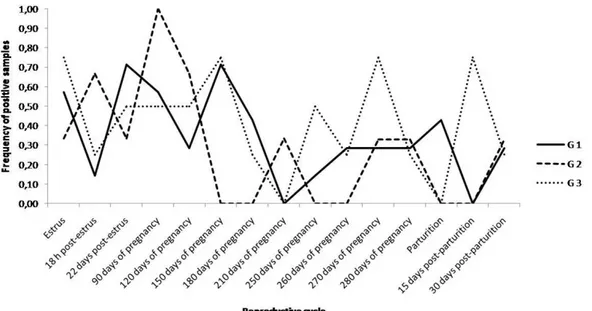
Considering the frequency of positive results with PCR for
detection of DNA of B. abortus B19 strain in urine samples,
over the total of urine samples collected during the whole
reproductive cycle studied it was observed a higher frequency
(50 to 54.2%) of positive samples from estrus to 150 days of
Pacheco, W.A. et al. B. abortus vaccine
days of pregnancy and then rising at parturition (41.1 to 53.1%)
(Tables 2 and 3). It means that B19 excretion occurred until
150 days of pregnancy and at post-parturition, with a
significant difference concerning the intermediary period for
the groups G1 (p = 0.024) and G2 (p = 0.034) (Tables 2 and 3
and Figure 1). G3 did not present statistical difference,
however, when the three group animals were analyzed, a
significant difference was observed (p = 0.0008).
Figure 1. Distribution of B19 strain excretion in urine detected by PCR during a reproductive cycle (estrus, pregnancy and
parturition) of cows vaccinated against brucellosis from 3 to 8 month age.
Table 1. Frequency of positive samples in Ring Test in milk and in PCR for detection of B. abortus B19 strain in milk and urine
from cows vaccinated from 3 to 8 month age.
Ring test Milk PCR Urine PCR
Groups No. of positive
samples/total %
No. of positive
samples/total %
No. of positive
samples/total %
G1 8/105 7.6 9/105 8.6 36/105 34.8
G2 1/45 2.2 1/45 2.2 13/45 28.8
G3 4/60 6.7 2/60 3.3 25/60 41.6
Total 13/210 6.2 12/210 5.7 74/210 35.2
Table 2. Frequency of positive urinary excretion of B. abortus B19 strain during a reproductive cycle, detected by PCR, in cows
vaccinated with B19 vaccine from 3 to 8 month age.
Pregnancy period (days) Post-parturition
period (days
Groups Estrus 18h
post-estrus
22o day post-estrus
90 120 150 180 210 250 260 270 280
Parturition
15 30
G1 4/7 1/7 5/7 4/7 2/7 5/7 3/7 0/7 1/7 2/7 2/7 2/7 3/7 0/7 2/7
G2 1/3 2/3 1/3 3/3 2/3 0/3 0/3 1/3 0/3 0/3 1/3 1/3 0/3 0/3 1/3
G3 3/4 1/4 2/4 2/4 2/4 3/4 1/4 0/4 2/4 1/4 3/4 1/4 0/4 3/4 1/4
Pacheco, W.A. et al. B. abortus vaccine
Table 3. Frequency of positive urinary excretion of B. abortus B19 strain, detected by PCR, according to the different phases of a
reproductive cycle in cows vaccinated with B19 vaccine from 3 to 8 month age.
Estrus to 150 days of pregnancy Between 180 and 280 days of
pregnancy Parturition
Groups
No. of positive
samples/total %
No. of positive
samples/total %
No. of positive
samples/total %
G1 21/42 50.0 13/49 26.5 23/56 41.1
G2 9/18 50.0 3/21 14.3 10/24 41.7
G3 13/24 54.2 8/28 28.6 17/32 53.1
DISCUSSION AND CONCLUSION
National Program for Control and Eradication of
Brucellosis and Tuberculosis (PNCEBT) in Brazil recommends
vaccination of all heifers from 3 to 8 months old with B19
vaccine. This instruction is essential since vaccination will
show similar responses and antibodies persistence will last for
a short period, that will interfere in the current serodiagnosis
for no longer than 18 months (10, 20).
Persistence and fluctuation of serum titers are related to
the capacity of the vaccinated animal to eliminate the vaccine
microorganism. Bacteriological studies in free brucellosis
herds revealed that the antibody titers persistence is due to B19
strain reactivation. The antibody titers variations with no
apparent cause can also occur, however, they are not enough to
interfere in the serological diagnosis. Concerning this
phenomenon, it is hypothesized that stress factors, due to
inadequate management practices, would be the reason for this
fluctuation (20).
All the 14 cows studied were serologically negative during
the monitoring period, and confirmed that it is a brucellosis
free herd. The PNCEBT recommends the use of RT, which
reveals IgA antibodies in milk adhered to the fat molecules, in
order to monitor dairy herds. Only 13 of 210 milk samples
responded positively to the RT throughout the experiment. G1
with three to four years old animals presented the highest
number of positive samples, but without any association with
age or even with the reproductive stage.
In this study, Brucella spp. or B. abortus B19 strain were
not isolated from urine and milk samples. The failure of
Brucella spp. isolation could be associated with freezing of
samples, which can interfere in the bacteria survival, and also
due to the sample contamination by microorganisms less
fastidious than Brucella, which would compete in the nutrient
utilization consuming them rapidly, thus releasing toxic
products from their metabolism that change pH of the medium
that could prevent Brucella survival (7, 9). The expressive
frequency of B. abortus (86/240; 20.5%) with specific B19
strain primers by Multiplex-PCR carried out in milk and urine
samples during the experiment (Table 2), confirms the capacity
of persistence of the vaccine strain during 9 years or along the
bovine female life even when they were vaccinated early in
life. In these conditions, the higher frequency of urinary
excretion (74/210; 35.2%) when compared to the milk (12/210,
5.7%), allows to conclude that the organism was viable and
able to multiply in the host not only in recognized sites as
spleen, mammary gland and lymph nodes, but possibly in the
urogenital system, mainly kidneys.
The concentration of circulating B.abortus B19 strain was
probably reduced to be achieved by bacteriological culture as
well as for inducing serological response along the life of the
vaccinated animal, thus it was not revealed by the usual
diagnostic tests, and these results agree with the observations
described by Manthei (1952) (16) and Meyer and Nelson
(1969) (19) that detected the short period of B19 persistence of
infection by means of microbiological culture. In this study,
PCR was a fundamental tool for this elucidation.
Pacheco, W.A. et al. B. abortus vaccine
has occurred during a whole reproductive cycle, from the estrus
synchronization, artificial insemination, pregnancy and
parturition of all animals. Although the mechanism of B19
strain infection is similar to that of B. abortus, it is not fully
understood which substances can stimulate its multiplication in
the host organism. During the estrous cycle, a sequence of
hormonal events results in greater resistance of the female
reproductive tract to injuries and/or contamination by
pathogens. If other substances act in order to enhance the
multiplication of Brucella spp., such as steroid hormones,
especially estradiol, since the great majority of abortions due to
brucellosis occurs in the last months of pregnancy (3, 21, 27);
in this experiment this has not been verified. The stradiol 17β
concentrations increases considerably during the last week of
pregnancy in cows, while progesterone concentrations slightly
decreases at the same period, and then abruptly decrease close
to the parturition; at this moment, a peak of the estrogen takes
place for a new ovulation (32). In case of estrogen to stimulate
the proliferation of B19 strain during the bovine female life,
the higher frequency of excretion should be limited to the
postpartum and the beginning of a new ovulatory cycle, during
the estrus, until about 40 days of pregnancy when the
placentation occurs, with production of progesterone.
It was observed that the higher frequency of positive
samples occurred from estrus (estrogen phase) to 150 days of
pregnancy (progesterone phase); decreasing between 180 and
280 days of pregnancy and then rising at parturition
(decreasing of progesterone followed by estrogen peak), so it
was not demonstrated a dependency on the hormonal period.
There was no difference among the three animal groups, so
there was no association with the animal age and the
reproductive cycle phase (Table 2).
The erythritol metabolism is the great difference between
virulent B. abortus and B19 strain (29). In the vaccinal strain,
ery gene (702 bp) is deleted, and this would be the explanation
for its sensitivity to erythritol, and make it unable to survive in
its presence. Erythritol is a polyalcohol found in the pregnant
uterus, which has its maximum concentration around 150 days
of pregnancy when achieve a stable level until the parturition
proximity (15).
Since B19 is sensitive to high erythritol concentration, this
strain disappears from circulation in this period, returning after
parturition (24). As exposed in Table 2, B19 excretion occurred
mainly until 150 days of pregnancy, with a marked decrease in
the period from 180 to 270 days of pregnancy, returning to
increase with the parturition proximity. The persistence of
some positive results in the intermediary period of the
reproductive cycle after 150 days of pregnancy until the
parturition can be due to different concentrations of circulating
erythritol. Some B19 strains can be tolerant to certain erythritol
concentrations and maybe this is one of the causes of persistent
infection (5, 31).
The impact of the presence of B19 strain in the
environment, the possibility of its transmission between cattle,
especially to bulls, or even to other animal species,
communicant and susceptible hosts including man, still need
more studies. Miyashiro et al. (2007) (20) reported the
detection of Brucella spp. DNA by multiplex- PCR in 37/192
fresh cheese; 30 were classified as been B19 strain, showing
the real risk to public health.
In the veterinary medicine practice, the B19 vaccine
handling requires very special attention to its zoonotic risk,
even during management of vaccinated animals that can be
potential renal carriers, as well as to the consumption of raw
milk and its sub-products provided from B19 vaccinated
animals.
REFERENCES
1. Acha, P.N.; Szyfres, B. (2001). Zoonosis y enfermedades transmisibles comunes al hombre y a los animales: bacteriosis y micosis. Organización Panamericana de la Salud, Washington.
Pacheco, W.A. et al. B. abortus vaccine
3. Anderson, T.D.; Meador, V.P.; Cheville, N.F. (1986). Pathogenesis of placentitis in the goat inoculated with Brucella abortus. II. Ultrastructural studies. Vet. Pathol. 23, 227–239.
4. Baily, G.G.; Krahn, J.B.; Drasar, B.W.; Stoker, N.G. (1992). Detection of Brucella melitensis and Brucella abortus by DNA amplification.J. Trop. Med. Hyg. 95, 271-275.
5. Bishop, G.C.; Bosman, P.P.; Herr, S. (1994). Bovine brucellosis. In:
Coetzer, J.A.N.; Homson, G.R.; Tustin, R.C. (eds). Infectious diseases of livestock. 2nd ed. A & M University Press, Austin, p. 1053-1066 6. Bricker BJ, Halling SM (1995) Enhancement of the Brucella AMOS
PCR assay for differentiation of Brucella abortus vaccine strains S19 and RB51. J Clin Microbiol 33:1640-1642
7. Carter, G.R.; Chengappa, M.M. (1991) Brucella. In: Carter, G.R.; Chengappa, M.M. (eds). Essentials of veterinary bacteriology and mycology. 4th ed.. Lea & Febiger, Philadelphia, p. 196-201
8. Chomczynski, P. (1993). A reagent for the single-step simultaneous isolation of RNA, DNA and proteins from cells and tissue samples.
Biotech. 15, 532-537.
9. Cortez, A.; Scarcelli, E.; Soares, R.M.; Heinemann, M.B.; Sakamoto, S.M.; Genovez, M.E.; Ferreira, F.; Richtzenhain, L.J. (2001). Detection of Brucella DNA from aborted bovine foetuses by polimerase chain reaction. Aust. Vet. J. 79, 500-501.
10. Costa, G.M.; Abreu, V.L.V.; Lobato, F.C.F.; Silva, J.A.; Martins, N.E. (1999). Avaliação do teste de imunodifusão mediante emprego do polissacarídeo “O” no diagnóstico da brucelose bovina. Arq. Bras. Med. Vet. Zootec. 51, 317-322.
11. Fekete, A.; Bantlet, J.A.; Halling, S.M. (1992). Detection of Brucella by polymerase chain reaction in bovine fetal and maternal tissues. J. Vet. Invest. 4, 79-83.
12. Gallien, P.; Dorn, C.; Alban, G.; Staal, C.; Protz, D. (1998). Detection of
Brucella species in organs of naturally infected cattle by polymerase chain reaction. Vet. Rec. 142, 512-514.
13. Genovez, M.E.; Scarcelli, E.; Rojas, S.; Giorgi, W.; Kaneto, C.N. (1993). Isolamentos bacterianos de fetos abortados bovinos examinados no Instituto Biológico de São Paulo, no período de 1985 a 1992. Braz. J. Vet. Res. Anim. Sci. 30, 107-112.
14. Holt, J.G.; Krieg, N.R.; Sneath, P.H.A.; Staley, J.T.; Williams, S.T. (1994). Bergey´s manual of determinative bacteriology. Williams & Wilkins, Baltimore.
15. Keppie, J.; Williams, A.E.; Witt, K.; Smith, H. (1964). The role of erytritol in the tissue localization of the brucellae. Br. J. Exp. Pathol. 46, 104-108.
16. Leal-Klevezas, D.S.; Martínez-Vázquez, I.O.; López-Mérino, A.; Martínez-Soriano, I.P. (1995). Single-step PCR for detection of Brucella
spp. from blood and milk of infected animals. J. Clin. Microbiol. 12, 3087-3090.
17. Manthei, C.A. (1952). Evaluation of vaccinal methods and doses of Brucella abortus strain 19. In: Proceedings of the 56th Annual Meeting of Livestock Sanitation Association, p. 115-125.
18. Matar, G.M.; Khneisser, I.A.; Abdelnioor, A.M. (1996). Rapid laboratory confirmation of human brucellosis by PCR analysis of a target sequence on the 31-killodalton Brucella antigen DNA. J. Clin. Microbiol. 34, 477-478.
19. Meyer, M.E.; Nelson, C.J. (1969). Persistence of Brucella abortus, strain 19 infection in immunized cattle. Proceedings - 73rd Annual Meeting of
the United States Animal Health Association 53, p. 159-165
20. Miyashiro, S.; Scarcelli, E.; Piatti, R.M.; Campos, F.R.; Vialta, A.; Keid, L.B.; Dias, R.A.; Genovez, M.E. (2007). Detection of Brucella abortus
DNA in illegal cheese from São Paulo and Minas Gerais and differentiation of B19 vaccinal strain by means of the polymerase chain reaction (PCR). Braz. J. Microbiol. 38, 17-22.
21. Neta, A.V.C.; Mol, J.P.S.; Xavier, M.N.; Paixão, T.A.; Lage, A.P.; Santos, R.L. (2010). Pathogenesis of bovine brucellosis, Vet. J. 184, 146–155.
22. Ornela Santos, P.P.; Araujo, R.F.; Reis, R.; Moreira, E.C.; Fiqueiredo, J.B.; Viana, F.C.; Barros, D.G.; Jardim, O.M. (1975). Brucelose bovina. I. Persistência da aglutinação pós-vacinal em bezerras de raças zebuínas vacinadas com amostra B19. Arq. Esc. Vet. UFMG 27, 363-373. 23. Paulin, L.M.; Ferreira Neto, J.S. (2003). A experiência brasileira no
combate à brucelose bovina. Funep, Jaboticabal.
24. Poester, F.P.; Gonçalves, V.S.P.; Lage, A.P. (2002). Brucellosis in Brazil. Vet. Microbiol. 90, 55-62.
25. Queipo-Ortuño, M.I.; Morata, P.; Ocón, P.; Manchado, P.; Comevero, J.D. (1997). Rapid diagnosis of human brucellosis by peripheral-blood PCR assay. J. Clin. Microbiol. 35, 2927-2930.
26. Romero, C.; Gamazo, C.; Pardo, M.; López-Goñi, I. (1995). Specific detection of Brucella DNA by PCR. J. Clin. Microbiol. 33, 615-617. 27. Samartino, L.E.; Traux, R.E.; Enright, F. (1994). Invasion and replication
of Brucella abortus in three different trophoblastic cell lines. Zent. fur Vet. B 41, 229–236.
28. Sambrook, J.; Fritsch, E.F.; Maniatis, T. (1989). Molecular cloning: a laboratory manual. Cold Spring Harbor Press, New York.
29. Sangari, F.J.; Agüero, J.; Garcia-Lobo, J.M. (2000). The genes for erythritol catabolism are organized as an inducible operon in Brucella abortus. Microbiol. 146, 487-495.
30. Sangari, F.J.; García-Lobo, J.M.; Agüero, J. (1994). The Brucella abortus vaccine strain B19 carries a deletion in the erythritol catabolic genes. FEMS Microbiol. Let. 121, 337-342.
Pacheco, W.A. et al. B. abortus vaccine
32. Senger, P.L. (2003). Placentation, the endocrinology of gestation and parturition. In: Senger, P.L. (ed). Pathways to pregnancy and parturition, 2nd ed. Current Conceptions, Washington, p. 304-325.
33. Zar, J.H. (1999). Biostatistical analysis. Prentice Hall, Upper Saddle River.