Coordinated Degradation of Replisome Components
Ensures Genome Stability upon Replication Stress in the
Absence of the Replication Fork Protection Complex
Laura C. Roseaulin1, Chiaki Noguchi1, Esteban Martinez1, Melissa A. Ziegler1, Takashi Toda2, Eishi Noguchi1*
1Department of Biochemistry and Molecular Biology, Drexel University College of Medicine, Philadelphia, Pennsylvania, United States of America,2Laboratory of Cell Regulation, Cancer Research UK, London Research Institute, Lincoln’s Inn Field Laboratories, London, United Kingdom
Abstract
The stabilization of the replisome complex is essential in order to achieve highly processive DNA replication and preserve genomic integrity. Conversely, it would also be advantageous for the cell to abrogate replisome functions to prevent inappropriate replication when fork progression is adversely perturbed. However, such mechanisms remain elusive. Here we report that replicative DNA polymerases and helicases, the major components of the replisome, are degraded in concert in the absence of Swi1, a subunit of the replication fork protection complex. In sharp contrast, ORC and PCNA, which are also required for DNA replication, were stably maintained. We demonstrate that this degradation of DNA polymerases and helicases is dependent on the ubiquitin-proteasome system, in which the SCFPof3ubiquitin ligase is involved. Consistently,
we show that Pof3 interacts with DNA polymerasee. Remarkably, forced accumulation of replisome components leads to abnormal DNA replication and mitotic catastrophes in the absence of Swi1. Swi1 is known to prevent fork collapse at natural replication block sites throughout the genome. Therefore, our results suggest that the cell elicits a program to degrade replisomes upon replication stress in the absence of Swi1. We also suggest that this program prevents inappropriate duplication of the genome, which in turn contributes to the preservation of genomic integrity.
Citation:Roseaulin LC, Noguchi C, Martinez E, Ziegler MA, Toda T, et al. (2013) Coordinated Degradation of Replisome Components Ensures Genome Stability upon Replication Stress in the Absence of the Replication Fork Protection Complex. PLoS Genet 9(1): e1003213. doi:10.1371/journal.pgen.1003213
Editor:Nick Rhind, University of Massachusetts Medical School, United States of America
ReceivedDecember 2, 2011;AcceptedNovember 15, 2012;PublishedJanuary 17, 2013
Copyright:ß2013 Roseaulin et al. This is an open-access article distributed under the terms of the Creative Commons Attribution License, which permits unrestricted use, distribution, and reproduction in any medium, provided the original author and source are credited.
Funding:This work was supported by NIH grant (GM077604 to EN) and the American Recovery and Reinvestment Act of 2009 (R01GM077604-S1 to EN). TT is supported by Cancer Research UK. The funders had no role in study design, data collection and analysis, decision to publish, or preparation of the manuscript.
Competing Interests:The authors have declared that no competing interests exist. * E-mail: [email protected]
Introduction
Initiation of DNA replication is directed by the formation of the pre-replication complex (pre-RC) at the origin of replication [1]. The pre-RC includes a number of essential replication proteins such as origin recognition complex (ORC), Cdc6, Cdt1, and the mini-chromosome maintenance (MCM) DNA helicase complex. However, to initiate actual DNA synthesis, additional factors are needed to facilitate the unwinding of origins and generation of replication forks. These factors include Cdc45, go-ichi-ni-san (GINS), replication protein A (RPA), proliferating cell nuclear antigen (PCNA), and other accessory factors prior to the loading of DNA polymerases. Together, these factors form the replisome complex at the replication fork [1]. However, how the cell maintains the integrity of the replisome is not well understood.
In response to replication stress, cells activate the DNA replication checkpoint to allow time for DNA repair. Central to this system are protein kinases such as human ATM and ATR, fission yeast Rad3, and budding yeast Mec1 [2–6]. These kinases are required for activation of downstream effector kinases by phosphorylation. In the fission yeast Schizosaccharomyces pombe, Rad3 activates Cds1 and Chk1 kinases in response to replication stress or DNA damage, facilitating DNA repair and recombination
multiple genome maintenance processes at the replication fork is not well understood.
Replication checkpoint studies have typically used chemical agents to stall replication forks. However, emerging evidence indicates that there are a number of chromosome regions that present obstacles to DNA replication. These include programmed fork blocking sites, DNA-binding proteins such as the transcription machinery, and DNA secondary structures caused by repeat sequences. These sites are considered to be difficult to replicate, causing arrest of replication forks or even fork breakage [30–35]. Fork arrest at difficult-to-replicate genome sites can promote both genome instability and stability depending on the circumstances. For example, polar fork pausing at rDNA loci stimulates recombination-dependent rDNA repeat expansion and contrac-tion, which can lead to rDNA instability. On the contrary, this polar fork pausing is also required to coordinate directionality of replication and transcription at rDNA loci, preventing genome instability due to head-on collisions of the replisome and transcriptional machinery [36,37]. Interestingly, studies found that FPC-related proteins are required for a number of fork arrest events, which are mediated by DNA–protein complexes. These include fork pausing at the rDNA loci, the fission yeast mating-type locus, tRNA loci, and highly transcribed RNA polymerase II genes [14,29,38–41]. At rDNA loci, loss of FPC causes hyper recombination, leading to contraction of rDNA repeats [17,40,42]. Similarly, the high rate of transcription and the presence of DNA-binding factors increase the chances of the replisome colliding with a transcription fork. Indeed, studies in fission yeast revealed that Swi1 is required to prevent DNA damage and hyper recombina-tion activity at these natural obstacles scattered throughout the genome [43–45].
In addition to these DNA–protein complex-mediated fork barriers, repeat DNA sequences themselves also cause genome instability in the absence of FPC-related proteins. At these sites, instead of promoting fork stalling, FPC appears to prevent or reduce the rate of fork stalling when the fork encounters DNA secondary structures caused by repeat sequences. Therefore, in the absence of FPC, fork stalling results in elevated levels of ssDNA
exposed at the replication fork, which appear to cause genome instability due to expansion and contraction at DNA structure-based impediments [46–51]. Thus, the mechanisms of the FPC-dependent fork regulation at repeat regions and at DNA–protein complex-mediated fork barriers are different. However, accumu-lated evidence indicates that FPC proteins are required for smooth passage of replication forks and for suppression of replication stresses at these natural impediments [14].
Therefore, in this study, we used swi1D as a model to understand replication stress response mechanisms. Strikingly, we have found that replicative DNA polymerases and helicases are highly unstable in the absence of Swi1. Our investigation revealed that this degradation is mediated by the ubiquitin-proteasome system, in which the SCFPof3(Skp1/Cul1/F-box) ubiquitin ligase complex is involved. In the absence of Pof3,swi1Dcells undergo mitotic catastrophes, suggesting the importance of proteasome-dependent replisome regulation in preserving genomic integrity. Considering that swi1D cells accumulate replication stress at difficult-to-replicate regions throughout the genome, our findings suggest that ubiquitin-dependent degradation of replisome com-ponents play a critical role in genome duplication in response to replication stresses.
It is widely understood that checkpoint proteins stabilize replication forks and replisomes in response to replication stress. However, our findings suggest an alternative mechanism that cells abrogate replisome functions when the fork encounters obstacles. Therefore, our study proves new mechanistic insights into the understanding of the replication stress response. In addition, although a number of studies have focused on the processes of replication initiation and regulation of fork progression, how the replisome itself is regulated is still largely unknown. Therefore, our findings also fill the knowledge gap in the regulation of replisome components in the DNA replication program.
Results
Replisome components are unstable inswi1Dcells Recent studies have shown that fork progression is impaired in the absence of FPC orthologues [24,27,28,38,52]. We also found a similar defect inS. pombe swi1Dcells (Figure S1), suggesting that FPC might regulate replisome stability. To test this possibility, we investigated the stability of various replication proteins in cells treated with cycloheximide (CHX), a compound that blocks the synthesis of new proteins and allows for the examination of protein stability. First, we examined the stability of the catalytic subunits of major essential replicative DNA polymerases. For this purpose, we employed S. pombe cells expressing Pol2-FLAG (the catalytic subunit of DNA polymerase e, required for leading-strand synthesis) [53] and Pol3-FLAG (the catalytic subunit of DNA polymerase d, required for lagging-strand synthesis) [54] from their genomic loci. Pol2-FLAG showed significant degradation in wild-type cells, whereas, Pol3-FLAG was relatively stable (Figure 1A and 1B). Intriguingly, Pol2 displayed even faster degradation when swi1 was deleted. In addition, Pol3 showed dramatic instability inswi1Dcells (Figure 1A and 1B). Next, we examined the stability of MCM helicase components.S. pombecells expressing Mcm2-GFP or Mcm6-GFP from their genomic loci were used, and Mcm4 was detected by the anti-Mcm4 antibody. In wild-type cells, Mcm2-GFP, Mcm4, and Mcm6-GFP were stable and did not undergo significant degradation throughout the CHX treatment (Figure 1C and 1D). In contrast, these helicase subunits were rapidly degraded inswi1Dcells (Figure 1C and 1D). To determine whether such degradation is specific to certain replication proteins, we also assessed the stability of Orc1 (an
Author Summary
Figure 1. Swi1 prevents degradation of DNA polymerases and helicases.Exponentially growing cells were treated with 0.1 mg/ml CHX at 25uC. (A) Cellular amounts of Pol2-FLAG and Pol3-FLAG were examined from 0 to 3 h of CHX treatment. The anti-FLAG M2 antibody was used to detect Pol2 and Pol3. Western blotting of tubulin was also performed as a loading control. (B) Stability of Pol2-FLAG and Pol3-FLAG shown inAwas quantified by NIH ImageJ. Relative intensity of protein bands at 0 h was set to 1 in each experiment. Error bars correspond to standard deviation of three independent experiments.wt, in blue;swi1D, in red. (C) Cellular amounts of Mcm2-GFP, Mcm6-GFP, Mcm4, Mrc1, Orc1-FLAG, and PCNA were determined from 0 to 4 h of CHX treatment. Anti-FLAG, anti-GFP, anti-Mcm4, anti-Mrc1, and anti-PCNA antibodies were used for Western blotting. (D) Stability of Mcm2-GFP, Mcm6-GFP, Mcm4, and Mrc1, shown inC, was quantified as described inB. Error bars represent average deviation (n= 2) or standard deviation (n= 3). (E) Replisome components in a chromatin-enriched fraction were degraded in response to CHX. Chromatin-free (Triton-soluble) and chromatin-enriched (Triton-in(Triton-soluble) fractions were prepared fromS. pombecells treated with CHX for 0 and 4 h. The fractions were analyzed by Western blotting using antibodies to detect the indicated proteins.
doi:10.1371/journal.pgen.1003213.g001
ORC subunit), Mrc1 (a mediator of S-phase checkpoints), and PCNA. Although the steady-state levels of Orc1-FLAG and PCNA before the addition of CHX were somewhat lower inswi1Dcells, their cellular amounts were maintained throughout the 4 h of CHX treatment in both wild-type andswi1Dcells (Figure 1C). As previously reported [55], Mrc1 was unstable and shows rapid degradation in the presence of CHX, although this degradation was not strengthened by the deletion ofswi1(Figure 1C and 1D). Thus, we concluded that Swi1 is involved in preventing rapid degradation of Pol2 and Pol3, as well as helicase components. Since Swi1 is involved in the suppression of fork collapse at difficult-to-replicate regions in fission yeast [43–45], it is possible that chromatin-bound replisome components are susceptible to degradation. Therefore, we fractionated cells into Triton-X-100-soluble (cytosol and nucleoplasm) and Triton-X-100-inTriton-X-100-soluble (enriched with chromatin- and nuclear matrix-bound proteins) fractions as described in previous studies (Figure 1E) [55,56]. Tubulin and histone H3 were exclusively fractionated into the Triton-soluble and Triton-insoluble fractions, respectively, indi-cating that fractionation was successful. Pol2 was mainly fractionated into the Triton-insoluble fraction, while approximate-ly 20% and 30% of Pol3 and Mcm4 were recovered into the Triton-insoluble fraction, respectively. Importantly, degradation of Pol2, Pol3 and Mcm4 was observed in the Triton-insoluble fraction when cells were treated with CHX, suggesting that the chromatin fraction of replisome components undergoes degrada-tion (Figure 1E). Therefore, our results are consistent with the notion that cells promote a fast turnover of replisome components bound to chromatin in response to the accumulation of fork collapse.
Swi1 protects the replisome components from proteasome-dependent degradation
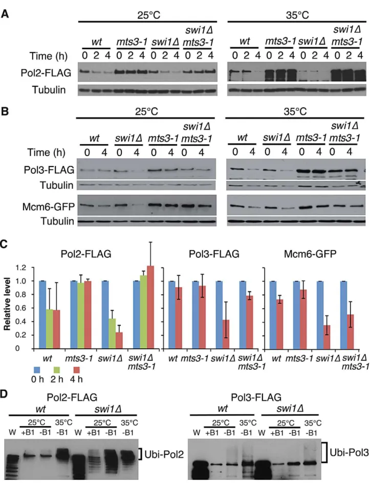
To understand the mechanisms of replisome degradation in response to unstable forks in the absence of Swi1, we determined whether the proteasome is responsible for degradation of DNA helicases and polymerases. Themts3-1temperature-sensitive allele, which has a mutation in a subunit of the 26S proteasome machinery, was used to inactivate the proteasome [57,58]. It is estimated that proteasome activity ofmts3-1cells is about 50% and 30% of the wild-type enzyme at 25uC and 35uC, respectively [58]. Cells were grown at 25uC or 35uC for 2 h, and then treated with CHX for 2 to 4 h. Strikingly, degradation of Pol2 was substantially inhibited inmts3-1andswi1Dmts3-1cells even at 25uC (Figure 2A and 2C). We observed similar stabilization of Pol3 and Mcm6 in
mts3-1 and swi1D mts3-1 cells (Figure 2B and 2C). At 35uC, degradation of these replisome components was accelerated both in wild type and swi1D cells probably due to increased cell metabolism (Figure 2A and 2B). However, degradation of these replisome components was abolished inmts3-1andswi1Dmts3-1
cells at 35uC (Figure 2A and 2B). Thus, our data indicate that Swi1 prevents proteasome-dependent degradation of replisome components.
Ubiquitin moieties (Ub) are conjugated to most of the proteins degraded by the proteasome [59,60]. Therefore, aforementioned data suggest that replisome core components (polymerases and helicases) are ubiquitinated. To test this possibility and further understand the mechanism of replisome degradation, we investi-gated whether replisome components were ubiquitinated. Cells harboring FLAG-tagged versions of Pol2 and Pol3 were engineered to express hexahistidine-fused ubiquitin (6xHis–Ub peptide) under the control of the thiamine (B1)-repressible nmt1
promoter. They were first cultured in the presence of thiamin (B1) to repress thenmt1 promoter and then grown in the absence of
thiamine for 22 h at 25uC, allowing cells to express 6xHis–Ub peptide. After the 22 h incubation, cultures were divided and further incubated at 25uC or 35uC for 2 h. Ubiquitinated proteins were purified with nickel agarose beads and analyzed by immunoblotting using antibodies against the FLAG-tag (Figure 2B). As shown in Figure 2D, Pol2-FLAG species with slower gel mobility were clearly detected in both wild type and
swi1Dcells, indicating that Pol2 is ubiquitinated. We also observed precipitation of non-ubiquitinated Pol2 with nickel agarose as previously reported for other proteins [61]. In addition, multiple Pol2 bands, which are probably products of degraded Pol2, were detected inswi1Dcells (Figure 2D), suggesting that Pol2 is more susceptible to degradation in the absence of Swi1. Similarly, ubiquitinated forms of Pol3-FLAG were detected in wild type and
swi1Dcells (Figure 2D). However, with our methods, we were not able to observe ubiquitinated forms of Mcm proteins (data not shown). Considering that Mcm proteins are stabilized inmts3-1
cells (Figure 2A), it is possible that the ubiquitination and degradation processes of Mcm proteins are too rapid to be detected.
Pol2 degradation occurs during S phase and is dependent on SCFPof3
Swi1 and its orthologues are involved in DNA replication, and their defects cause replication stress at difficult-to-replicate genome regions [14,15,18,21,22,24,25,27,28,43–45]. Thus, our results suggest that replisome core degradation occurs during S phase in the absence of Swi1. To test this possibility, wild type andswi1D
cells were synchronized at the G1/S boundary in the presence of 12 mM hydroxyurea (HU) and released into S phase in fresh medium supplemented with CHX. FACS analysis showed that the addition of CHX did not perturb cell cycle progression through S phase after the removal of HU (Figure 3A). There was no significant Pol2 degradation in both wild type andswi1Dcells in the absence of CHX. In contrast, the level of Pol2-FLAG dramatically dropped between 30 and 45 min after CHX addition in the absence of Swi1 (Figure 3B and 3C), when cells are in S phase (Figure 3A). In contrast, wild-type cells displayed only a mild decrease in the level of Pol2-FLAG (Figure 3B and 3C). We also used thecdc25-22temperature sensitive allele to synchronize cells at the G2/M boundary at the restrictive temperature (35uC), and cells were released into the cell cycle at permissive temperature (25uC). As determined by the increase of septation index, cells synchronously entered S phase after the release in the absence of CHX (Figure S2A). In this condition, Pol2 levels were maintained throughout the experiments in bothcdc25-22andcdc25-22 swi1D
cells (Figure S2B and S2C). When cells were treated with CHX, Pol2-FLAG levels gradually decreased incdc25-22 swi1Dcells but not incdc25-22cells (Figure S2B and S2C). This mild degradation is probably because cells were unable to synchronously progress through S phase in the presence of CHX (Figure S2A), although our data indicate that Pol2-FLAG is unstable in swi1D cells. Interestingly, Mcm4 showed rapid degradation ascdc25-22 swi1D
cells progress through S phase in the absence of CHX (Figure S2B and S2C), indicating that Mcm4 is degraded during replication. Mcm4 degradation incdc25-22 swi1Dcells was further exacerbated in the presence of CHX. In contrast, there is no significant Mcm4 degradation in cdc25-22 cells with or without CHX treatment (Figure S2B and S2C). Taken together, we concluded that degradation of replisome core components occurs during DNA replication in the absence of Swi1.
Skp1, a major component of SCF ubiquitin ligases inS. pombe[63]. Strikingly, Pol2-FLAG was significantly stabilized when skp1-94
cells were incubated at 35uC, indicating the involvement of SCF ubiquitin ligases in Pol2 degradation (Figure 3D; Figure S3B). SCF ligases contain F-box subunits, which are responsible for substrate specificity. Therefore, we examined Pol2 stability in a series of mutants defective for F-box proteins (Figure S3). Among the eleven F-box mutants we tested, we found that Pol2 becomes most stable in the absence of Pof3 (Figure 3E; Figure S3), an F-box protein that has been suggested to be involved in the preservation of genomic integrity [64]. Thus, our data suggest that Pol2 degradation is in part mediated by the SCFPof3ubiquitin ligase.
To further understand the mechanism of Pol2 degradation, we examined whether Pof3 interacts with Pol2, using immunoprecip-itation assays. Cells expressing Pol2-FLAG proteins were engi-neered to express Pof3-Myc from its genomic locus. As shown in Figure 3F, Pol2-FLAG coprecipitated with Pof3-Myc in the absence of Swi1, indicating that SCFPof3interacts with Pol2. The Pol2–Pof3 interaction was not detectable in wild-type cells even in the presence of a protein crosslinker dithio-bis succinimidyl propionate (DSP) (Figure 3F). Therefore, our data suggest that SCFPof3–Pol2 interaction is transient in wild-type cells but is enhanced when Pol2 degradation is accelerated in the absence of Swi1.
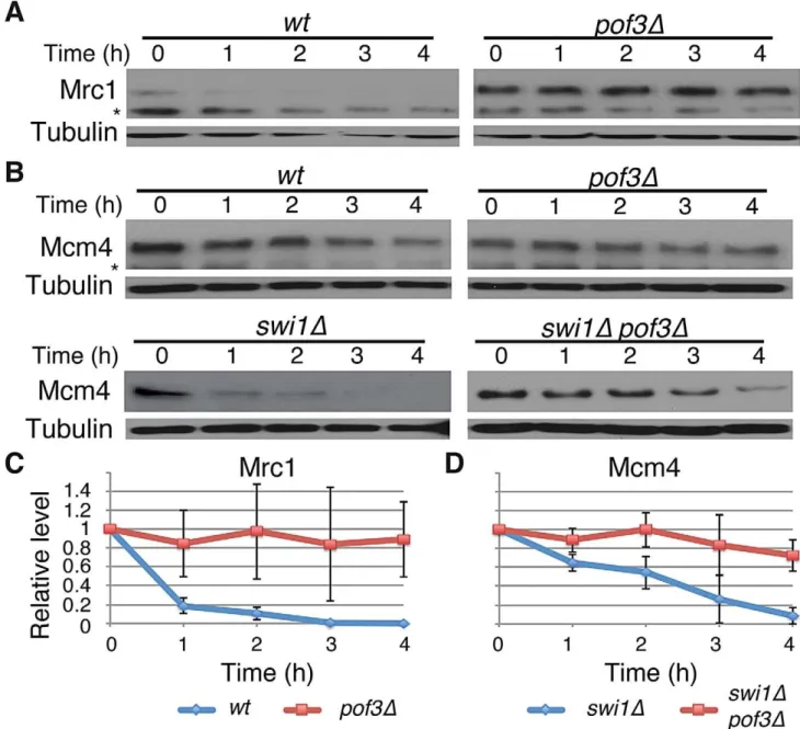
SCFPof3 is involved in degradation of Mcm4 and Mrc1 SCFPof3 has been shown to interact with fission yeast Mcl1, a DNA polymeraseaaccessory factor related to budding yeast Ctf4 [64–66]. Moreover, in budding yeast, Dia2 (Pof3-related protein) is recruited to the replication fork [67,68] and is involved in the ubiquitination of Mrc1 [69]. Therefore, SCFPof3-dependent Pol2 degradation suggests that SCFPof3may also target other replisome components for degradation. We first sought to determine whether Pof3 is also involved in degradation of Mrc1 inS. pombe. As shown in Figure 4, Mrc1 became highly stable inpof3Dcells under CHX treatment (Figure 4A and 4C). We then examined whether Mcm4 degradation in swi1D cells is inhibited by the inactivation of SCFPof3(Figure 4B and 4D). Intriguingly, Mcm4 was significantly more stable inpof3Dswi1Dcells than in swi1D cells after CHX treatment. This result suggests that SCFPof3also targets Mcm4 for proteasome-dependent degradation in response to replication stress provoked byswi1deletion. Taken together, our results are consistent with the notion that SCFPof3is involved in degradation of multiple replisome components.
Replisome degradation prevents mitotic catastrophes in
swi1Dcells
In order to understand the physiological importance of replisome core degradation in the absence of Swi1, we investigated cellular phenotypes of swi1D, pof3D and swi1D pof3D double mutant cells. For this purpose, cells were stained with DAPI to visualize nuclear DNA. As shown in Figure 5A and 5B,swi1Dand
pof3D cells displayed an increased level of mitotic catastrophes (including chromosome missegregation, aneuploidy, fragmented nuclei, hypercondensed nuclei, ‘‘cut’’ and other aberrant pheno-types, which are shown by arrows) compared to wild-type cells. Importantly, this phenotype was further exacerbated in swi1D
pof3D cells even in the absence of exogenous genotoxic agents (Figure 5A and 5B). We then used HU and camptothecin (CPT) to
introduce S phase specific genotoxic stress. HU depletes the dNTP pool and causes an arrest of replication fork progression, while CPT traps topoisomerase I on DNA and induces replication fork breakage. HU or CPT treatment further enhanced the aberrant mitotic phenotypes (Figure 5A and 5B). Consistently,swi1Dpof3D
cells were more sensitive to HU and CPT than either single mutant (Figure 5C). In the presence of HU or CPT,swi1Dcells accumulate DNA damage due to failure in the completion of DNA replication, which causes activation of the DNA damage checkpoint, leading to abnormal cell cycle arrest and a cell elongation phenotype [17,70]. As expected, HU or CPT treatment caused cell elongation inswi1D cells (Figure 5A). However, this elongation phenotype was abolished in swi1D pof3D cells (Figure 5A), suggesting that the stabilization of replisome components attenuated cell cycle arrest inswi1Dpof3Dcells. This attenuation of cell cycle arrest could have caused a growth advantage, leading to the rather weak increase in the HU and CPT sensitivity ofswi1Dpof3D cells (Figure 5C), although these cells show strong mitotic catastrophes (Figure 5A).
Next, we examined the ability of cells to recover DNA replication after CPT-dependent replication fork breakage. Exponentially growing cells (Log) were exposed to a low dose of CPT (5mM) for 3 h and returned to fresh medium for 2 and 4 h (Figure 5D). Chromosome samples were then analyzed by pulsed-field gel electrophoresis (PFGE), which permits only fully replicated chromosomes to migrate into the gel. In contrast, chromosomes with replication intermediates stay in the well of the gel, allowing us to determine the rate of replication recovery. There was no detectable DNA replication defect in wild-type cells throughout the experiment (Figure 5D, Log, CPT, 2 h), indicating that the low dose of CPT used in this assay did not cause major replication problems in wild-type cells. Although chromosomes fromswi1Dcells migrated into the gel immediately after the CPT exposure (CPT), we observed a marked reduction in chromosome intensity at 2 h after CPT treatment (Figure 5D). This result indicates that the low dose of CPT caused replication problems in
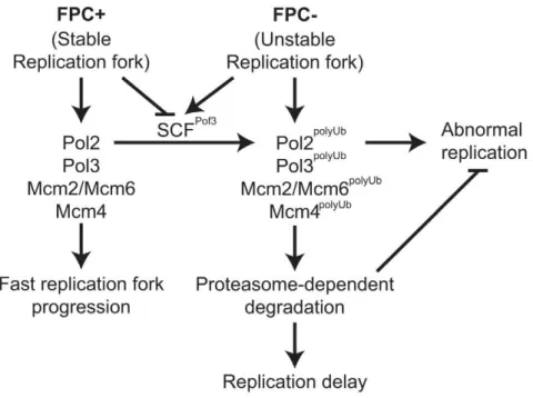
swi1D cells, which is consistent with previous studies [70]. However, there was a significant recovery at 4 h after the CPT removal due to the completion of DNA synthesis. A similar replication recovery was also observed inpof3Dcells. In contrast, there was no DNA replication recovery inswi1Dpof3Dcells during the course of the experiment (Figure 5D), indicating that these cells experience further difficulties in replication and/or repair of broken replication forks when treated with a replication-stressing agent. Interestingly, we repeatedly observed much less appearance of chromosomes I and II in the gel forswi1Dpof3Dcells (Figure 5D; Figure S4), suggesting that these cells experience major problems in DNA replication and chromosome maintenance. Consistently, there was an increased level of mitotic catastrophes in these cells (Figure 5A and 5B). Considering that pof3 deletion stabilizes replisome components (Figure 3E; Figure 4), our results suggest that programmed replisome degradation represents a mechanism to prevent catastrophic DNA replication in response to replication stress caused byswi1deletion (Figure 6). Similar replication and mitotic phenotypes were observed inswi1Dmts3-1cells, which are defective in proteasome functions (Figure S5), strengthening the idea that replisome degradation plays a critical role in the maintenance of genomic integrity.
Discussion
Accurate transmission of genetic information is one of the major tasks cells need to achieve in order to preserve the species and prevent genetic diseases. Accordingly, eukaryotic cells have
developed a variety of genome maintenance mechanisms. In response to DNA damage or replication stress, cells activate checkpoint pathways to coordinate cell cycle arrest with DNA repair activities. It is also known that the replication checkpoint functions to stabilize replication forks by preserving replisome and
Figure 3. Pol2 degradation occurs in S phase and is SCFPof3dependent.Exponentially growing cells (AS) were synchronized at the G
1/S (time zero) boundary in the presence of 12 mM HU for 3 h, and were released into fresh YES medium with or without CHX. Cells were collected and processed for DNA content analysis inA, and for Western blotting inB. (A) Cells were fixed at the indicated times, and DNA contents were analyzed by flow cytometry. (B) Pol2 is degraded during S phase inswi1mutants. Cellular amounts of Pol2-FLAG were determined at the indicated times. Tubulin levels were also monitored as a loading control. Representative results of repeat experiments are shown. (C) Stability of Pol2-FLAG during the CHX treatment shown inBwas quantified as described in Figure 1. For each strain, relative intensity of the Pol2-FLAG band at 0 h was set to 1. (D) Pol2 is stabilized in theskp1-94mutants. Exponentially growingskp1-94cells were treated with CHX at 25uC and 35uC. Cellular amounts of Pol2-FLAG were examined from 0 to 4 h of CHX treatment. (E)pof3deletion stabilizes Pol2. As inD, Pol2-FLAG levels were examined during the CHX treatment of wild-type andpof3Dcells. (F) Pof3 interacts with Pol2. Cells expressing the indicated fusion proteins (Pol2-FLAG and/or Pof3-Myc) were harvested in the presence or absence of DSP, and protein extracts were prepared. Pof3-Myc was immunoprecipitated, and associated proteins were probed with anti-FLAG antibody. Representative results of repeat experiments are shown. IP, immunoprecipitation; WB, Western blotting; WCE, whole cell extract. doi:10.1371/journal.pgen.1003213.g003
DNA structures. In this study, we have described an alternative cellular mechanism in response to replication stress. Our studies suggest that cells facilitate proteasome-dependent degradation of replisome components in response to replication stress to preserve genomic integrity.
Proteasome-dependent degradation of replisome components preserves genomic integrity
Swi1 and its orthologues are known to be involved in the stabilization of replication forks to prevent genetic instability during DNA replication. Genetic analyses have suggested that FPC is involved in coordinating leading- and lagging-strand DNA synthesis. It is also suggested that the FPC couples polymerase and
helicase activities at stalled forks [14,15]. Thus, the functions of Swi1 would become even more important to maintain the integrity of the replication fork when it encounters difficult-to-replicate sites or programmed fork pausing sites that are scattered throughout the genome. Consistently, FPC plays a critical role in programmed fork pausing and replication termination events near the mating-type (mat1) locus and at fork pausing sites in rDNA repeats and tRNA loci in yeast [16,17,21,29,38,39]. Importantly, recent studies indicated thatswi1Dcells experience fork collapse at these difficult-to-replicate regions [43–45]. Therefore, inactivation of Swi1 causes defects in replication fork stabilization at natural impediments, leading to general replication stress at the replication fork.
Figure 4. Pof3-dependent degradation of Mrc1 and Mcm4.(A) Cellular amount of Mrc1 was determined inwtandpof3Dcells, from 0 to 4 h of CHX treatment. Western blotting of tubulin was performed as a loading control. (B) As inA, cellular amount of Mcm4 was determined inwt,pof3D, swi1D, andpof3Dswi1Dcells. The asterisks indicate non-specific bands. (C) Stability of Mrc1 during the CHX treatment shown inAwas quantified as described in Figure 1. For each strain, relative intensity of the Mrc1 band at 0 h was set to 1. (D) Stability of Mcm4 inswi1Dandpof3Dswi1Dshown in Bwas quantified as described inC. Samples for Mrc1 or Mcm4 blots were derived from the same experiment and processed in parallel. Representative results of repeat experiments are shown.
It is well known that Cdt1 and Cdc6 undergo rapid proteasome-dependent degradation to restrict replication licens-ing once per cell cycle [71,72]. However, how replisome degradation contributes to DNA replication process is largely unknown. In this report, we show that DNA polymerases and helicases undergo rapid degradation upon replication stress in the absence of Swi1 (Figure 1). This degradation is dependent on the ubiquitin-proteasome system (Figure 2). In the absence of Swi1, cells experience unstable replication forks that lead to an increased level of replication-dependent DNA damage and hyper-recombination [16,17,43]. Such replication stress appears to cause replisome degradation in order to prevent abnormal DNA replication and mitotic catastrophes (Figure 5; Figure S5). These results suggest that replisome degradation functions to maintain genomic integrity during DNA replication in response to replication stress (Figure 6). Similar mechanisms have been described in the transcription-coupled DNA repair (TCR), which is activated by transcription blockage in response to genotoxic agents [73,74]. In this mechanism, the Cockayne syndrome B protein (budding yeast Rad26) interacts with Def1 to regulate
ubiquitination of Rpb1, the large subunit of RNA polymerase II (RNAPII), which results in proteasome-dependent degradation of RNAPII [75,76]. Ubiquitination of Rbp1 is achieved by the Rsp5/Nedd4 ubiquitin ligase, which promotes DNA-damage induced degradation of RNAPII in budding yeast and human cells [77–79]. RNAPII degradation appears to be an alternative mechanism to TCR. Studies indicate that the loss of both TCR and RNAPII degradation pathways renders cells hypersensitive to DNA damage, thus Def1 promotes proteolysis of RNAPII when the lesion cannot be rapidly repaired by TCR [75,80–82]. Therefore, analogous to the DNA damage-induced RNAPII degradation pathway, our present findings suggest that the cell elicits a replisome degradation program when the replication fork is adversely blocked. We speculate that, depending on the degree of replication problems, re-building and re-loading new repli-somes might be advantageous to the cell, rather than re-using existing replisome components that are compromised. Therefore, we suggest that replisome degradation is an alternative mecha-nism to replisome stabilization and prevents DNA synthesis by compromised replisomes.
Figure 5. Forced accumulation of replisome components in swi1D cells causes catastrophic DNA replication and mitotic abnormalities.(A)swi1Dpof3Dcells have increased levels of mitotic catastrophes. Exponentially growing cells were treated with or without the indicated drugs (12 mM HU or 20mM CPT for 6 h), fixed in ethanol, and stained with 49,6-diamidino-2-phenylindole (DAPI). Representative images of observed nuclear phenotypes are shown. Representative mitotic failures are shown by arrows. Arrows were omitted from the images ofswi1Dpof3D
cells because a large numbers of cells showed mitotic catastrophes. The scale bar represents 10mM. (B) Quantification of cells with defective mitosis including chromosome missegregation, aneuploidy, cut and other aberrant phenotypes. More than 200 cells were counted for each strain. Error bars correspond to standard deviations obtained from three experiments. (C) DNA damage sensitivity ofswi1Dmutants is increased bypof3deletion. Five-fold serial dilutions of cells were incubated on YES agar medium supplemented with the indicated drugs (2 mM HU or 1mM CPT) for 2 to 3 days at 32uC. (D)pof3deletion exacerbates replication recovery defects ofswi1Dmutants. Exponentially growing cells (Log) were incubated in the presence of 5mM CPT for 3 h at 30uC (CPT), then washed and returned into fresh medium for 2 h or 4 h (2 h, 4 h). Chromosome samples were examined by PFGE. Representative results of repeat experiments are shown.swi1Dcells have shorter chromosome III due to hyper recombination at rDNA repeats [42,70,101].
doi:10.1371/journal.pgen.1003213.g005
Roles of Pol2 degradation in replisome dynamics We also found that Pol2 (Pole) is significantly unstable even in wild-type cells (Figure 1A and 1B), while Pol3 (Pold) is relatively stable (Figure 1A and 1B). The high rate of Pol2 turnover may suggest that Pol2 needs to be re-loaded during leading-strand synthesis. Since Pol2 is suggested to work continuously on the leading-strand [53], one might think that the high turnover of Pol2 poses a disadvantage to the cells. However, it is possible that the polymerases fall off the chromatin every time the fork arrives at programmed pausing sites or difficult-to-replicate regions. In addition, Pol2 may undergo degradation once it falls off the chromatin. Such a degradation mechanism would also be advantageous for the cell to refresh Pol2 enzymes by efficiently reloading newly synthesized Pol2 at the moving replication fork. On the other hand, the discontinuous nature of Pol3-dependent lagging-strand synthesis would be sufficient to keep Pol3 refreshed at the fork in order to avoid replication-dependent errors or mutations. Another possibility is that this mechanism might simply maintain the coupling of leading- and lagging-strand synthesis. Thus, in addition to the role of replisome degradation in preventing genomic instability described above, polymerase degradation may function to eliminate non-functional replisomes and serve as a mechanism to maintain active DNA polymerases at the replication fork.
Our investigation also revealed that Pol2 and Mcm4 undergo rapid degradation in the presence of CPT (Figure S6), which breaks replication forks. However, in this condition, the Mcm6 level was maintained (Figure S6), although it was highly unstable inswi1Dcells (Figure 1C and D). It is possible that some replisome components remain stable on the chromatin in the presence of CPT. Interestingly, Trenz et al. reported that polymerases fall off the chromatin in response to CPT, whereas Mcm7 persists [83]. Therefore, swi1 deletion generates a situation distinct from a simple mechanical breakage of the fork caused by DNA damaging agents, where the replisome cannot continue replicating DNA. Importantly, Swi1 functions as an ancillary component of the replisome by interacting with various replisome components, coupling polymerase and helicase activities and coordinating semi-discontinuous DNA synthesis [14,15]. It is also reported that Swi1 protects replication forks at difficult-to-replicate sites [43–45]. Therefore, we suggest that the loss of Swi1 results in unstable replisome structures at the moving replication fork during ongoing DNA synthesis, allowing us to examine replisome degradation pathways during DNA replication.
The FPC–dependent stabilization of replisome components
The FPC moves with the replication fork and interacts with replisome components [16,18,21–25,27,28,84–87]. Surprisingly, Pol2, Pol3, and MCM subunits are rapidly degraded inswi1Dcells (Figure 1). Consistently, replication fork progression is compro-mised in FPC deficient cells (Figure S1) [27,52]. These results suggest that Swi1 prevents degradation of replisome components to maintain efficient progression of replication forks. In wild-type cells, multiple activities required for DNA synthesis are coupled to form a large replisome complex, resulting in efficient progression of the replication fork. However, in the absence of Swi1, DNA replication-related activities are probably uncoupled especially at naturally difficult-to-replicate regions. This uncoupling generates unstable replisome structures, which may expose degradation signals of various replisome components to a ubiquitin ligase(s) associated with the replication fork. Importantly, swi1D pof3D
double mutants showed catastrophic DNA replication and mitosis, suggesting that Pof3-dependent degradation of replisome
compo-nents prevents genomic instability. However, we cannot exclude the possibility that mitotic catastrophe phenotypes are caused by stabilization of other Pof3 targets. For example, Pof3-dependent proteolysis of Ams2 is responsible for cell cycle-dependent transcriptional activation of core histone genes inS. pombe[88]. Indeed, defects in Ams2 degradation leads to accumulation of histones and alteration of centromere structures [88]. Such dysregulation of histone homeostasis during S phase could also lead to abnormal DNA replication, leading to mitotic problems. However, Dia2, a Pof3-related F-box protein, is associated with the replisome and regulates replication forks in budding yeast. Dia2 is involved in ubiquitination of budding yeast Mrc1, which is a component of the replisome [67–69]. Moreover, Tof1 (Swi1 orthologue) collaborates with Dia2 to maintain genomic integrity [89]. These findings suggest that Pof3/Dia2 acts as a part of the replisome. Consistently, we found in fission yeast that SCFPof3is largely responsible for degradation of some replisome components (Figure 3 and Figure 4). Therefore, Pof3-mediated ubiquitination of replisome components may be prevented by Swi1-dependent replisome stabilization, which may mask potential degradation signals of multiple replisome components (Figure 6). Since many SCF ubiquitin ligases are known to recognize phosphorylated degradation signals (phospho-degrons), it is also possible that replisome components undergo phosphorylation in the absence of Swi1. Therefore, Swi1 might have direct functions in inhibiting SCFPof3 ligase possibly by inhibiting Pof3 or inhibiting potential kinases. In this regard, it is interesting to note that Mrc1 contains a potential phospho-degron, and that the Hsk1 kinase is required for efficient degradation of Mrc1 [55]. Consistently, our present study shows that Pof3 is involved in Mrc1 degradation (Figure 4). Therefore, it is possible that Hsk1-dependent phosphorylation creates Pof3-targeted phospho-degrons on multiple replisome components. However, Mrc1 degradation is independent of replication stress (Figure 1C), raising the possibility that other kinases are responsible for replisome degradation upon replication stress. Further investigation of proteasome-dependent replisome degradation would identify detailed pathways in the regulation of the replisome.
Materials and Methods
General techniques
The methods used for genetic and biochemical analyses of fission yeast have been described previously [90,91]. Drug sensitivity assays, Western blotting, pulsed-field gel electrophoresis (PFGE) and 49,6-diamidino-2-phenylindole (DAPI) staining of nuclear DNA were performed as described [70,92]. Flow cytometry of DNA content has been described [93,94].
S. pombestrains
S. pombe strains used in this study were constructed using standard techniques [91], and their genotypes and sources are listed in Table S1.swi1D (swi1::hphMX6 and swi1::natMX6) and
pof3D(pof3::ura4MX6) were generated by a two-step PCR method [95], to replaceswi1andpof3open reading frames with selection marker genes. The two-step PCR method was also used to construct a GFP or 13Myc tag at the C terminus ofmcm2,mcm6
and pof3, generating mcm2-GFP:hphMX6 (mcm2-GFP), mcm6-GFP:hphMX6 (mcm6-GFP), and pof3-13Myc:hphMX6 (pof3-13Myc), respectively. Oligonucleotide primers used in the two-step PCR method described above are listed in Table S2. A temperature-sensitive skp1-94 mutation was isolated using error-prone PCR methods [63].
Mutations and epitope-tagged genes have been described for
orc1-5FLAG [96], pol2-5FLAG, pol3-5FLAG [97], swi1::kanMX6
[17],cdc45-5FLAG[98],cdc25-22[99] andmts3-1[57].
mcm2-GFP, mcm6-GFP, pof3-13MYC, orc1-5FLAG, pol2-5FLAG,
pol3-5FLAG, andcdc45-5FLAGcells show normal growth pheno-type and were not abnormally sensitive to HU, CPT and MMS, indicating that the tagged version of these proteins are functional.
Cell extract preparation for Western blotting
To examine protein stability, exponentially growing cells were treated with 0.1 mg/ml of cycloheximide (CHX) for the indicated times and collected. Whole-cell extracts were prepared as described [100]. Briefly, cells were washed in STOP buffer (150 mM NaCl, 50 mM NaF, 10 mM EDTA, and 1 mM NaN3) and lysed by glass beads in lysis buffer U (50 mM Tris-HCl pH 6.8, 2% SDS, 2 mM EDTA, 10% glycerol, and 4 M urea) using a FastPrep Cell disruptor (Qbiogene, Irvine, CA) for 40 seconds at speed 6. Protein extract was clarified by centrifugation at 13,000 rpm in an Eppendorf microcentrifuge 5415R for 10 min at 4uC, and the protein concentration was determined using BCA protein Assay Reagent (Thermo Fisher Scientific, Waltham, MA). Immediately after the protein concentration assay, protein extracts were boiled in the presence of 5% beta-mercaptoethanol and stored at220uC. For immunoblotting, Myc, GFP, and FLAG fusion proteins were probed with the anti-c-Myc 9E10 antibody (Covance, Princeton, NJ), anti-GFP antibody (Roche, Indianapolis, IN), and anti-FLAG M2 (Sigma-Aldrich) antibody, respectively. The anti-tubulin TAT-1 (gift from Dr. K. Gull), anti-Mcm4 (gift from Drs. S. Kearsey, Z. Lygerou, and H. Nishitani), anti-Mrc1 (gift from Dr. K. Tanaka), and anti-PCNA (gift from Dr. T. Tsurimoto) antibodies were used to detect the corresponding proteins. Quantification of protein bands was performed using NIH ImageJ software.
Fractionation of cells into soluble and chromatin-enriched fractions
Cell fractionation was performed as described elsewhere [55,56] with modifications. Exponentially growing cells were harvested in 0.01% sodium azide by centrifugation and washed sequentially with STOP buffer, water, and 1.2 M sorbitol, at 4uC. Cells were resuspended in CB1 buffer (50 mM sodium citrate, 40 mM EDTA, 1.2 M sorbitol) and treated with 2.5 mg/ml of Zymolyase for approximately 20 min at 32uC. When cell lysis reached approxi-mately 95%, cell wall digestion by Zymolyase was terminated by adding equal volume of ice-cold CB2 buffer (1.2 M sorbitol, 10 mM Tris-HCl ph7.5), and resulting spheroplasts were washed twice with 1.2 M Sorbitol. Spheroplasts were then incubated in Lysis buffer T (50 mM potassium acetate, 2 mM MgCl2, 20 mM HEPES-KOH pH 7.4, 10 mM EDTA, 1 M Sorbitol, 1% Triton X-100) supple-mented with Halt protease inhibitor cocktail (Thermo Fisher Scientific) for 10 min at 4uC. Subsequently, extracts were fraction-ated into soluble and pellet fractions by centrifugation for 10 min at 4uC. Supernatants (Triton X-100-soluble fraction) were removed, boiled with a one-third volume of 36SDS-PAGE loading buffer (150 mM Tris-HCl pH 6.8, 6% SDS, 6 mM EDTA, 30% glycerol, 15% beta-mercaptoethanol), and stored at 220uC. The pellets (Triton X-100-insoluble fraction) were washed once with Lysis buffer (without Triton X-100), suspended in Lysis buffer, boiled with a one-third volume of 36SDS-PAGE loading buffer, and stored at220uC.
Immunoprecipitation and detection of ubiquitinated proteins
Immunoprecipitation was performed using the anti-myc 9E10 (Covance) antibody with protein G sepharose beads as described
[70]. Proteins associated with the anti-myc antibody were analyzed by Western blotting. For detection of ubiquitinated protein, S. pombecells expressing a hexahistidine-ubiquitin (6xHis-Ub) peptide [61] were lysed in lysis buffer G (6 M guanidine hydrochloride, 100 mM sodium phosphate pH 8.0, and 50 mM Tris-HCl pH 8.0). Hexahistidine-ubiquitinated proteins were purified with Ni-NTA agarose beads (Qiagen, Valencia, CA), eluted in the presence of 4 M urea, and analyzed by Western blotting.
Supporting Information
Figure S1 Replisome progression is delayed in the absence of Swi1. (A) Diagram of the replication origin 2004 (ori2004) region used in chromatin immunoprecipitation. Protein association was monitored atori2004and a position 30 kb away fromori2004as described [16,97]. (B) Septation index. cdc25-22 cells were synchronized at the G2/M boundary at 35uC for 3 hours and released at 25uC. The septation index was determined to monitor cell cycle progression.swi1D(green) cells have a 20 min delay in the increase of septation index when compared to wild-type (swi1+
; red) cells. InS. pombe, an increase in septation index coincides with the onset of S-phase [91]. In order to remove this difference in our analysis, we set the point of septation increase (40 min inswi1+and
60 min inswi1D) to 0 (S) min as the onset of S-phase inCandD. (C,D) Relative enrichment of replication proteins at theori2004
region during 120 min from the onset of S-phase (0 (S) min). (C) Association of Cdc45-FLAG with chromatin was monitored at
ori2004(blue) and at a position 30 kb (green) away fromori2004, to evaluate their translocation through theori2004region in wild-type (top panel) andswi1Dcells (bottom panel). (D) As inC, chromatin recruitment of Pol2-FLAG was monitored through the ori2004
region in wild-type (top panel) and swi1D cells (bottom panel). Representative results of repeat experiments are shown.
(EPS)
Figure S2 Stability of Pol2 and Mcm4 during the cell cycle.
cdc25-22 cells were synchronized at the G2/M boundary by incubation at 35uC for 3 h and then released into fresh YES medium with or without CHX at 25uC. (A) An increase in the septation index indicates the onset of S-phase. Cells entered S phase synchronously in the absence of CHX. However CHX affected cell cycle progression after the release from G2/M. (B) Cellular amounts of Pol2-FLAG and Mcm4 were determined at the indicated times after the release from G2/M. (C) Stability of Pol2-FLAG and Mcm4 shown in B was quantified as described in Figure 1. Pol2-FLAG was unstable inswi1Dcells in response to CHX. Mcm4 showed rapid degradation during S-phase with or without CHX.
(EPS)
Figure S3 Quantification of Pol2-FLAG stability in F-box mutants. (A) Pol2 degradation was examined in wild-type (wt) and eleven F-box mutants (pof). Cells of the indicated genotypes were incubated in YES supplemented with 0.1 mg/ml of CHX for the indicated times at 30uC. The anti-FLAG (M2) antibody was used for Western blotting. Tubulin levels were also monitored as a loading control. (B) Relative Pol2-FLAG amounts at 0 and 4 h of CHX treatment shown inAwere quantified using EZQuant-Gel 2.1. Relative intensity of protein bands at 0 h was set to 1 in each experiment. Relative Pol2-FLAG levels inskp1-94cells at 25 and 35uC shown in (Figure 3D) were also quantified.
(EPS)
chromosomes I and II are much lower than that of chromosome III.
(EPS)
Figure S5 Proteasome defect provokes mitotic catastrophes and replication defects in swi1D cells. (A) Mitotic phenotypes of wt,
swi1D,mts3-1, and swi1Dmts3-1 cells with or without genotoxic agents were investigated. Exponentially growing cells were shifted to 30uC for 3 h and fixed in ethanol and stained with DAPI.swi1D
mts3-1cells undergo mitotic catastrophes. Quantification of cells with defective chromosome segregation was performed. More than 300 cells were counted at each time point. (B) Representative images of observed nuclear phenotypes inAare shown. The scale bar represents 10mm. (C) DNA damage sensitivity of swi1D
mutant is increased bymts3-1mutation. Five-fold serial dilutions of cells were incubated on YES agar medium supplemented with the indicated amounts of HU and CPT for 4 to 5 days at 25uC. (D) The checkpoint-dependent cell elongation phenotype ofswi1Dis abolished by mts3-1 mutation. Cells of the indicated genotypes were incubated on YES agar medium containing 5 mM HU or 5mM CPT for 2 days at 25uC and photographed. The scale bar represents 10mm. (E) mts3-1 mutation exacerbates replication recovery defects of swi1D mutants. Cells were incubated in the presence of 10mM CPT for 4 h at 30uC, then washed and returned into fresh medium. Chromosome samples were examined by PFGE. Representative results of repeat experiments are shown. The size of chromosome III (ch.3) varies betweenS. pombestrains due to recombination at rDNA repeats. (F) Quantification of DNA replication recovery shown inE. Chromosome band intensity was quantified using EZQuant-Gel 2.1. Average intensities of the three chromosomes are shown. Error bars correspond to standard deviations obtained from the band intensities of the three
chromosomes. Relative average intensity of chromosome bands of mid-log-phase cells (Log) was set to 1 in each cell line. (EPS)
Figure S6 Stability of Pol2 and Mcm subunits in the presence of camptothecin. CPT causes degradation of Pol2 and Mcm4, but not of Mcm6. Thirty minutes before CHX treatment, cells were exposed to 10mM CPT. Cellular levels of Pol2-Flag, Mcm6-GFP and Mcm4 were determined at the indicated times after CHX treatment. Tubulin levels were also monitored as a loading control. Representative results of repeat experiments are shown. (EPS)
Table S1 S. pombestrains used in this study. (DOCX)
Table S2 Primers used in this study. (DOCX)
Acknowledgments
We thank members of the Noguchi laboratory and Drs. Benoit Arcangioli, Antony Carr, Alexander Mazin, Mauricio Reginato, and Mitsuhiro Yanagida for their support, comments, and encouragement. We thank Drs. Keith Gull, Zoi Lygerou, Stephen Kearsey, Hisao Masukata, Hideo Nishitani, Katsunori Tanaka, and Toshiki Tsurimoto for antibodies; Alan Lehmann for plasmids; Paul Russell forS. pombestrains.
Author Contributions
Conceived and designed the experiments: LCR EN. Performed the experiments: LCR EN CN EM MAZ. Analyzed the data: LCR EN TT. Contributed reagents/materials/analysis tools: LCR EN CN EM MAZ TT. Wrote the paper: LCR EN.
References
1. Bell SP, Dutta A (2002) DNA replication in eukaryotic cells. Annu Rev Biochem 71: 333–374.
2. Boddy MN, Russell P (2001) DNA replication checkpoint. Curr Biol 11: R953– R956.
3. Osborn AJ, Elledge SJ, Zou L (2002) Checking on the fork: the DNA-replication stress-response pathway. Trends Cell Biol 12: 509–516. 4. Nyberg KA, Michelson RJ, Putnam CW, Weinert TA (2002) TOWARD
MAINTAINING THE GENOME: DNA Damage and Replication Check-points. Annu Rev Genet 36: 617–656.
5. Abraham RT (2001) Cell cycle checkpoint signaling through the ATM and ATR kinases. Genes Dev 15: 2177–2196.
6. Rouse J, Jackson SP (2002) Interfaces between the detection, signaling, and repair of DNA damage. Science 297: 547–551.
7. Carr AM (2002) DNA structure dependent checkpoints as regulators of DNA repair. DNA Repair (Amst) 1: 983–994.
8. Lopes M, Cotta-Ramusino C, Pellicioli A, Liberi G, Plevani P, et al. (2001) The DNA replication checkpoint response stabilizes stalled replication forks. Nature 412: 557–561.
9. Paciotti V, Clerici M, Scotti M, Lucchini G, Longhese MP (2001) Characterization ofmec1kinase-deficient mutants and of new hypomorphic
mec1alleles impairing subsets of the DNA damage response pathway. Mol Cell Biol 21: 3913–3925.
10. Sogo JM, Lopes M, Foiani M (2002) Fork reversal and ssDNA accumulation at stalled replication forks owing to checkpoint defects. Science 297: 599–602. 11. Tercero JA, Diffley JF (2001) Regulation of DNA replication fork progression
through damaged DNA by the Mec1/Rad53 checkpoint. Nature 412: 553– 557.
12. Tercero JA, Longhese MP, Diffley JF (2003) A central role for DNA replication forks in checkpoint activation and response. Mol Cell 11: 1323–1336. 13. Lee BS, Grewal SI, Klar AJ (2004) Biochemical interactions between proteins
andmat1cis-acting sequences required for imprinting in fission yeast. Mol Cell Biol 24: 9813–9822.
14. Leman AR, Noguchi E (2012) Local and global functions of Timeless and Tipin in replication fork protection. Cell Cycle 11: 3945–3955.
15. McFarlane RJ, Mian S, Dalgaard JZ (2010) The many facets of the Tim-Tipin protein families’ roles in chromosome biology. Cell Cycle 9: 700–705. 16. Noguchi E, Noguchi C, McDonald WH, Yates JR, 3rd, Russell P (2004) Swi1
and Swi3 are components of a replication fork protection complex in fission yeast. Mol Cell Biol 24: 8342–8355.
17. Noguchi E, Noguchi C, Du LL, Russell P (2003) Swi1 prevents replication fork collapse and controls checkpoint kinase Cds1. Mol Cell Biol 23: 7861– 7874.
18. Gotter AL, Suppa C, Emanuel BS (2007) Mammalian TIMELESS and Tipin are evolutionarily conserved replication fork-associated factors. J Mol Biol 366: 36–52.
19. Noguchi E (2010) The DNA Replication Checkpoint and Preserving Genomic Integrity During DNA Synthesis. Nature Education 3: 46.
20. Sabatinos SA (2010) Replication Fork Stalling and the Fork Protection Complex. Nature Education 3: 40.
21. Calzada A, Hodgson B, Kanemaki M, Bueno A, Labib K (2005) Molecular anatomy and regulation of a stable replisome at a paused eukaryotic DNA replication fork. Genes Dev 19: 1905–1919.
22. Chou DM, Elledge SJ (2006) Tipin and Timeless form a mutually protective complex required for genotoxic stress resistance and checkpoint function. Proc Natl Acad Sci U S A 103: 18143–18147.
23. Errico A, Costanzo V, Hunt T (2007) Tipin is required for stalled replication forks to resume DNA replication after removal of aphidicolin inXenopusegg extracts. Proc Natl Acad Sci U S A 104: 14929–14934.
24. Katou Y, Kanoh Y, Bando M, Noguchi H, Tanaka H, et al. (2003) S-phase checkpoint proteins Tof1 and Mrc1 form a stable replication-pausing complex. Nature 424: 1078–1083.
25. Leman AR, Noguchi C, Lee CY, Noguchi E (2010) Human Timeless and Tipin stabilize replication forks and facilitate sister-chromatid cohesion. J Cell Sci 123: 660–670.
26. Tanaka H, Kubota Y, Tsujimura T, Kumano M, Masai H, et al. (2009) Replisome progression complex links DNA replication to sister chromatid cohesion inXenopusegg extracts. Genes Cells 14: 949–963.
27. Unsal-Kacmaz K, Chastain PD, Qu P-P, Minoo P, Cordeiro-Stone M, et al. (2007) The Human Tim/Tipin Complex Coordinates an Intra-S Checkpoint Response to UV That Slows Replication Fork Displacement. Mol Cell Biol 27: 3131–3142.
28. Yoshizawa-Sugata N, Masai H (2007) Human Tim/Timeless-interacting protein, Tipin, is required for efficient progression of S phase and DNA replication checkpoint. J Biol Chem 282: 2729–2740.
29. Dalgaard JZ, Klar AJ (2000)swi1andswi3perform imprinting, pausing, and termination of DNA replication inS. pombe. Cell 102: 745–751.
30. Aguilera A, Gomez-Gonzalez B (2008) Genome instability: a mechanistic view of its causes and consequences. Nat Rev Genet 9: 204–217.
31. Azvolinsky A, Dunaway S, Torres JZ, Bessler JB, Zakian VA (2006) TheS. cerevisiae Rrm3p DNA helicase moves with the replication fork and affects replication of all yeast chromosomes. Genes Dev 20: 3104–3116.
32. Lemoine FJ, Degtyareva NP, Lobachev K, Petes TD (2005) Chromosomal translocations in yeast induced by low levels of DNA polymerase: A model for chromosome fragile sites. Cell 120: 587–598.
33. Mirkin EV, Mirkin SM (2007) Replication fork stalling at natural impediments. Microbiol Mol Biol Rev 71: 13–35.
34. Pearson CE, Nichol Edamura K, Cleary JD (2005) Repeat instability: mechanisms of dynamic mutations. Nat Rev Genet 6: 729–742.
35. Raveendranathan M, Chattopadhyay S, Bolon YT, Haworth J, Clarke DJ, et al. (2006) Genome-wide replication profiles of S-phase checkpoint mutants reveal fragile sites in yeast. EMBO J 25: 3627–3639.
36. Kobayashi T, Horiuchi T (1996) A yeast gene product, Fob1 protein, required for both replication fork blocking and recombinational hotspot activities. Genes Cells 1: 465–474.
37. Takeuchi Y, Horiuchi T, Kobayashi T (2003) Transcription-dependent recombination and the role of fork collision in yeast rDNA. Genes Dev 17: 1497–1506.
38. Hodgson B, Calzada A, Labib K (2007) Mrc1 and Tof1 regulate DNA replication forks in different ways during normal S phase. Mol Biol Cell 18: 3894–3902.
39. Krings G, Bastia D (2004) swi1- and swi3-dependent and independent replication fork arrest at the ribosomal DNA ofSchizosaccharomyces pombe. Proc Natl Acad Sci U S A 101: 14085–14090.
40. Mohanty BK, Bairwa NK, Bastia D (2006) The Tof1p-Csm3p protein complex counteracts the Rrm3p helicase to control replication termination of
Saccharomyces cerevisiae. Proc Natl Acad Sci U S A 103: 897–902.
41. Sanchez-Gorostiaga A, Lopez-Estrano C, Krimer DB, Schvartzman JB, Hernandez P (2004) Transcription termination factor reb1p causes two replication fork barriers at its cognate sites in fission yeast ribosomal DNA in vivo. Mol Cell Biol 24: 398–406.
42. Sommariva E, Pellny TK, Karahan N, Kumar S, Huberman JA, et al. (2005)
Schizosaccharomyces pombeSwi1, Swi3, and Hsk1 are components of a novel S-phase response pathway to alkylation damage. Mol Cell Biol 25: 2770–2784. 43. Pryce DW, Ramayah S, Jaendling A, McFarlane RJ (2009) Recombination at DNA replication fork barriers is not universal and is differentially regulated by Swi1. Proc Natl Acad Sci U S A 106: 4770–4775.
44. Rozenzhak S, Mejia-Ramirez E, Williams JS, Schaffer L, Hammond JA, et al. (2010) Rad3ATR
decorates critical chromosomal domains withcH2A to protect genome integrity during S-Phase in fission yeast. PLoS Genet 6: e1001032. doi:10.1371/journal.pgen.1001032
45. Sabouri N, McDonald KR, Webb CJ, Cristea IM, Zakian VA (2012) DNA replication through hard-to-replicate sites, including both highly transcribed RNA Pol II and Pol III genes, requires theS. pombePfh1 helicase. Genes Dev 26: 581–593.
46. Cherng N, Shishkin AA, Schlager LI, Tuck RH, Sloan L, et al. (2011) Expansions, contractions, and fragility of the spinocerebellar ataxia type 10 pentanucleotide repeat in yeast. Proc Natl Acad Sci U S A 108: 2843–2848. 47. Leman AR, Dheekollu J, Deng Z, Lee SW, Das MM, et al. (2012) Timeless
preserves telomere length by promoting efficient DNA replication through human telomeres. Cell Cycle 11: 2337–2347.
48. Liu G, Chen X, Gao Y, Lewis T, Barthelemy J, et al. (2012) Altered replication in human cells promotes DMPK (CTG)(n). (CAG)(n) repeat instability. Mol Cell Biol 32: 1618–1632.
49. Razidlo DF, Lahue RS (2008) Mrc1, Tof1 and Csm3 inhibit CAG.CTG repeat instability by at least two mechanisms. DNA Repair (Amst) 7: 633–640. 50. Shishkin AA, Voineagu I, Matera R, Cherng N, Chernet BT, et al. (2009)
Large-scale expansions of Friedreich’s ataxia GAA repeats in yeast. Mol Cell 35: 82–92.
51. Voineagu I, Narayanan V, Lobachev KS, Mirkin SM (2008) Replication stalling at unstable inverted repeats: interplay between DNA hairpins and fork stabilizing proteins. Proc Natl Acad Sci U S A 105: 9936–9941.
52. Tourriere H, Versini G, Cordon-Preciado V, Alabert C, Pasero P (2005) Mrc1 and Tof1 promote replication fork progression and recovery independently of Rad53. Mol Cell 19: 699–706.
53. Pursell ZF, Isoz I, Lundstrom EB, Johansson E, Kunkel TA (2007) Yeast DNA polymeraseeparticipates in leading-strand DNA replication. Science 317: 127– 130.
54. Nick McElhinny SA, Gordenin DA, Stith CM, Burgers PM, Kunkel TA (2008) Division of labor at the eukaryotic replication fork. Mol Cell 30: 137–144. 55. Shimmoto M, Matsumoto S, Odagiri Y, Noguchi E, Russell P, et al. (2009)
Interactions between Swi1–Swi3, Mrc1 and S phase kinase, Hsk1 may regulate cellular responses to stalled replication forks in fission yeast. Genes Cells 14: 669–682.
56. Kai M, Tanaka H, Wang TS (2001) Fission yeast Rad17 associates with chromatin in response to aberrant genomic structures. Mol Cell Biol 21: 3289– 3301.
57. Gordon C, McGurk G, Wallace M, Hastie ND (1996) A conditional lethal mutant in the fission yeast 26 S protease subunitmts3+
is defective in metaphase to anaphase transition. J Biol Chem 271: 5704–5711.
58. Seeger M, Gordon C, Ferrell K, Dubiel W (1996) Characteristics of 26 S proteases from fission yeast mutants, which arrest in mitosis. J Mol Biol 263: 423–431.
59. Fu H, Lin YL, Fatimababy AS (2010) Proteasomal recognition of ubiquitylated substrates. Trends Plant Sci 15: 375–386.
60. Schrader EK, Harstad KG, Matouschek A (2009) Targeting proteins for degradation. Nat Chem Biol 5: 815–822.
61. Frampton J, Irmisch A, Green CM, Neiss A, Trickey M, et al. (2006) Postreplication repair and PCNA modification inSchizosaccharomyces pombe. Mol Biol Cell 17: 2976–2985.
62. Nakayama KI, Nakayama K (2006) Ubiquitin ligases: cell-cycle control and cancer. Nat Rev Cancer 6: 369–381.
63. Lehmann A, Katayama S, Harrison C, Dhut S, Kitamura K, et al. (2004) Molecular interactions of fission yeast Skp1 and its role in the DNA damage checkpoint. Genes Cells 9: 367–382.
64. Katayama S, Kitamura K, Lehmann A, Nikaido O, Toda T (2002) Fission yeast F-box protein Pof3 is required for genome integrity and telomere function. Mol Biol Cell 13: 211–224.
65. Mamnun YM, Katayama S, Toda T (2006) Fission yeast Mcl1 interacts with SCF(Pof3) and is required for centromere formation. Biochem Biophys Res Commun 350: 125–130.
66. Williams DR, McIntosh JR (2002) mcl1+
, the Schizosaccharomyces pombe
homologue of CTF4, is important for chromosome replication, cohesion, and segregation. Eukaryot Cell 1: 758–773.
67. Koepp DM, Kile AC, Swaminathan S, Rodriguez-Rivera V (2006) The F-box protein Dia2 regulates DNA replication. Mol Biol Cell 17: 1540–1548. 68. Morohashi H, Maculins T, Labib K (2009) The amino-terminal TPR domain
of Dia2 tethers SCF(Dia2) to the replisome progression complex. Curr Biol 19: 1943–1949.
69. Mimura S, Komata M, Kishi T, Shirahige K, Kamura T (2009) SCF(Dia2) regulates DNA replication forks during S-phase in budding yeast. EMBO J 28: 3693–3705.
70. Rapp JB, Noguchi C, Das MM, Wong LK, Ansbach AB, et al. (2010) Checkpoint-dependent and -independent roles of Swi3 in replication fork recovery and sister chromatid cohesion in fission yeast. PLoS ONE 5: e13379. doi:10.1371/journal.pone.0013379
71. Hook SS, Lin JJ, Dutta A (2007) Mechanisms to control rereplication and implications for cancer. Curr Opin Cell Biol 19: 663–671.
72. O’Connell BC, Harper JW (2007) Ubiquitin proteasome system (UPS): what can chromatin do for you? Curr Opin Cell Biol 19: 206–214.
73. Hammond-Martel I, Yu H, Affar EB (2012) Roles of ubiquitin signaling in transcription regulation. Cell Signal 24: 410–421.
74. Svejstrup JQ (2007) Contending with transcriptional arrest during RNAPII transcript elongation. Trends Biochem Sci 32: 165–171.
75. Woudstra EC, Gilbert C, Fellows J, Jansen L, Brouwer J, et al. (2002) A Rad26-Def1 complex coordinates repair and RNA pol II proteolysis in response to DNA damage. Nature 415: 929–933.
76. Ratner JN, Balasubramanian B, Corden J, Warren SL, Bregman DB (1998) Ultraviolet radiation-induced ubiquitination and proteasomal degradation of the large subunit of RNA polymerase II. Implications for transcription-coupled DNA repair. J Biol Chem 273: 5184–5189.
77. Huibregtse JM, Yang JC, Beaudenon SL (1997) The large subunit of RNA polymerase II is a substrate of the Rsp5 ubiquitin-protein ligase. Proc Natl Acad Sci U S A 94: 3656–3661.
78. Beaudenon SL, Huacani MR, Wang G, McDonnell DP, Huibregtse JM (1999) Rsp5 ubiquitin-protein ligase mediates DNA damage-induced degradation of the large subunit of RNA polymerase II inSaccharomyces cerevisiae. Mol Cell Biol 19: 6972–6979.
79. Anindya R, Aygun O, Svejstrup JQ (2007) Damage-induced ubiquitylation of human RNA polymerase II by the ubiquitin ligase Nedd4, but not Cockayne syndrome proteins or BRCA1. Mol Cell 28: 386–397.
80. Lommel L, Bucheli ME, Sweder KS (2000) Transcription-coupled repair in yeast is independent from ubiquitylation of RNA pol II: implications for Cockayne’s syndrome. Proc Natl Acad Sci U S A 97: 9088–9092. 81. Somesh BP, Reid J, Liu WF, Sogaard TM, Erdjument-Bromage H, et al.
(2005) Multiple mechanisms confining RNA polymerase II ubiquitylation to polymerases undergoing transcriptional arrest. Cell 121: 913–923.
82. Somesh BP, Sigurdsson S, Saeki H, Erdjument-Bromage H, Tempst P, et al. (2007) Communication between distant sites in RNA polymerase II through ubiquitylation factors and the polymerase CTD. Cell 129: 57–68.
83. Trenz K, Smith E, Smith S, Costanzo V (2006) ATM and ATR promote Mre11 dependent restart of collapsed replication forks and prevent accumu-lation of DNA breaks. EMBO J 25: 1764–1774.
84. Errico A, Cosentino C, Rivera T, Losada A, Schwob E, et al. (2009) Tipin/ Tim1/And1 protein complex promotes Polachromatin binding and sister chromatid cohesion. EMBO J 28: 3681–3692.
85. Matsumoto S, Ogino K, Noguchi E, Russell P, Masai H (2005) Hsk1-Dfp1/ Him1, the Cdc7-Dbf4 kinase inSchizosaccharomyces pombe, associates with Swi1, a component of the replication fork protection complex. J Biol Chem 280: 42536–42542.
86. Nedelcheva MN, Roguev A, Dolapchiev LB, Shevchenko A, Taskov HB, et al. (2005) Uncoupling of unwinding from DNA synthesis implies regulation of MCM helicase by Tof1/Mrc1/Csm3 checkpoint complex. J Mol Biol 347: 509–521.
88. Takayama Y, Mamnun YM, Trickey M, Dhut S, Masuda F, et al. (2010) Hsk1-and SCF(Pof3)-dependent proteolysis of S. pombe Ams2 ensures histone homeostasis and centromere function. Dev Cell 18: 385–396.
89. Bairwa NK, Mohanty BK, Stamenova R, Curcio MJ, Bastia D (2011) The intra-S phase checkpoint protein Tof1 collaborates with the helicase Rrm3 and the F-box protein Dia2 to maintain genome stability inSaccharomyces cerevisiae. J Biol Chem 286: 2445–2454.
90. Moreno S, Klar A, Nurse P (1991) Molecular genetic analysis of fission yeast
Schizosaccharomyces pombe. Methods Enzymol 194: 795–823.
91. Alfa C, Fantes P, Hyams J, McLeod M, Warbrick E (1993) Experiments with Fission Yeast: A laboratory course manual. Cold Spring Harbor Laboratory Press, Cold Spring Harbor, NY.
92. Noguchi C, Noguchi E (2007) Sap1 promotes the association of the replication fork protection complex with chromatin and is involved in the replication checkpoint inSchizosaccharomyces pombe. Genetics 175: 553–566.
93. Rhind N, Russell P (1998) TheSchizosaccharomyces pombeS-phase checkpoint differentiates between different types of DNA damage. Genetics 149: 1729– 1737.
94. Noguchi E, Shanahan P, Noguchi C, Russell P (2002) CDK phosphorylation of Drc1 regulates DNA replication in fission yeast. Curr Biol 12: 599–605.
95. Krawchuk MD, Wahls WP (1999) High-efficiency gene targeting in
Schizosaccharomyces pombe using a modular, PCR-based approach with long tracts of flanking homology. Yeast 15: 1419–1427.
96. Ogawa Y, Takahashi T, Masukata H (1999) Association of fission yeast Orp1 and Mcm6 proteins with chromosomal replication origins. Mol Cell Biol 19: 7228–7236.
97. Moser BA, Subramanian L, Chang YT, Noguchi C, Noguchi E, et al. (2009) Differential arrival of leading and lagging strand DNA polymerases at fission yeast telomeres. EMBO J 28: 810–820.
98. Yabuuchi H, Yamada Y, Uchida T, Sunathvanichkul T, Nakagawa T, et al. (2006) Ordered assembly of Sld3, GINS and Cdc45 is distinctly regulated by DDK and CDK for activation of replication origins. EMBO J 25: 4663–4674. 99. Fantes P (1979) Epistatic gene interactions in the control of division in fission
yeast. Nature 279: 428–430.
100. Masai H, Miyake T, Arai K (1995)hsk1+
, aSchizosaccharomyces pombe gene related to Saccharomyces cerevisiae CDC7, is required for chromosomal replication. EMBO J 14: 3094–3104.
101. Ansbach AB, Noguchi C, Klansek IW, Heidlebaugh M, Nakamura TM, et al. (2008) RFCCtf18
and the Swi1–Swi3 complex function in separate and redundant pathways required for the stabilization of replication forks to facilitate sister chromatid cohesion inSchizosaccharomyces pombe. Mol Biol Cell 19: 595–607.