Comparative pathogenicity of Coxsackievirus
A16 circulating and noncirculating strains
in vitro
and in a neonatal mouse model
L. Huang
1,3*, X. Liu
1*, J.L. Li
1, J.L. Chang
1, G.C. Liu
1, X.F. Yu
1,2and W.Y. Zhang
11Institute of Virology and AIDS Research, The First Hospital of Jilin University, Changchun, China 2Department of Molecular Microbiology and Immunology, Johns Hopkins Bloomberg School of Public Health, Baltimore, MD, USA 3The 208th Hospital of PLA, Changchun, China
Abstract
An enterovirus 71 (EV71) vaccine for the prevention of hand, foot, and mouth disease (HMFD) is available, but it is not known whether the EV71 vaccine cross-protects against Coxsackievirus (CV) infection. Furthermore, although an inactivated circulating CVA16 Changchun 024 (CC024) strain vaccine candidate is effective in newborn mice, the CC024 strain causes severe lesions in muscle and lung tissues. Therefore, an effective CV vaccine with improved pathogenic safety is needed. The aim of this study was to evaluate thein vivosafety andin vitroreplication capability of a noncirculating CVA16 SHZH05 strain. The replication capacity of circulating CVA16 strains CC024, CC045, CC090 and CC163 and the noncirculating SHZH05 strain was evaluated by cytopathic effect in different cell lines. The replication capacity and pathogenicity of the CC024 and SHZH05 strains were also evaluated in a neonatal mouse model. Histopathological and viral load analyses demonstrated that the SHZH05 strain had anin vitroreplication capacity comparable to the four CC strains. The CC024, but not the SHZH05 strain, became distributed in a variety of tissues and caused severe lesions and mortality in neonatal mice. The differences in replication capacity andin vivopathogenicity of the CC024 and SHZH05 strains may result from differences in the nucleotide and amino acid sequences of viral functional polyproteins P1, P2 and P3. Our findings suggest that the noncirculating SHZH05 strain may be a safer CV vaccine candidate than the CC024 strain.
Key words: Coxsackievirus A16; Pathogenicity; Circulating strain; Non-circulating strain
Introduction
Hand, foot, and mouth disease (HFMD) mainly affects infants and children, and occasionally occurs in adults worldwide. Several major outbreaks have occurred in Southeast Asia in recent decades (1). Coxsackievirus A16 (CVA16) and enterovirus 71 (EV71), members of the genus Enterovirusand familyPicornaviridae, have been identified as the first and second most frequent causes of HFMD, respectively (2). CVA16 infections can lead to severe complications, such as aseptic meningitis, encephalitis, lethal myocarditis, and pneumonia (3-6). Coinfection with CVA16 and EV71 increases genetic recombination between the two viruses, making control of HFMD epidemics even more complex and difficult (7). Such a coinfection may have been responsible for an HFMD outbreak in Fuyang, Anhui Province, China in 2008 (1).
Currently, no antiviral treatment is available for HFMD infection; however, a recently developed EV71 vaccine
was consistently immunogenic and provided protection against mild-to-severe disease in a phase III trial (8). An effective vaccine against HFMD should protect against both EV71 and CV infection, but it is not known whether the EV71 vaccine is cross-protective. Maternal vaccina-tion with an inactivated CVA16 Changchun 024 (CC024) strain (a circulating strain that is epidemic in the Changchun city region of China) protected neonatal mice from a series of CVA16 strain challenges. However, the CC024 strain caused severe lesions in the muscle and lung tissues of the newborn mice (9). Therefore, a CV candidate vaccine that protects against HFMD infection and is both effective and pathogenically safe is needed.
In 2010, multiple circulating CVA16 CC strains were isolated from hospitalized patients with HFMD in Jilin Province of Changchun (9,10). A CVA16 SHZH05 strain was also isolated from the city of Shenzhen in Guangdong
Correspondence: Wenyan Zhang:,[email protected].; Xiao-Fang Yu:,[email protected]..
*These authors contributed equally to this study.
Province (11). The SHZH05 strain is considered to be a noncirculating CVA16 strain because it shares the same recombination pattern and forms a close cluster with the CVA16 circulating CC strains, but is not closely related to the prototype CVA16 G10 (10,12). It is not known why infection with only some CVA16 strains results in neurological complications and even death. It is also unclear whether the CVA16 circulating and noncirculating strains differ in replication capacity and pathogenesis. In this study, we aimed to determine whether the noncircu-lating SHZH05 strain is safe for newborn mice while maintaining a replication capacity similar to that of the circulating CC strains in different cell lines.
Material and Methods
Ethics statement
Approval for this study was obtained from the Ethics Committee at the First Hospital of Jilin University. Written informed consent was obtained from the parents of all the children involved in the study. All animal protocols were approved by the Ethics Committee of Jilin University Institute of Animal Care and Use. All specimens were confirmed to be positive for the CVA16 VP1 conserved region by a Coxsackievirus A16 polymerase chain reaction (PCR) kit (DAAN Gene Co., Ltd. of Sun Yat-Sen University, China) (13).
Cells and viruses
The C6 cell line was a gift from the Tumor Center of First Hospital of Jilin University, Chanagchun, China. Vero, baby hamster kidney (BHK), C6 and L929 cell lines were purchased from the American Type Culture Collection (ATCC, USA) and cultured in Dulbecco’s modified Eagle’s medium (DMEM; Gibco, Invitrogen, USA) supplemented with 10% fetal bovine serum (FBS; Gibco, Invitrogen) at 376C in an atmosphere containing 5% CO2. The CVA16 strains CC024, CC045, CC090 and CC163 that were isolated in 2010 from throat swabs of hospitalized HFMD patients in Changchun were propagated in a Vero cell line. Viral samples were diluted in DMEM and passed through a 0.22-mm filter before infection of Vero cells. Viruses were harvested and continuously passaged until a cytopathic effect (CPE) was observed. The SHZH05 strain (GenBank accession No. EU262658.1) was a gift from Professor Qi Jin of the Institute of Pathogen Biology, Chinese Academy of Medical Sciences and Peking Union Medical College, Beijing, China.
Isolation and primary neonatal mouse lung cell culture
Lung tissue of 1-day-old specific pathogen-free (SPF) neonatal ICR mice from the Experimental Animal Center, College of Basic Medicine, Jilin University, was minced and kept overnight in 2 mL of 0.25% trypsin-EDTA at 46C. The suspension was diluted in 2 mL of DMEM with 15%
FBS and filtered through several layers of gauze to remove tissue pieces. The suspension was then centri-fuged at 200g for 5 min to collect the cells, which were then resuspended in DMEM with 15% FBS in a cell culture flask at 376C in a humidified atmosphere contain-ing 5% CO2. The cells obtained from this primary culture were named ML-1.
Virus titration
Virus titers were expressed as the median tissue culture infectious dose (TCID50) obtained by end-point dilution. Serially diluted viruses were added to Vero cells grown in 96-well plates (n=8) that were incubated for 7 days at 356C. The TCID50 values were measured by determining the CPE in infected Vero cells and calculated by the Reed-Muench method.
In vitroinfection
Monolayers of Vero, BHK, C6, L929, and ML-1 cells cultured in 24-well plates were infected with the SHZH05, CC024, CC045, CC090 or CC163 strains or mock-infected with DMEM media. The mock-infected cells were maintained at 376C in a 5% CO2atmosphere for 96 h.
Neonatal mouse infection
One-day-old SPF ICR neonatal mice weighing 1.8-2.0 g were randomly allocated to 3 groups of 8-10 mice from single litters for infection with: a) 105.5 CCID
50/mL CC024 strain, b) 105.5CCID50/mL SHZH05 strain, or c) mock infection with DMEM. The neonatal mice were intracerebrally inoculated with 20mL of 10-fold serially
diluted virus or mock inoculated with DMEM. The severity of clinical disease was scored as: 0, healthy; 1, lethargy and inactivity; 2, wasting; 3, limb-shaking weakness; 4, hind limb paralysis; 5, moribund or dead. Body weight, activity, occurrence of limb paralysis, morbidity, and death were monitored and recorded until 21 days post-infection. The median lethal dose (LD50) was calculated by the Reed-Muench method.
Histopathological analysis
At 21 days post-infection, after disease severity scoring, samples of brain, lung, spinal and hind-limb muscle, liver, kidney, spleen, heart, and intestine tissue were examined. Tissue was obtained from a) 3 dead mice infected with 105.5 CCID
50/mL CC024 strain, b) 3 mice infected with 105.5 CCID50/mL SHZH05 strain, and c) three mock-infected mice. Tissue samples were fixed in 10% formalin for 3-5 days, dehydrated through an ethanol gradient, embedded in paraffin, sectioned at 4mm, and stained with hematoxylin and eosin. Histopathological analysis was performed by light microscopy (CKX-31, Olympus, Japan).
Viral loads in infected neonatal mouse tissue
spinal and hind-limb muscle tissue and blood were collected from 3 mice in each group on days 2, 3, 4 and 5 post-infection. The tissue samples were weighed, homogenized in sterile phosphate buffered saline, dis-rupted by freeze-thawing, and centrifuged at 7500g for 10 min. All samples were treated with TRIZOL (Invitrogen) for RNA extraction, and the viral load was determined by quantitative real-time polymerase chain reaction (qRT-PCR) and expressed as log10 copies/mg tissue or log10copies/mL blood.
RNA extraction and qRT-PCR
For qRT-PCR, viral RNA was extracted from fresh tissue homogenates using TRIZOL (Invitrogen), and cDNA was generated using a high-capacity cDNA reverse transcription kit (Applied Biosystems, Inc., USA) and oligo-d(T)18 primers according to the supplier’s instructions. Primers designed according to the VP1 conserved region sequences of CVA16 were: CVA16-F1, CATGCAGCGC TTGTGCTT; CVA16-F2, CATGCAACGACTGTGCTTTC; R1, CACACAATTCCCCCGTCTTAC; and CVA16-R2, CATAATTCGCCCGTTTTGCT. The SYBR Green-based qRT-PCR was carried out on a Mx3005P machine
(Agilent Technologies Stratagene, USA) using the double-strand DNA-binding dye method with SYBR Green PCR Master Mix (Applied Biosystems, Inc.). Each 20mL reaction mixture contained 10mL of SYBR Premix, 0.2mL each of F1, R1, F2, and R2 (all 10mM), 7.2mL of double-distilled H2O, and 2mL of cDNA template. Cycling conditions were: 506C for 2 min, then 956C for 10 min, followed by 50 cycles of 956C for 15 s and 606C for 1 min. The melting curve analysis was performed at 906C for 1 min, then 556C for 30 s, and 956C for 30 s.
The copy number of the target cDNA in the qRT-PCR was determined by a standard curve of 10-fold serially diluted nonlinearized plasmid DNAs containing the target VP1 sequence (ranging from 102 to 109 copies). Absolute RNA copy numbers were calculated by stan-dard dilution curves of the plasmids containing the target sequence. The sensitivity of the assay (i.e., limit of detection) was determined as the lowest copy number that was amplified consistently within the linear portion of the standard curve.
Gene sequence alignment
circulating CC024 and noncirculating SHZH05 strains were aligned using the DNAMAN software (Version 6.0, USA).
Statistical analysis
Data for the viral loads and clinical scores were
compared using a nonparametric one-way analysis of variance (ANOVA). Survival rates were evaluated by a log-rank test. Results are reported as means±SE. P,0.05 was considered to be statistically significant.
Results
Circulating CC and noncirculating SHZH05 strain replicationin vitro
Vero, BHK, C6, L929, and ML-1 cells were infected with one of the four CC strains or with the SHZH05 strain at 105.5 CCID
50/mL. The results demonstrated that the SHZH05 strain and the CC024, CC045, CC090, and CC163 strains induced similar CPEs in Vero and BHK cells at 96 h post-infection (Figure 1). However, no CPE was observed in C6, L929 and ML-1 cells infected by any of the virus strains (Figure 1). The primary mouse ML-1 cells were used as a control to rule out possible resistance to CVA16 infection by immortalized cells and to test the sensitivity of the ML-1 cells to CVA16.
Circulating CC strains, but not the noncirculating SHZH05 strain, induced lethal symptoms and mortality in neonatal mice
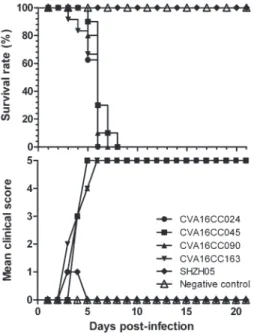
To test whether the circulating (CC024) and non-circulating (SHZH05) strains differed in virulence, the disease scores and the mortality rates of newborn mice were recorded until 21 days post-infection. All mice infected with the CC024, CC045, CC090 or CC163 strain became sick by day 3 post-infection, had a mean clinical score of grade 1, and mortality reached 100% by day 6 (Figure 2). As expected, the mock-infected mice had clinical scores of grade 0 and a 100% survival rate.
The mice challenged with 20mL of the CC024 strain at 105.5CCID
50/mL showed signs of illness on days 3 to 10 Figure 2.Clinical scores and survival of neonatal mice infected
with circulating Coxsackievirus A16 (CVA16) Changchun (CC) strains CC024, CC045, CC090, or CC163, or the noncirculating CVA16 SHZH05 strain. Neonatal mice (n=8-10 per litter) were intracerebrally infected with 20mL of 105.5CCID50/mL of each of the CVA16 virus strains, or mock infected with DMEM. The mean clinical scores and survival rates were monitored and recorded daily for 21 days postinfection.
(clinical scores of grade 1 to 5) and 100% mortality on day 7 (Figures 2 and 3A). The circulating CC024 strain demonstrated dose-dependent disease symptoms and mortality; dose-dependent mortality was also observed with the other circulating CC strains (CC045, CC090, and CC163). Mice infected with 103.5 CCID50/mL strains CC163, CC045, or CC090 started to die on days 4 or 5, with 100% mortality on days 9, 10, and 11, respectively (Figure 3B,C,D). Mice infected with circulating CC strains at 104.5CCID50/mL or 105.5CCID50/mL exhibited increas-ing morbidity on days 3 to 5, with 100% mortality on days 7 to 8 (Figure 3B,C,D). All the CC strain infections were lethal, but SHZH05 strain infections failed to induce illness or death in the neonatal mice (data not shown).
Pathological analysis of the SHZH05 and CC024 strain infections in neonatal mice
Pathological analysis was carried out in brain, lung, spinal and hind-limb muscle, liver, kidney, spleen, heart and intestine tissues from mice infected with the CC024 strain, which was considered representative of the four CC strains, and the SHZH05 strain. CC024 infection caused obvious lung tissue lesions, including severe alveolar shrinkage (Figure 4B), scattered areas of pulmonary fibrosis (Figure 4B), pulmonary edema, vas-cular dilation and congestion, severe necrosis of skeletal muscle, muscle bundle fracture, and dissolution of muscle fibers (Figure 4E and H). No pathological changes were observed in tissues of mice infected with the SHZH05 Figure 4.Histological analysis of tissues from Coxsackievirus A16 (CVA16)-infected neonatal mice. One-day-old SPF ICR mice were infected intracerebrally with 20mL of 105.5CCID50/mL of the CC024 or the SHZH05 strain, or mock infected with DMEM. No histological change was observed in the lung tissue, hind limb muscle, and spinal skeletal muscle of the SHZH05 strain-infected (C, F, and I) or mock-infected mice (A, D, andG). Mice with grade 5 clinical symptoms infected with the CC024 strain virus exhibited severe alveolar shrinkage and vascu-lar congestion (arrow) in the lung tissue (B), severe necrosis and loose muscle fibers in the hind limb muscle (E) and spinal skeletal muscle (H). Magnification 4006. All experiments were repeated three times.
Figure 5. Mean viral loads in tissues of Coxsackievirus A16 (CVA16) CC024 or CVA16 SHZH05 strain-infected neonatal mice with 20mL of 105.5CCID
strain (Figure 4C,F,I) or of mock infected neonatal mice (Figure 4A,D,G). These results demonstrate that the CC024 strain had a strong tropism for muscle and lung tissues and was responsible for the severe lesions in these tissues, but that the SHZH05 strain did not have a muscle tropism or cause any lesions in this neonatal mouse model.
Kinetics of viral replication in various tissues of the SHZH05 or CC024 strain-infected neonatal mice
To further understand the replication and distribution of the SHZH05 and CC024 strains in infected mice, we determined the viral loads in various tissues on different days postinfection. At 2 days postinfection, viral loads were detected only in the heart (102.652copies/mg), brain (103.322 copies/mg) and blood (103.052 copies/mL) of CC024 strain-infected mice (Figure 5). The viral loads were increased in the heart, lung, brain, intestine, spinal and hind-limb muscle, and blood of the CC024 strain-infected mice at 3 and 4 days postinfection (Figure 5). However, no viruses were detected in any tissues of SHZH05 strain-infected or mock-infected mice.
Genetic sequence alignment
Gene sequence analysis by DNAMAN software revealed a 3.76% diversity between the SHZH05 and CC024 strains in the 59 untranslated regions (UTR). Approximately 6.54, 6.34, and 7.04% diversity was seen in the viral genome nucleotide sequences of viral functional polyproteins P1, P2, and P3, respectively. Diversities of 0.7, 1.21 and 1.73% were observed in the corresponding P1, P2, and P3 amino acid sequences (P1: R51K, K52R, T295A, H364R, N464S, R850K; P2: S37T, M165V, S180N, K191R, V345T, K355R, R529K; P3: N11S, S48P, S299P, A320V, Y322H, S328N, T335S, I383T, Y397H, F434L, R570K, I595V, R637K), respectively (Table 1).
Discussion
An enterovirus 17 (EV71) vaccine has been developed (8), but whether this vaccine candidate cross-protects against CV infection remains unknown. Furthermore, a CV vaccine candidate developed in a mouse model was found to cause severe lesions in muscle and lung tissue (9). We evaluatedin vitroviral replication in different cell lines and pathogenicity in a neonatal mouse model to determine the pathogenic safety and replication capacity of the noncirculating CVA16 SHZH05 and circulating CVA16 Changchun (CC) strains. We found that the SHZH05 strain had a replication capability similar to the four CC strainsin vitro. However, the SHZH05 strain was less pathogenic than the CC strains in the neonatal mouse model. All four CC strains, but not the SHZH05 strain, caused tissue-specific pathological changes and lethal infections.
We found that the SHZH05 strain had similar replica-tion capacities in each of the cell lines, including the ML-1 cell primary cultures. However, histopathological analysis and viral load measurements confirmed that it failed to replicate in neonatal mouse tissues. The capacity to replicate in vitro and the improved safety indicate that the SHZH05 strain may be a better CV candidate than the CC024 strain vaccine (9). It is still not clear why the circulating CC024, but not the noncirculating SHZH05 virus strain, caused death in the neonatal mice. One reason might be that some genetic changes have occurred within the SHZH05 genome, resulting in a failure of the receptors on the surface of neonatal mouse cells to recognize the SHZH05 strain. In fact, the CVA16 and EV71 strains are unable to infect some mouse-derived cell lines, such as L929 and Ltr246, because there are no CVA16- or EV71-related receptors on the cell surface (14). It should be noted that comparative analysis of the SHZH05 and CVA16 CC024 strains revealed differences Table 1. Genetic differences between CVA16 CC024 and SHZH05 strains.
Gene segments Differences
Nucleotides Amino acids
59UTR 3.76%: G26A, T29C, G67A, A89G, C96T, C97T, T100C, C104T, T120C, G122A, T123C, A149G, T158C, T169C, C199T, G226A, C241T, T271C, C332T, C424T, C448T, G491A, C579T, T580C,
T584C, A660G, G675A, G742A
P1 6.54% 0.7%, changes are: R51K, K52R, T295A, H364R,
N464S, R850K
P2 6.34% 1.21%, changes are: S37T, M165V, S180N,
K191R, V345T, K355R, R529K
P3 7.04% 1.73%, changes are: N11S, S48P, S299P,
in the nucleotide and amino acid sequences of functional polyproteins P1, P2 and P3 (Table 1). These changes might account for the difference in pathogenicity of the SHZH05 and CC024 strains. Further study of the lack of infectivity of the SHZH05 strain in neonatal mice is important for the development of a CV vaccine.
The pathological analysis of infected neonatal mouse tissues indicated a CC024 strain tropism toward lung and muscle, which is in line with the findings of a previous study (9). Furthermore, viral load analysis demonstrated that the CC024 strain distributed among all of the tissues examined, but that the SHZH05 strain was not found in any of the tissues. These results further indicate that the noncirculating SHZH05 strain might be a safer CV vaccine candidate, the efficacy of which would be worth testing in a further study.
The noncirculating CVA16 SHZH05 strain had a replication capacity similar to the four circulating CVA16 CC strainsin vitro, but unlike the circulating CC024 strain, it failed to replicate or cause adverse pathological changes and mortality in vivo. These differences might
result from differences in nucleotide and amino acid sequences of the functional P1, P2 and P3 polyproteins in the two strains. Our findings demonstrated that the CVA16 SHZH05 strain may be a potentially safer CV vaccine candidate for prevention of HMFD than other available strains.
Acknowledgments
The authors thank Professor Qi Jin from the Institute of Pathogen Biology, Chinese Academy of Medical Sciences for the CVA16 SHZH05 strain. The research was supported in part by the National Natural Science Foundation of China (No. 31270202), the Chinese M i n i s t r y o f S c i e n c e a n d T e c h n o l o g y ( N o . 2012CB911102 and No. 2013ZX0001-005), the Health and Family Planning Commission of Jilin Province (No. 2013Z066), the Chinese Ministry of Education (No. IRT1016) and the Key Laboratory of Molecular Virology of Jilin Province (No. 20102209).
References
1. Mao Q, Wang Y, Yao X, Bian L, Wu X, Xu M, et al. Coxsackievirus A16: Epidemiology, diagnosis, and vaccine.
Hum Vaccin Immunother2014; 10: 360-367, doi: 10.4161/ hv.27087.
2. Repass GL, Palmer WC, Stancampiano FF. Hand, foot, and mouth disease: Identifying and managing an acute viral syndrome. Cleve Clin J Med 2014; 81: 537-543, doi: 10.3949/ccjm.81a.13132.
3. Goto K, Sanefuji M, Kusuhara K, Nishimura Y, Shimizu H, Kira R, et al. Rhombencephalitis and coxsackievirus A16.
Emerg Infect Dis 2009; 15: 1689-1691, doi: 10.3201/ eid1510.090594.
4. Wang CY, Li LF, Wu MH, Lee CY, Huang LM. Fatal coxsackievirus A16 infection.Pediatr Infect Dis J2004; 23: 275-276, doi: 10.1097/01.inf.0000115950.63906.78. 5. Legay F, Leveque N, Gacouin A, Tattevin P, Bouet J,
Thomas R, et al. Fatal coxsackievirus A-16 pneumonitis in adult.Emerg Infect Dis2007; 13: 1084-1086, doi: 10.3201/ eid1307.070295.
6. Eyckmans T, Wollants E, Janssens A, Schoemans H, Lagrou K, Wauters J, et al. Coxsackievirus A16 Encephalitis during Obinutuzumab Therapy, Belgium, 2013. Emerg Infect Dis2014; 20: 913-915, doi: 10.3201/eid2005.131766. 7. Wang X, Zhu C, Bao W, Zhao K, Niu J, Yu XF, et al. Characterization of full-length enterovirus 71 strains from severe and mild disease patients in northeastern China.PLoS One2012; 7: e32405, doi: 10.1371/journal.pone.0032405. 8. Zhu FC, Xu WB, Xia JL, Liang ZL, Liu Y, Zhang XF, et al.
Protection from lethal challenge in a neonatal mouse model by circulating recombinant form coxsackievirus A16 vaccine candidates.J Gen Virol2014; 95: 1083-1093, doi: 10.1099/ vir.0.065003-0.
9. Li J, Chang J, Liu X, Yang J, Guo H, Wei W, et al. Protection from lethal challenge in a neonatal mouse model by circulating recombinant form coxsackievirus A16 vaccine candidates.J Gen Virol2014; 95: 1083-1093, doi: 10.1099/ vir.0.063560-0.
10. Wei W, Guo H, Li J, Ren S, Wei Z, Bao W, et al. Circulating HFMD-associated coxsackievirus A16 is genetically and phenotypically distinct from the prototype CV-A16. PLoS One2014; 9: e94746, doi: 10.1371/journal.pone.0094746. 11. Wu Z, Yang F, Zhao R, Zhao L, Guo D, Jin Q. Identification
of small interfering RNAs which inhibit the replication of several Enterovirus 71 strains in China. J Virol Methods
2009; 159: 233-238, doi: 10.1016/j.jviromet.2009.04.002. 12. Zhao K, Han X, Wang G, Hu W, Zhang W, Yu XF.
Circulating coxsackievirus A16 identified as recombinant type A human enterovirus, China.Emerg Infect Dis2011; 17: 1537-1540.
13. Hagiwara A, Tagaya I, Yoneyama T. Epidemic of hand, foot and mouth disease associated with enterovirus 71 infection.
Intervirology1978; 9: 60-63, doi: 10.1159/000148922. 14. Yamayoshi S, Koike S. Identification of a human SCARB2