Human papillomavirus and genome instability:
from productive infection to cancer
Bruna Prati, Bruna Marangoni, Enrique Boccardo*
Departamento de Microbiologia, Instituto de Ciencias Biomedicas, Universidade de Sao Paulo, Sao Paulo, SP, BR.
Prati B, Marangoni B, Boccardo E. Human papillomavirus and genome instability: from productive infection to cancer. Clinics. 2018;73(suppl 1):e539s
*Corresponding author. E-mail: [email protected]
Infection with high oncogenic risk human papillomavirus types is the etiological factor of cervical cancer and
a major cause of other epithelial malignancies, including vulvar, vaginal, anal, penile and head and neck
carci-nomas. These agents affect epithelial homeostasis through the expression of specific proteins that deregulate
important cellular signaling pathways to achieve efficient viral replication. Among the major targets of
viral proteins are components of the DNA damage detection and repair machinery. The activation of many of
these cellular factors is critical to process viral genome replication intermediates and, consequently, to sustain
faithful viral progeny production. In addition to the important role of cellular DNA repair machinery in the
infective human papillomavirus cycle, alterations in the expression and activity of many of its components are
observed in human papillomavirus-related tumors. Several studies from different laboratories have reported
the impact of the expression of human papillomavirus oncogenes, mainly E6 and E7, on proteins in almost all
the main cellular DNA repair mechanisms. This has direct consequences on cellular transformation since it
causes the accumulation of point mutations, insertions and deletions of short nucleotide stretches, as well as
numerical and structural chromosomal alterations characteristic of tumor cells. On the other hand, it is clear
that human papillomavirus-transformed cells depend on the preservation of a basal cellular DNA repair
activity level to maintain tumor cell viability. In this review, we summarize the data concerning the effect
of human papillomavirus infection on DNA repair mechanisms. In addition, we discuss the potential of
exploiting human papillomavirus-transformed cell dependency on DNA repair pathways as effective
anti-tumoral therapies.
KEYWORDS:
HPV; Genomic Instability; E6; E7; Cervical Cancer; Synthetic Lethality.
Human papillomaviruses and cancer
Human papillomaviruses (HPVs) are small, nonenveloped
viruses with a genome composed of double-stranded circular
DNA of approximately 8 kbp (1,2). HPVs belong to the
Papillomaviridae
family and, to date, over 200 different types
have been identified and their genomes completely
sequen-ced (1). The HPV genome can be divided in three functional
regions. The upstream regulatory region (URR) or long
control region (LCR) is a noncoding portion encompassing
almost an eighth of the viral genome and is involved in the
regulation of viral replication and transcription. In addition,
there are two coding regions. The early (E) region harbors the
E1, E2, E4, E5, E6 and E7 early genes that are critical for
regulating viral genome replication, viral gene expression,
immune evasion and genome persistence. The late (L) region
encodes the major and minor capsid proteins from the L1
and L2 genes, respectively. These viruses display a marked
tropism for stratified epithelial tissues and are transmitted by
direct contact with infected skin and mucosa. As such, HPV
infections are associated with a variety of benign
hyperpro-liferative conditions, such as common and genital warts.
Approximately 40 HPV types infect the mucosa and skin
of the anogenital tract. These HPV types are further
sub-classified as low or high risk according to their association
with the development of cervical carcinoma and its precursor
lesions. Low-risk HPV types, such as HPV6 and HPV11, are
associated with genital warts and low-grade cervical
intra-epithelial neoplasia (CIN) (3,4). On the other hand, high-risk
HPV types, for example, HPV16, HPV18, HPV31 and HPV45,
are associated with the development of high-grade CIN and
cervical cancer. In addition, they are associated with a
signif-icant proportion of vulvar, vaginal, anal, penile and head and
neck carcinomas (5).
To establish a productive infection, HPVs have to reach
cells from the basal layer of the epithelium. These are the
only keratinocytes of the tissue that proliferate and express
the DNA replication machinery. Therefore, they provide a
permissive environment for initial viral genome replication.
The virus can reach these cells through microwounds caused
during sexual intercourse or at specific anatomical sites were
the basal layer is readily accessible, such as the
squamoco-lumnar junction (SCJ) of the cervix (6). Once in the cell
DOI:10.6061/clinics/2018/e539s
Copyright&2018CLINICS–This is an Open Access article distributed under the terms of the Creative Commons License (http://creativecommons.org/licenses/by/ 4.0/) which permits unrestricted use, distribution, and reproduction in any medium or format, provided the original work is properly cited.
No potential conflict of interest was reported.
Received for publication on December 27, 2017. Accepted for publication onMay 16, 2018
nucleus, the HPV genome remains in the episomal state
(100-500 copies/per cell) and replicates together with the cellular
genome (7). The rest of the viral cycle, including vegetative
genome amplification, structural protein expression, virion
mounting and release, requires infected keratinocytes to
migrate through the different layers of the epithelium and
complete their differentiation program (8).
In high-grade CIN and HPV-associated tumors, viral DNA
is frequently integrated into the host cell genome. Viral
integration is an accidental event that usually disrupts the
region were the E1 and E2 genes are located, leading to
the loss of their expression. This constitutes a critical step in
HPV-associated carcinogenesis since the loss of E2 leads to
the upregulated expression of the main viral oncoproteins,
namely, E6 and E7 (9). The sustained expression of E6 and E7
ensures the existence of a cellular milieu favorable for HPV
replication in the suprabasal layers of the epithelium and is
critical in the process of HPV-mediated cell transformation
(see below) (10).
It is estimated that approximately 291 million women are
infected by genital HPV types worldwide. Each year, nearly
500,000 new cases of cervical cancer are diagnosed and over
270,000 women die from this disease around the world.
Importantly, most of the cases (
B
86%) and deaths (88%) occur
in developing countries (1). Collectively, cervical cancer and
the other HPV-associated malignancies represent 5.2% of all
tumors affecting humans (11).
DNA damage repair pathways and the HPV cycle
DNA contains the essential information for the formation
and function of an organism, and its preservation is critical
for survival. DNA is a fairly stable macromolecule; however,
it suffers constant attacks from extracellular and intracellular
factors that jeopardize its integrity. These factors include
chemical and physical agents, infectious agents, highly
reactive metabolic byproducts, and replication errors (12).
In fact, DNA damage is a common event in human cells. It is
estimated that each human cell suffers approximately one
million lesions in its DNA every day (13). Therefore, if DNA
damage is not efficiently repaired, the integrity of the genome
is compromised, causing the accumulation of mutations that
ultimately may lead to premature aging, cancer development
and death (14,15).
DNA repair is the process by which alterations in DNA
are reverted. Usually, this involves the action of nucleases,
helicases, ligases and DNA polymerases. Different
specia-lized DNA repair mechanisms exist in which complex
arrays of cellular proteins detect and repair different kinds
of DNA lesions (16). These mechanisms include base
exci-sion repair (BER), nucleotide exciexci-sion repair (NER), DNA
mismatch repair (MMR), homologous recombination repair
(HRR) and nonhomologous end joining (NHEJ). Hereditary
or acquired defects in any of these pathways may lead
to genome instability with severe consequences for the
individual (17).
The DNA damage response (DDR) is composed of different
signal transduction pathways that include damage sensors,
signal transducers and effectors that act in a coordinated fashion
to maintain genome integrity. Some DNA lesions activate
spe-cific sensors, such as the MRN complex (Mre11-RAD50-NSB1),
replication protein A (RPA) or ATR-interacting protein (ATRIP),
that trigger signaling pathways by the phosphorylation of
apical protein kinases, such as ataxia telangiectasia mutated
(ATM), ataxia telangiectasia and Rad3 related (ATR) and
DNA-dependent protein kinase (DNA-PK). Different types of
DNA lesions activate different kinases (18,19). For instance,
double-stranded DNA breaks may activate DNA-PK or ATM.
On the other hand, single-stranded breaks preferentially
acti-vate ATR. The signaling cascade triggered by ATM and ATR
then activates downstream kinases CHK2 (checkpoint kinase
2) and CHK1 (checkpoint kinase 1), respectively. However, the
ATR-CHK1 and ATM-CHK2 pathways are not completely
independent since they exhibit a high degree of redundancy
and overlap. These pathways may regulate the phosphatase
Cdc25 and activate the transcription factor p53, leading to
cyclin-dependent kinase (CDK) inhibition and blocking cell
cycle progression by the activation of specific checkpoints.
Consequently, DNA damage effectors may mediate cell cycle
arrest to allow DNA repair or, when the amount of damage is
too high, induce cell death (14,17).
Many viruses, especially those with oncogenic properties,
express proteins that affect the cell cycle and DNA damage
repair regulatory pathways. Several studies have shown that
during genome amplification, HPV proteins interact with
different components of the cellular DNA repair machinery
to activate or downregulate the expression or activity of
factors of the ATM and ATR pathways. In 2009, Moody and
coworkers showed that ATM activation is required for
the productive genomic replication of HPV31 but not for
episomal maintenance (20). On the other hand, it was
obser-ved that CHK1 inhibition causes an important reduction in
the number of HPV episomes in differentiated cells (21). In
addition, the knockdown of DNA topoisomerase 2-binding
protein 1 (TopBP1), a protein that acts upstream of ATR,
sup-presses HPV31 replication (22). In line with these
observa-tions, it has been reported that E2 protein from HPV16
interacts with TopBP1 and that this interaction improves
E2-mediated viral transcription and replication (23). Moreover,
ATM and ATR kinases are constitutively activated in
HPV-positive keratinocytes, and ATR/CHK1 blockade is
asso-ciated with the downregulation of HPV productive
replica-tion and the reduced expression of late genes (22).
Importantly, many proteins from ATM and ATR pathways
colocalize with HPV replication sites, further supporting the
role of these factors in the HPV life cycle. Different studies
have shown that E1 and E2 early proteins may activate the
DDR, recruiting factors involved in DNA repair to process
replication intermediates during viral genome
amplifica-tion. Identified proteins at centers of HPV replication include
53BP1, ATRIP, OPbp1, p-ATM, p-H2AX, p-p53, ATR, CHK1,
CHK2, PCNA, RPA, NBS1, BRCA1, RAD51, MRE11 and
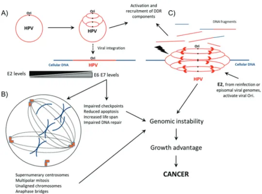
Ku70/80 (24-27) (Figure 1A). Collectively, these studies
indicate that HPV may modulate different signaling
path-ways involved in the DDR to warrant effective viral
tran-scription, faithful genome replication and, ultimately, the
production of a large number of infective virions (10,28,29).
HPV oncoproteins pave the road to genome
instability
accom-plished by the action of viral proteins, mainly E6 and E7,
on cellular factors involved in cell cycle regulation. E6 and
E7 are the major HPV transforming proteins, and their
sustained expression is required to maintain the oncogenic
properties of HPV-transformed cells in most scenarios
(31-33). These proteins collaborate to immortalize primary
cells and can independently abrogate the mitotic
check-point and p53-mediated cell cycle arrest in response to
DNA damage (34,35).
Among the main targets of E6 is the tumor suppressor
protein p53. E6 forms a ternary complex with E6-associated
protein (E6AP) and p53, altering p53
’
s functional capacity
and inducing the degradation of this cellular protein by the
ubiquitin-mediated proteolysis pathway (36-38). In addition,
E6 from HPV16 binds the transcriptional coactivator
CREB-binding protein/p300 (CBP/p300) and downregulates its
ability to activate p53-responsive elements in the promoters
of several p53-regulated genes (39). E6 protein also
upregu-lates the expression of the catalytic subunit of telomerase
(hTERT) in primary cells and delays cell senescence (40).
Moreover, E6 targets other cellular factors, including those
regulating cell polarity (PDZ proteins), apoptosis and cell
differentiation (40,41). Similarly, E7 from high-risk HPV
types binds and induces the degradation of members of the
retinoblastoma (pRb) tumor suppressor proteins pRb1
p110,
p107 and p130 (pRb2). This interrupts the interaction of pRb
proteins with members of the E2F transcription factor family.
Once free from inhibition, E2F activates the transcription of
several genes involved in S phase induction and progression,
including cyclins A and E (42,43). E7 also promotes cell
cycle progression by downregulating the activity of
cyclin-dependent kinase inhibitors (CKIs) p21 and p27 (44,45). In
addition, E7 of high-risk HPV16 and HPV31 interact with
histone deacetylases 1 and 2 (HDAC1 and HDAC2),
affect-ing the gene expression pattern of infected cells. Together,
these data show that E6 and E7 affect the proliferation and
differentiation of human keratinocytes by extending the
lifespan of these cells (46,47).
Due to their impact on different cellular pathways, HPV
oncoproteins may also promote the accumulation of genomic
alterations that contribute to malignant transformation
(48-54). For instance, Song and coworkers (55) demonstrated that
mouse cells expressing E6 and E7 from high-risk HPV types
continue to replicate DNA even in the presence of lesions
induced by ionizing radiation (IR) and accumulate large
numbers of alterations in the molecule. Similarly, HPV16 E7
expression is associated with the persistence of
g
H2AX and
Rad51 foci upon the exposure of head and neck cancer cells
to IR (56). Importantly, the expression of high-risk E6 and E7
proteins has been associated with the induction of DNA
breaks and a high frequency of foreign DNA integration into
host cells (35,57).
Figure 1 -Interplay between HPV and cellular DNA repair machinery during infective viral cycle and virus-mediated cell transformation.
Several studies have shown that ATM and ATR pathways
are targeted by HPV oncoproteins. For instance, Banerjee et
al. (30) observed that the oncoprotein E7 from HPV18
induces increased levels of phosphorylated ATM and the
downstream kinases CHK1, CHK2, and JNKs (c-Jun
N-terminal kinases). It was also reported that E7 from HPV31
binds ATM, inducing its phosphorylation and activating
CHK2 (20). Another study showed that E7 from HPV16
induces the degradation of claspin, a protein from the
ATR-CHK1 pathway, attenuating the DNA damage checkpoint
(58). In addition, the results from recent studies support the
involvement of HPV in skin cancer. In this context, it was
observed that the protein E6 from HPV5 and HPV8, two
important cutaneous HPV types, reduces ATM levels and
downregulates the p300/ATR signaling axis, leading to the
persistence of DNA lesions induced by UVB light (10,59).
Finally, it was observed that E6 and E7 from HPV16 interact
with breast cancer-associated protein 1 (BRCA1), inactivating
its function in the repair of double-stranded DNA breaks (60).
Other alterations in DNA repair systems associated with
HPVs have been described by different groups. One report
has shown that oral keratinocytes immortalized with HPV16
exhibit deficiencies in the NER system and, consequently, are
unable to remove cyclobutane pyrimidine dimers (CPDs)
induced by UV light (61). A similar observation was made in
fibroblasts expressing E7 from HPV16. These cells show
defective NER activity and display a marked delay in the
removal of CPDs induced by UVB light (62). Recently, it was
observed that cells derived from squamous carcinoma of the
head and neck positive for HPV are more sensitive to
radiotherapy than HPV-negative cell lines due to a defect in
BER in the the former cells (63). This finding is also in
agree-ment with observations showing that E7 delays the repair of
DNA lesions induced by IR in culture systems and
labo-ratory animal models (56).
The oncoprotein E6 also targets DNA repair pathways. For
instance, epithelial mammary cells expressing this protein
from HPV16 exhibit a reduced capacity for removing thymine
dimers after exposure to UV light (64,65). In addition, oral
fibroblasts expressing E6 from HPV16 exhibit an impaired
ability to repair double-stranded breaks by NHEJ.
Interest-ingly, E6 achieves this effect via either dependent or
p53-independent pathways (66). Moreover, it has been described
that E6 mediates O
6-methylguanine DNA methyltransferase
(MGMT) degradation via the ubiquitin/proteasome
path-way in a process that requires the interaction of E6 with
E6AP. The action of MGMT, which is impaired by E6,
pro-tects cells and tissues against the effects of alkylating agents
(67). Finally, it was reported that E6 from different HPV types
targets X-ray repair cross-complementing protein 1 (XRCC1)
and inhibits its ability to repair DNA lesions (68).
These observations show that HPV infection may increase
the frequency of DNA alterations in the host cell and delay
the removal of these alterations by targeting DNA repair
pathways. In addition, by impairing cell cycle checkpoints
and apoptosis, HPV oncoproteins cause sustained
prolifera-tion while preventing cell death. Collectively, these effects
may lead to cellular alterations that give rise to precursor
lesions with a tendency for malignant progression.
HPV-mediated genome instability and cancer
During tumor evolution, cells acquire genetic alterations
that may upregulate the expression of proto-oncogenes or
induce gain-of-function mutations in these genes. In
addi-tion, mutations, deletions and alterations in the
methyla-tion pattern of promoter sequences may downregulate the
activity of tumor suppressor genes. Cells harboring these
alterations may have proliferative advantages and acquire
other phenotypical alterations, such as the capacity to invade
other tissues and organs (69). Genome instability is a
defin-ing phenotype of most malignant tumors that comprises
numerical and structural chromosomal abnormalities, as
well as microdeletions, small insertions, the duplication of
short nucleotide stretches and the accumulation of point
mutations. Alterations in the sequences or expression level
of proteins involved in DNA damage repair may result in
the accumulation of genetic modifications important for
cancer development. Normal cells exhibit an impressive
array of mechanisms to detect and repair DNA defects.
How-ever, many of these mechanisms are altered in tumors cells
(70,71). Although HPV activates several DNA repair
path-ways to assist its genome replication, as described above,
HPV-induced tumors exhibit a high degree of genome
instability. This fact seems to be critical for HPV-mediated
carcinogenesis and suggests that DNA repair is attenuated
during the steps leading from cellular transformation to
cancer onset (7,35).
In hereditary cancers, mutations transmitted from
pro-genitors predispose the host to the development of certain
types of tumors and/or increase their sensitivity to
carcino-gens. This process may involve alterations in components of
different DNA repair pathways. Several studies have
sug-gested that mutations in this group of genes may act as
cofactors in the establishment and progression of
HPV-asso-ciated tumors. For instance, patients with Fanconi anemia
(FA) or individuals carrying mutations in the breast
cancer-associated (BRCA) gene exhibit a higher risk of developing
HPV-associated cancerous lesions (72). In fact, the expression
of HPV16 E7 in FA-deficient fibroblasts is associated with an
increased number of chromosome aberrations (73).
More-over, K14E7/FancD2(-/-) mice exhibit a significantly higher
incidence of head and neck squamous cell carcinoma than
animals expressing K14E7 on a normal FancD2 background
(
+
/
+
) (74). Using a similar approach, it was observed in
FA-deficient mice that cervical tumors persisted even in the
absence of HPV16 E7 expression, supporting the notion that
FA-deficient tumors may escape from their dependency on
the viral oncogene (32). Upregulation of the FA pathway is
a frequent event in cervical SCC.
In vitro
data indicate that
the activation of this pathway is mediated by E7 and is
characterized by the formation of large FANCD2 foci and the
recruitment of FANCD2 and FANCD1/BRCA2 to chromatin
(73). These observations suggest that FA pathway activation
plays a role in the HPV cycle. The malfunction of this
path-way may lead to the accumulation of HPV-mediated
geno-mic alterations and the promotion of tumor development in
FA patients. Of clinical relevance, in this context, where FA
facilitates the accumulation of mutations, sustained viral
oncogene expression may no longer be required to maintain
the transformed phenotype (32).
was downregulated in tumors and precursor lesions
com-pared to samples from control subjects (75). Recently, Seiwert
et al. (76) sequenced 617 cancer-associated genes in 120
matched head and neck squamous cell carcinoma/normal
samples, of which 42.5% were positive for HPV DNA. They
observed that HPV-positive tumors showed 5.8% of DNA
repair gene aberrations (including 7.8% of BRCA1/2
muta-tions). In a study conducted in our laboratory, we compared
the expression pattern of 135 genes involved in DNA
damage repair/signaling between normal human
keratino-cytes and cervical cancer-derived cell lines and observed that
the mRNA levels of 18 genes are altered in HPV-transformed
cell lines (77).
Genome instability, in the context of HPV infection, may
arise through different molecular pathways. A clear example
of this is represented by the results of a study conducted by
Kadaja et al. (25). The authors reported that the ectopic
expression of HPV18 E1 and E2 triggers HPV replication
from viral-integrated HPV genomes in SiHa and HeLa cells,
leading to the accumulation of chromosome defects.
Differ-ent from the cellular origin of replication, which is activated
once per cell cycle, the viral origin of replication can be
triggered several times during the same cell cycle, leading
to the
‘‘
onion skin
’’
type of DNA replication. This process
generates replication intermediates that stress the DNA
mole-cules, leading to double- or single-stranded breaks and the
recruitment of DNA repair machinery that ligates loose
DNA ends and promotes chromosomal rearrangement.
These observations raise the disturbing possibility that the
coexistence of integrated and episomal HPV genomes in the
same cell may induce chromosome aberrations arising from
the integrated viral DNA. Moreover, the de novo HPV
infection of cells harboring integrated DNA genomes may
also endanger the cellular genome integrity and favor cell
transformation (Figure 1C).
Chronic inflammation also plays a major role in cancer
biology (69). This is also true for tumors associated with HPV
infection (78). For instance, cells harboring HPV16 genomes
exhibit increased levels of nitric oxide (NO), which triggers
inflammation and promotes DNA breaks (79). In addition,
extracellular factors, mainly inflammatory mediators, may
affect HPV infection outcome. For instance, it has been
obser-ved that interferon
b
may induce the elimination of HPV16
episomes from naturally infected cervical keratinocytes by
selecting cells with integrated viral genomes (80). In
addi-tion, interferon may induce HPV genome integration (81).
These observations suggest that chronic inflammation may
potentiate viral persistence and, consequently, lesion
estab-lishment and progression (82). Importantly, these findings
indicate that HPV therapies should be evaluated for the
potential selection of cells with integrated genomes and
increased proliferative potential.
As previously stated, HPV oncogenes E6 and E7 play a
critical role in HPV-mediated carcinogenesis by affecting
major cellular processes. The consequences of HPV
oncopro-tein actions are reflected on cellular genome homeostasis.
For instance, inhibition of the postmitotic checkpoint by
high-risk HPV E6 and E7 is associated with the induction
of polyploidy in human keratinocytes (83-85). It was also
observed that HPV16 oncoprotein expression induce
super-numerary centrosomes and multipolar mitotic spindles that
may lead to aneuploidy (49). In addition, an independent
study showed that E6 oncoprotein causes centrosome
accumu-lation, while HPV16 E7 interferes with the centrosome
duplication cycle (86). Interestingly, while E7 from HPV16
may induce the delocalization of dynein from mitotic
spindles, this has not been correlated with mitotic defects
(87) (Figure 1B).
Alterations in the regulation of the cell cycle and the
inhibition of proapoptotic factors are important underlying
events in HPV-mediated genome instability. As expected,
genomic instability is an early event during HPV infection
that precedes viral integration, the development of
asso-ciated lesions and their eventual progression to cancer
(88-90). Genomic instability is crucial in the appearance of
aneuploid cells and lesion progression (88). This is
high-lighted by the fact that highly polyploid as well as
aneuploid cells are mainly detected in high-grade cervical
lesions (91). Of note, most of the alterations described are
restricted to cells infected with high-risk HPV types and
are not detected upon infection with low-risk types (92).
Studies conducted using clinical samples have shown that
cervical tumors exhibit a plethora of chromosomal
altera-tions, including gains in 1, 3q, 5p, 6p, 7, 8q, 9q, 16q, and 20
and losses in 2q, 3p, 4q, 6q, 11q, 13q, 16, and 17 (93-98).
Finally, genomic alterations associated with HPV infection
have also been observed in tumors from different
anato-mical locations, including oral, anal, laryngeal and head
and neck neoplasias (76,99-101).
Genome instability is probably not a consequence of
HPV-mediated malignant transformation. However, it may favor
the acquisition of genetic alterations that confer growing
advantages to cells expressing viral oncogenes. In addition,
the underlying mutator phenotype may allow
transfor-med cells to adapt rapidly to the harsh conditions of the
tumor microenvironment, contributing to tumor onset and
progression.
HPV-associated disease treatment: can we target
DNA repair systems?
The data discussed above further support the established
notion that the main mechanism by which HPV induces cell
transformation is the targeting of p53 and pRb by E6 and E7,
respectively (43,102-104). Therefore, it is assumed that the
downregulation of these tumor suppressor proteins by E6
and E7 mimics the effect of inactivating mutations observed
in p53 and pRb in tumors not associated with HPV
infec-tion. Consequently, it is believed that HPV-positive cancers,
including anogenital, oropharyngeal tract and anal canal
tumors, are less likely to present p53 and pRb mutations
(105,106). Conversely, different studies have shown that
HPV-transformed cells retain the ability to respond to
geno-toxic stress by inducing a p53-mediated response. Therefore,
p53 downregulation as a consequence of HPV oncogene
action is not functionally equivalent to p53 inactivation by
mutation (107-109). This may be at least one of the
under-lying molecular mechanisms explaining why the presence
of HPV is associated with a better response to therapy and
constitutes a positive prognostic factor for patients with
oropharyngeal tumors (110).
In addition, several examples of synthetic lethality
involv-ing genes from DNA damage repair systems and tumor
suppressor-regulated pathways have been described and
suggested as potential targets for cancer therapy (113).
The first case of synthetic lethality involving genes
asso-ciated with DNA damage repair was with poly(ADP-ribose)
polymerase (PARP) and breast and ovarian cancer
suscept-ibility genes BRCA1 and BRCA2 (114,115). PARP is a family
of proteins involved in several cellular processes,
includ-ing DNA repair and the maintenance of genome stability.
PARP1, the most studied member of the family, is rapidly
recruited to nicks and double-stranded breaks in cellular
DNA, where it gathers components of the DNA repair
machinery and promotes the removal of lesions (116). In fact,
cells with alterations in different DNA damage repair
path-ways exhibit increased susceptibility to the loss of PARP
activity. As expected, PARP inhibitors (iPARPs) prevent
DNA damage repair and are used in cancer therapy,
parti-cularly in tumors with germline or somatic mutations in
BRCA1/2 (117). The administration of iPARPs promotes cell
death by downregulating BER and promoting the
accumula-tion of DNA defects in the cell (116,118-121). Addiaccumula-tionally, by
preventing DNA repair, PARP inhibition increases the
sensitivity of cells to chemotherapeutical agents that promote
DNA lesions (122).
E6 and E7 form high-risk HPV types are pleotropic proteins
that target an increasing list of cellular factors affecting their
expression and function. As such, HPV-transformed cells
exhibit major alterations in important signaling pathways
and cellular processes, including several DNA damage repair
mechanisms. This fact has consequences of clinical relevance.
For instance, patients harboring HPV-associated head and
neck squamous cell carcinoma (HNSCC) have significantly
improved survival compared with patients affected by
HPV-negative tumors. Although the molecular mechanisms
underlying this difference are not completely understood, it
is accepted that impaired DNA repair abilities, probably
due to HPV oncoproteins action, play a major role (110,123).
In fact, HPV-positive HNSCC-derived cell lines accumulate
more double-stranded DNA breaks than HPV-negative
counterparts and exhibit higher radiosensitivity (63,124).
In conclusion, HPV-transformed cells exhibit major defects
in DNA repair. Nevertheless, we anticipate that
HPV-transformed cells depend on the preservation of a basal
level of DNA repair activity meditated by cellular
machi-nery to maintain the minimal genomic stability needed for
tumor cell viability. Therefore, a great effort should be
directed toward identification of the molecular pathways
necessary for the survival HPV-driven tumor cells. This
will certainly contribute to the development of more
effi-cient antitumor therapies.
’
ACKNOWLEDGMENTS
This research was supported by grants from the FAPESP (2010/20002-0) and CNPq (480552/2011-8), as well as a fellowship awarded to BP (CNPq 573799/2008-3; CAPES 1524553).
’
AUTHOR CONTRIBUTIONS
Prati B prepared the text describing the effect of HPV on DNA repair machinery. Marangoni B prepared the text describing HPV-associated disease treatment strategies. Boccardo E prepared, revised and corrected all the text.
’
REFERENCES
1. International Agency for Research on Cancer. Human Papillomaviruses, vol. 90; 2007 [Internet]. IARC Monographs on the evaluation of carci-nogenic risks to humans. Available from: http://monographs.iarc.fr/ ENG/Monographs/vol90/mono90-6.pdf%5Cnhttp://monographs.iarc. fr/ENG/Monographs/vol100B/mono100B-11.pdf
2. Bernard HU, Burk RD, Chen Z, van Doorslaer K, zur Hausen H, de Villiers EM. Classification of papillomaviruses (PVs) based on 189 PV types and proposal of taxonomic amendments. Virology. 2010;401(1): 70-9, http://dx.doi.org/10.1016/j.virol.2010.02.002.
3. zur Hausen H. Papillomaviruses in the causation of human cancers - a brief historical account. Virology. 2009;384(2):260-5, http://dx.doi.org/ 10.1016/j.virol.2008.11.046.
4. Bosch FX, de Sanjose S, Castellsague X. HPV and genital cancer: the essential epidemiology. In: Vaccines for the Prevention of Cervical Cancer [Internet]. Oxford University Press; 2008. Available from: http://oxfordmedicine.com/view/10.1093/med/9780199543458.001. 0001/med-9780199543458-chapter-4
5. Parkin DM, Bray F. Chapter 2: The burden of HPV-related cancers. Vaccine. 2006;24 Suppl 3: S3/11-25, http://dx.doi.org/10.1016/j.vaccine. 2006.05.111.
6. Herfs M, Yamamoto Y, Laury A, Wang X, Nucci MR, McLaughlin-Drubin ME, et al. A discrete population of squamocolumnar junction cells implicated in the pathogenesis of cervical cancer. Proc Natl Acad Sci U S A. 2012;109(26): 10516-21, http://dx.doi.org/10.1073/pnas. 1202684109.
7. Moody CA, Laimins LA. Human papillomavirus oncoproteins: path-ways to transformation. Nat Rev Cancer. 2010;10(8):550-60, http://dx. doi.org/10.1038/nrc2886.
8. Doorbar J. The papillomavirus life cycle. J Clin Virol. 2005;32 Suppl 1: S7-15, http://dx.doi.org/10.1016/j.jcv.2004.12.006.
9. Münger K, Baldwin A, Edwards KM, Hayakawa H, Nguyen CL, Owens M, et al. Mechanisms of human papillomavirus-induced oncogenesis. J Virol. 2004;78(21):11451-60, http://dx.doi.org/10.1128/JVI.78.21.11451-11460.2004.
10. Wallace NA, Gasior SL, Faber ZJ, Howie HL, Deininger PL, Galloway DA. HPV 5 and 8 E6 expression reduces ATM protein levels and attenu-ates LINE-1 retrotransposition. Virology. 2013;443(1):69-79, http://dx. doi.org/10.1016/j.virol.2013.04.022.
11. Tota JE, Chevarie-Davis M, Richardson LA, Devries M, Franco EL. Epi-demiology and burden of HPV infection and related diseases: implica-tions for prevention strategies. Prev Med. 2011;53 Suppl 1: S12-21, http:// dx.doi.org/10.1016/j.ypmed.2011.08.017.
12. Lindahl T. Instability and decay of the primary structure of DNA. Nat-ure. 1993;362(6422):709-15, http://dx.doi.org/10.1038/362709a0. 13. Branzei D, Foiani M. Regulation of DNA repair throughout the cell cycle.
Nat Rev Mol Cell Biol. 2008;9(4):297-308, http://dx.doi.org/10.1038/ nrm2351.
14. Sulli G, Di Micco R, d’Adda di Fagagna F. Crosstalk between chromatin state and DNA damage response in cellular senescence and cancer. Nat Rev Cancer. 2012;12(10):709-20, http://dx.doi.org/10.1038/nrc3344.
15. Sancar A, Lindsey-Boltz LA, Unsal-Kac¸maz K, Linn S. Molecular
mechanisms of mammalian DNA repair and the DNA damage check-points. Annu Rev Biochem. 2004;73:39-85, http://dx.doi.org/10.1146/ annurev.biochem.73.011303.073723.
16. Zhou BB, Elledge SJ. The DNA damage response: putting checkpoints inperspective. Nature. 2000;408(6811):433-9, http://dx.doi.org/10.1038/ 35044005.
17. Yang J, Yu Y, Hamrick HE, Duerksen-Hughes PJ. ATM, ATR and DNA-PK: initiators of the cellular genotoxic stress responses. Carcinogenesis. 2003;24(10):1571-80, http://dx.doi.org/10.1093/carcin/bgg137. 18. Abraham RT. Cell cycle checkpoint signaling through the ATM and ATR
kinases. Genes Dev. 2001;15(17):2177-96, http://dx.doi.org/10.1101/ gad.914401.
19. Lowndes NF, Murguia JR. Sensing and responding to DNA damage. Curr Opin Genet Dev. 2000;10(1):17-25, http://dx.doi.org/10.1016/ S0959-437X(99)00050-7.
20. Moody CA, Laimins LA. Human papillomaviruses activate the ATM DNA damage pathway for viral genome amplification upon differ-entiation. PLoS Pathog. 2009;5(10):e1000605, http://dx.doi.org/10.1371/ journal.ppat.1000605.
21. Edwards TG, Helmus MJ, Koeller K, Bashkin JK, Fisher C. Human papillomavirus episome stability is reduced by aphidicolin and con-trolled by DNA damage response pathways. J Virol. 2013;87(7):3979-89, http://dx.doi.org/10.1128/JVI.03473-12.
22. Hong S, Cheng S, Iovane A, Laimins LA. STAT-5 Regulates Transcription of the Topoisomerase IIb-Binding Protein 1 (TopBP1) Gene To Activate the ATR Pathway and Promote Human Papillomavirus Replication. MBio. 2015;6(6):e02006-15, http://dx.doi.org/10.1128/mBio.02006-15. 23. Boner W, Taylor ER, Tsirimonaki E, Yamane K, Campo MS, Morgan IM.
TopBP1. J Biol Chem. 2002;277(25):22297-303, http://dx.doi.org/10.1074/ jbc.M202163200.
24. Gillespie KA, Mehta KP, Laimins LA, Moody CA. Human papilloma-viruses recruit cellular DNA repair and homologous recombination factors to viral replication centers. J Virol. 2012;86(17):9520-6, http://dx. doi.org/10.1128/JVI.00247-12.
25. Kadaja M, Isok-Paas H, Laos T, Ustav E, Ustav M. Mechanism of genomic instability in cells infected with the high-risk human papillo-maviruses. PLoS Pathog. 2009;5(4):e1000397, http://dx.doi.org/10.1371/ journal.ppat.1000397.
26. Reinson T, Toots M, Kadaja M, Pipitch R, Allik M, Ustav E, et al. Engagement of the ATR-dependent DNA damage response at the human papillomavirus 18 replication centers during the initial amplification. J Virol. 2013;87(2):951-64, http://dx.doi.org/10.1128/JVI.01943-12. 27. Sakakibara N, Mitra R, McBride AA. The papillomavirus E1 helicase
activates a cellular DNA damage response in viral replication foci. J Virol. 2011;85(17): 8981-95, http://dx.doi.org/10.1128/JVI.00541-11. 28. Swindle CS, Zou N, Van Tine BA, Shaw GM, Engler JA, Chow LT.
Human papillomavirus DNA replication compartments in a transient DNA replication system. J Virol. 1999;73(2):1001-9.
29. Gillespie KA, Mehta KP, Laimins LA, Moody CA. Human papilloma-viruses recruit cellular DNA repair and homologous recombination factors to viral replication centers. J Virol. 2012;86(17):9520-6, http://dx. doi.org/10.1128/JVI.00247-12.
30. Banerjee NS, Wang HK, Broker TR, Chow LT. Human papillomavirus (HPV) E7 induces prolonged G2 following S phase reentry in differ-entiated human keratinocytes. J Biol Chem. 2011;286(17):15473-82, http://dx.doi.org/10.1074/jbc.M110.197574.
31. Goodwin EC, DiMaio D. Repression of human papillomavirus onco-genes in HeLa cervical carcinoma cells causes the orderly reactivation of dormant tumor suppressor pathways. Proc Natl Acad Sci U S A. 2000; 97(23):12513-8, http://dx.doi.org/10.1073/pnas.97.23.12513.
32. Park S, Park JW, Pitot HC, Lambert PF. Loss of Dependence on Con-tinued Expression of the Human Papillomavirus 16 E7 Oncogene in Cervical Cancers and Precancerous Lesions Arising in Fanconi Anemia Pathway-Deficient Mice. MBio. 2016;7(3): e00628-16, http://dx.doi.org/ 10.1128/mBio.00628-16.
33. Hoppe-Seyler K, Bossler F, Lohrey C, Bulkescher J, Rösl F, Jansen L, et al. Induction of dormancy in hypoxic human papillomavirus-positive can-cer cells. Proc Natl Acad Sci U S A. 2017;114(6):E990-E8, http://dx.doi. org/10.1073/pnas.1615758114.
34. Halbert CL, Demers GW, Galloway DA. The E6 and E7 genes of human papillomavirus type 6 have weak immortalizing activity in human epi-thelial cells. J Virol. 1992;66(4):2125-34.
35. Duensing S, Münger K. The human papillomavirus type 16 E6 and E7 oncoproteins independently induce numerical and structural chromo-some instability. Cancer Res. 2002;62(23):7075-82.
36. Crook T, Tidy JA, Vousden KH. Degradation of p53 can be targeted by HPV E6 sequences distinct from those required for p53 binding and trans-activation. Cell. 1991;67(3):547-56, http://dx.doi.org/10.1016/ 0092-8674(91)90529-8.
37. Lechner MS, Mack DH, Finicle AB, Crook T, Vousden KH, Laimins LA. Human papillomavirus E6 proteins bind p53 in vivo and abrogate p53-mediated repression of transcription. EMBO J. 1992;11(8):3045-52, http://dx.doi.org/10.1002/j.1460-2075.1992.tb05375.x.
38. Scheffner M, Huibregtse JM, Vierstra RD, Howley PM. The HPV-16 E6 and E6-AP complex functions as a ubiquitin-protein ligase in the ubi-quitination of p53. Cell. 1993;75(3):495-505, http://dx.doi.org/10.1016/ 0092-8674(93)90384-3.
39. Patel D, Huang SM, Baglia LA, McCance DJ. The E6 protein of human papillomavirus type 16 binds to and inhibits co-activation by CBP and p300. EMBO J. 1999;18(18):5061-72, http://dx.doi.org/10.1093/emboj/ 18.18.5061.
40. Klingelhutz AJ, Foster SA, McDougall JK. Telomerase activation by the E6 gene product of human papillomavirus type 16. Nature. 1996;380 (6569):79-82, http://dx.doi.org/10.1038/380079a0.
41. Pim D, Banks L. Interaction of viral oncoproteins with cellular target molecules: infection with high-risk vs low-risk human papillomaviruses. APMIS. 2010;118(6-7):471-93, http://dx.doi.org/10.1111/j.1600-0463.2010. 02618.x.
42. Barbosa MS, Edmonds C, Fisher C, Schiller JT, Lowy DR, Vousden KH. The region of the HPV E7 oncoprotein homologous to adenovirus E1a and Sv40 large T antigen contains separate domains for Rb binding and casein kinase II phosphorylation. EMBO J. 1990;9(1):153-60, http://dx. doi.org/10.1002/j.1460-2075.1990.tb08091.x.
43. Dyson N, Howley PM, Münger K, Harlow E. The human papilloma virus-16 E7 oncoprotein is able to bind to the retinoblastoma gene product. Science. 1989;243(4893):934-7, http://dx.doi.org/10.1126/science. 2537532.
44. Helt AM, Galloway DA. Destabilization of the retinoblastoma tumor suppressor by human papillomavirus type 16 E7 is not sufficient to overcome cell cycle arrest in human keratinocytes. J Virol. 2001;75(15): 6737-47, http://dx.doi.org/10.1128/JVI.75.15.6737-6747.2001.
45. Cho NH, Kim YT, Kim JW. Alteration of cell cycle in cervical tumor associated with human papillomavirus: cyclin-dependent kinase inhibi-tors. Yonsei Med J. 2002;43(6):722-8, http://dx.doi.org/10.3349/ymj. 2002.43.6.722.
46. Brehm A, Nielsen SJ, Miska EA, McCance DJ, Reid JL, Bannister AJ, et al. The E7 oncoprotein associates with Mi2 and histone deacetylase activity to promote cell growth. EMBO J. 1999;18(9):2449-58, http://dx.doi.org/ 10.1093/emboj/18.9.2449.
47. Longworth MS, Laimins LA. The binding of histone deacetylases and the integrity of zinc finger-like motifs of the E7 protein are essential for the life cycle of human papillomavirus type 31. J Virol. 2004;78 (7):3533-41, http://dx.doi.org/10.1128/JVI.78.7.3533-3541.2004. 48. Akerman GS, Tolleson WH, Brown KL, Zyzak LL, Mourateva E, Engin
TS, et al. Human papillomavirus type 16 E6 and E7 cooperate to increase epidermal growth factor receptor (EGFR) mRNA levels, overcoming mechanisms by which excessive EGFR signaling shortens the life span of normal human keratinocytes. Cancer Res. 2001;61(9):3837-43. 49. Duensing S, Lee LY, Duensing A, Basile J, Piboonniyom S, Gonzalez S,
et al. The human papillomavirus type 16 E6 and E7 oncoproteins cooperate to induce mitotic defects and genomic instability by uncou-pling centrosome duplication from the cell division cycle. Proc Natl Acad Sci U S A. 2000;97(18):10002-7, http://dx.doi.org/10.1073/pnas. 170093297.
50. Francis DA, Schmid SI, Howley PM. Repression of the integrated papillomavirus E6/E7 promoter is required for growth suppression of cervical cancer cells. J Virol. 2000;74(6):2679-86, http://dx.doi.org/ 10.1128/JVI.74.6.2679-2686.2000.
51. Nees M, Geoghegan JM, Munson P, Prabhu V, Liu Y, Androphy E, et al. Human papillomavirus type 16 E6 and E7 proteins inhibit differentia-tion-dependent expression of transforming growth factor-beta2 in cer-vical keratinocytes. Cancer Res. 2000;60(15):4289-98.
52. Sherman L, Itzhaki H, Jackman A, Chen JJ, Koval D, Schlegel R. Inhi-bition of serum- and calcium-induced terminal differentiation of human keratinocytes by HPV 16 E6: study of the association with p53 degra-dation, inhibition of p53 transactivation, and binding to E6BP. Virology. 2002;292(2):309-20, http://dx.doi.org/10.1006/viro.2001.1263. 53. Thomas JT, Laimins LA. Human papillomavirus oncoproteins E6 and E7
independently abrogate the mitotic spindle checkpoint. J Virol. 1998; 72(2): 1131-7.
54. McLaughlin-Drubin ME, Münger K. The human papillomavirus E7 oncoprotein. Virology. 2009;384(2):335-44, http://dx.doi.org/10.1016/ j.virol.2008.10.006.
55. Song S, Gulliver GA, Lambert PF. Human papillomavirus type 16 E6 and E7 oncogenes abrogate radiation-induced DNA damage responses in vivo through p53-dependent and p53-independent pathways. Proc Natl Acad Sci U S A. 1998;95(5):2290-5, http://dx.doi.org/10.1073/pnas. 95.5.2290.
56. Park JW, Nickel KP, Torres AD, Lee D, Lambert PF, Kimple RJ. Human papillomavirus type 16 E7 oncoprotein causes a delay in repair of DNA damage. Radiother Oncol. 2014;113(3):337-44, http://dx.doi.org/10.1016/ j.radonc.2014.08.026.
57. Kessis TD, Connolly DC, Hedrick L, Cho KR. Expression of HPV16 E6 or E7 increases integration of foreign DNA. Oncogene. 1996;13(2):427-31. 58. Spardy N, Covella K, Cha E, Hoskins EE, Wells SI, Duensing A, et al.
Human papillomavirus 16 E7 oncoprotein attenuates DNA damage checkpoint control by increasing the proteolytic turnover of claspin. Cancer Res. 2009;69(17):7022-9, http://dx.doi.org/10.1158/0008-5472. CAN-09-0925.
59. Wallace NA, Robinson K, Howie HL, Galloway DA. HPV 5 and 8 E6 abrogate ATR activity resulting in increased persistence of UVB induced DNA damage. PLoS Pathog. 2012;8(7):e1002807, http://dx.doi.org/ 10.1371/journal.ppat.1002807.
60. Zhang Y, Fan S, Meng Q, Ma Y, Katiyar P, Schlegel R, et al. BRCA1 interaction with human papillomavirus oncoproteins. J Biol Chem. 2005;280(39):33165-77, http://dx.doi.org/10.1074/jbc.M505124200. 61. Rey O, Lee S, Park NH. Impaired nucleotide excision repair in
UV-irradiated human oral keratinocytes immortalized with type 16 human papillomavirus genome. Oncogene. 1999;18(50):6997-7001, http://dx. doi.org/10.1038/sj.onc.1203180.
62. Therrien JP, Drouin R, Baril C, Drobetsky EA. Human cells compromised for p53 function exhibit defective global and transcription-coupled nucleotide excision repair, whereas cells compromised for pRb function are defective only in global repair. Proc Natl Acad Sci U S A. 1999; 96(26):15038-43, http://dx.doi.org/10.1073/pnas.96.26.15038. 63. Nickson CM, Moori P, Carter RJ, Rubbi CP, Parsons JL. Misregulation of
DNA damage repair pathways in HPV-positive head and neck squa-mous cell carcinoma contributes to cellular radiosensitivity. Oncotarget. 2017;8(18):29963-75, http://dx.doi.org/10.18632/oncotarget.16265. 64. El-Mahdy MA, Hamada FM, Wani MA, Zhu Q, Wani AA.
65. Giampieri S, Storey A. Repair of UV-induced thymine dimers is com-promised in cells expressing the E6 protein from human papilloma-viruses types 5 and 18. Br J Cancer. 2004;90(11):2203-9, http://dx.doi. org/10.1038/sj.bjc.6601829.
66. Shin KH, Ahn JH, Kang MK, Lim PK, Yip FK, Baluda MA, et al. HPV-16 E6 oncoprotein impairs the fidelity of DNA end-joining via p53-dependent and-inp53-dependent pathways. Int J Oncol. 2006;28(1):209-15, http://dx.doi.org/10.3892/ijo.28.1.209.
67. Srivenugopal KS, Ali-Osman F. The DNA repair protein, O(6)-methyl-guanine-DNA methyltransferase is a proteolytic target for the E6 human papillomavirusoncoprotein. Oncogene. 2002;21(38):5940-5, http://dx. doi.org/10.1038/sj.onc.1205762.
68. Iftner T, Elbel M, Schopp B, Hiller T, Loizou JI, Caldecott KW, et al. Interference of papillomavirus E6 protein with single-strand break repair byinteraction with XRCC1. EMBO J. 2002;21(17):4741-8, http://dx.doi. org/10.1093/emboj/cdf443.
69. Hanahan D, Weinberg RA. Hallmarks of cancer: the next generation. Cell. 2011;144(5):646-74, http://dx.doi.org/10.1016/j.cell.2011.02.013. 70. Frazer IH. Interaction of human papillomaviruses with the host immune
system: a well evolved relationship. Virology. 2009;384(2):410-4, http:// dx.doi.org/10.1016/j.virol.2008.10.004.
71. Stanley MA, Pett MR, Coleman N. HPV: from infection to cancer. Biochem Soc Trans. 2007;35(Pt 6):1456-60, http://dx.doi.org/10.1042/ BST0351456.
72. Moldovan GL, D’Andrea AD. How the fanconi anemia pathway guards the genome. Annu Rev Genet. 2009;43:223-49, http://dx.doi.org/10.1146/ annurev-genet-102108-134222.
73. Spardy N, Duensing A, Charles D, Haines N, Nakahara T, Lambert PF, et al. The human papillomavirus type 16 E7 oncoprotein activates the Fanconi anemia (FA) pathway and causes accelerated chromosomal instability in FA cells. J Virol. 2007;81(23):13265-70, http://dx.doi.org/ 10.1128/JVI.01121-07.
74. Park JW, Pitot HC, Strati K, Spardy N, Duensing S, Grompe M, et al. Deficiencies in the Fanconi anemia DNA damage response pathway increase sensitivity to HPV-associated head and neck cancer. Cancer Res. 2010;70(23):9959-68, http://dx.doi.org/10.1158/0008-5472.CAN-10-1291. 75. Bajpai D, Banerjee A, Pathak S, Jain SK, Singh N. Decreased expression
of DNA repair genes (XRCC1, ERCC1, ERCC2, and ERCC4) in squa-mous intraepithelial lesion and invasive squasqua-mous cell carcinoma of the cervix. Mol Cell Biochem. 2013;377(1-2):45-53, http://dx.doi.org/10.1007/ s11010-013-1569-y.
76. Seiwert TY, Zuo Z, Keck MK, Khattri A, Pedamallu CS, Stricker T, et al. Integrative and comparative genomic analysis of HPV-positive and HPV-negative head and neck squamous cell carcinomas. Clin Cancer Res. 2015;21(3):632-41, http://dx.doi.org/10.1158/1078-0432.CCR-13-3310. 77. Prati B. Expressão de genes de vias de reparo de dano ao DNA em células
infectadas por papilomavírus humano (HPV). Dissertac¸ão de Mestrado.
São Paulo. Depto. de Microbiologia, Instituto de Ciências Biomédicas, USP, 2014, http://dx.doi.org/10.11606/D.42.2014.tde-12082014-175455. 78. Boccardo E. HPV-mediated genome instability: at the roots of cervical
carcinogenesis. Cytogenet Genome Res. 2010;128(1-3):57-65, http://dx. doi.org/10.1159/000290657.
79. Wei L, Gravitt PE, Song H, Maldonado AM, Ozbun MA. Nitric oxide induces early viral transcription coincident with increased DNA damage and mutation rates in human papillomavirus-infected cells. Cancer Res. 2009;69(11):4878-84, http://dx.doi.org/10.1158/0008-5472.CAN-08-4695. 80. Herdman MT, Pett MR, Roberts I, Alazawi WO, Teschendorff AE, Zhang
XY, et al. Interferon-beta treatment of cervical keratinocytes naturally infected with human papillomavirus 16 episomes promotes rapid reduc-tion in episome numbers and emergence of latent integrants. Carcino-genesis. 2006;27(11):2341-53, http://dx.doi.org/10.1093/carcin/bgl172. 81. Lace MJ, Anson JR, Haugen TH, Dierdorff JM, Turek LP. Interferon
treatment of human keratinocytes harboring extrachromosomal, persis-tent HPV-16 plasmid genomes induces de novo viral integration. Car-cinogenesis. 2015;36(1):151-9, http://dx.doi.org/10.1093/carcin/bgu236. 82. Bodily J, Laimins LA. Persistence of human papillomavirus infection:
keys to malignant progression. Trends Microbiol. 2011;19(1):33-9, http:// dx.doi.org/10.1016/j.tim.2010.10.002.
83. Heilman SA, Nordberg JJ, Liu Y, Sluder G, Chen JJ. Abrogation of the postmitotic checkpoint contributes to polyploidization in human papil-lomavirus E7-expressing cells. J Virol. 2009;83(6):2756-64, http://dx.doi. org/10.1128/JVI.02149-08.
84. Liu Y, Heilman SA, Illanes D, Sluder G, Chen JJ. p53-independent abrogation of a postmitotic checkpoint contributes to human papillo-mavirus E6-induced polyploidy. Cancer Res. 2007;67(6):2603-10, http:// dx.doi.org/10.1158/0008-5472.CAN-06-3436.
85. Southern SA, Noya F, Meyers C, Broker TR, Chow LT, Herrington CS. Tetrasomy is induced by human papillomavirus type 18 E7 gene expression in keratinocyte raft cultures. Cancer Res. 2001;61(12):4858-63. 86. Duensing A, Spardy N, Chatterjee P, Zheng L, Parry J, Cuevas R, et al. Centrosome overduplication, chromosomal instability, and human papillomavirus oncoproteins. Environ Mol Mutagen. 2009;50(8):741-7, http://dx.doi.org/10.1002/em.20478.
87. Nguyen CL, McLaughlin-Drubin ME, Münger K. Delocalization of the microtubule motor Dynein from mitotic spindles by the human papillomavirus E7 oncoprotein is not sufficient for induction of multi-polar mitoses. Cancer Res. 2008;68(21):8715-22, http://dx.doi.org/10.1158/ 0008-5472.CAN-08-1303.
88. Duensing S, Münger K. Centrosome abnormalities, genomic instability and carcinogenic progression. Biochim Biophys Acta. 2001;1471(2):M81-8, http://dx.doi.org/10.1016/S0304-419X(00)00025-1.
89. Alazawi W, Pett M, Strauss S, Moseley R, Gray J, Stanley M, et al. Genomic imbalances in 70 snap-frozen cervical squamous intraepithelial lesions: associations with lesion grade, state of the HPV16 E2 gene and clinical outcome. Br J Cancer. 2004;91(12):2063-70, http://dx.doi.org/ 10.1038/sj.bjc.6602237.
90. Stanley MA, Browne HM, Appleby M, Minson AC. Properties of a non tumorigenic human cervical keratinocyte cell line. Int J Cancer. 1989;43 (4):672-6, http://dx.doi.org/10.1002/ijc.2910430422.
91. Méhes G, Speich N, Bollmann M, Bollmann R. Chromosomal aberrations accumulate in polyploid cells of high-grade squamous intraepithelial lesions (HSIL). Pathol Oncol Res. 2004;10(3):142-8, http://dx.doi.org/ 10.1007/BF03033742.
92. Rihet S, Lorenzato M, Clavel C. Oncogenic human papillomaviruses and ploidy in cervical lesions. J Clin Pathol. 1996;49(11):892-6, http://dx.doi. org/10.1136/jcp.49.11.892.
93. Pett MR, Alazawi WO, Roberts I, Dowen S, Smith DI, Stanley MA, et al. Acquisition of high-level chromosomal instability is associated with integration of human papillomavirus type 16 in cervical keratinocytes. Cancer Res. 2004;64(4):1359-68, http://dx.doi.org/10.1158/0008-5472. CAN-03-3214.
94. Arias-Pulido H, Narayan G, Vargas H, Mansukhani M, Murty VV. Mapping common deleted regions on 5p15 in cervical carcinoma and their occurrence in precancerous lesions. Mol Cancer. 2002;1:3, http:// dx.doi.org/10.1186/1476-4598-1-3.
95. Rao PH, Arias-Pulido H, Lu XY, Harris CP, Vargas H, Zhang FF, et al. Chromosomal amplifications, 3q gain and deletions of 2q33-q37 are the frequent genetic changes in cervical carcinoma. BMC Cancer. 2004;4:5, http://dx.doi.org/10.1186/1471-2407-4-5.
96. Sokolova I, Algeciras-Schimnich A, Song M, Sitailo S, Policht F, Kipp BR, et al. Chromosomal biomarkers for detection of human papillomavirus associated genomic instability in epithelial cells of cervical cytology specimens. J Mol Diagn. 2007;9(5):604-11, http://dx.doi.org/10.2353/ jmoldx.2007.070007.
97. Wilting SM, Smeets SJ, Snijders PJ, van Wieringen WN, van de Wiel MA, Meijer GA, et al. Genomic profiling identifies common HPV-associated chromosomal alterations in squamous cell carcinomas of cervix and head and neck. BMC Med Genomics. 2009;2:32, http://dx.doi.org/10.1186/ 1755-8794-2-32.
98. Wistuba II, Montellano FD, Milchgrub S, Virmani AK, Behrens C, Chen H, et al. Deletions of chromosome 3p are frequent and early events in the pathogenesis of uterine cervical carcinoma. Cancer Res. 1997;57(15):3154-8. 99. Cattani P, Hohaus S, Bellacosa A, Genuardi M, Cavallo S, Rovella V, et al.
Association between cyclin D1 (CCND1) gene amplification and human papillomavirus infection in human laryngeal squamous cell carcinoma. Clin Cancer Res. 1998;4(11):2585-9.
100. Gagne SE, Jensen R, Polvi A, Da Costa M, Ginzinger D, Efird JT, et al. High-resolution analysis of genomic alterations and human papilloma-virus integration in anal intraepithelial neoplasia. J Acquir Immune Defic Syndr. 2005;40(2):182-9, http://dx.doi.org/10.1097/01.qai.0000179 460.61987.33.
101. Steenbergen RD, Hermsen MA, Walboomers JM, Joenje H, Arwert F, Meijer CJ, et al. Integrated human papillomavirus type 16 and loss of heterozygosity at 11q22 and 18q21 in an oral carcinoma and its deriva-tive cell line. Cancer Res.1995;55(22):5465-71.
102. Scheffner M, Werness BA, Huibregtse JM, Levine AJ, Howley PM. The E6 oncoprotein encoded by human papillomavirus types 16 and 18 promotes the degradation of p53. Cell. 1990;63(6):1129-36, http://dx.doi. org/10.1016/0092-8674(90)90409-8.
103. Münger K, Phelps WC, Bubb V, Howley PM, Schlegel R. The E6 and E7 genes of the human papillomavirus type 16 together are necessary and sufficient for transformation of primary human keratinocytes. J Virol. 1989;63(10):4417-21.
104. Boyer SN, Wazer DE, Band V. E7 protein of human papilloma virus-16 induces degradation of retinoblastoma protein through the ubiquitin-proteasome pathway. Cancer Res. 1996;56(20):4620-4.
105. Tommasino M, Accardi R, Caldeira S, Dong W, Malanchi I, Smet A, et al. The role of TP53 in Cervical carcinogenesis. Hum Mutat. 2003;21(3): 307-12, http://dx.doi.org/10.1002/humu.10178.
106. Hong A, Zhang X, Jones D, Veillard AS, Zhang M, Martin A, et al. Relationships between p53 mutation, HPV status and outcome in oro-pharyngeal squamous cell carcinoma. Radiother Oncol. 2016;118(2): 342-9, http://dx.doi.org/10.1016/j.radonc.2016.02.009.
108. Butz K, Geisen C, Ullmann A, Spitkovsky D, Hoppe-Seyler F. Cellular responses of HPV-positive cancer cells to genotoxic anti-cancer agents: repression of E6/E7-oncogene expression and induction of apoptosis. Int J Cancer. 1996;68(4):506-13, http://dx.doi.org/10.1002/(SICI)1097-0215
(19961115)68:4o506::AID-IJC1743.0.CO;2-2.
109. Butz K, Shahabeddin L, Geisen C, Spitkovsky D, Ullmann A, Hoppe-Seyler F. Functional p53 protein in human papillomavirus-positive can-cer cells. Oncogene.1995;10(5):927-36.
110. Ang KK, Harris J, Wheeler R, Weber R, Rosenthal DI, Nguyen-Tân PF, et al. Human papillomavirus and survival of patients with orophar-yngeal cancer. N Engl J Med. 2010 Jul 1;363(1):24-35, http://dx.doi.org/ 10.1056/NEJMoa0912217.
111. Monk BJ, Sill MW, Burger RA, Gray HJ, Buekers TE, Roman LD. Phase II trial of bevacizumab in the treatment of persistent or recurrent squamous cell carcinoma of the cervix: a gynecologic oncology group study. J Clin Oncol. 2009;27(7):1069-74, http://dx.doi.org/10.1200/ JCO.2008.18.9043.
112. Zighelboim I, Wright JD, Gao F, Case AS, Massad LS, Mutch DG, et al. Multicenter phase II trial of topotecan, cisplatin and bevacizumab for recurrent or persistent cervical cancer. Gynecol Oncol. 2013;130(1):64-8, http://dx.doi.org/10.1016/j.ygyno.2013.04.009.
113. Reinhardt HC, Jiang H, Hemann MT, Yaffe MB. Exploiting synthetic lethal interactions for targeted cancer therapy. Cell Cycle. 2009;8 (19):3112-9, http://dx.doi.org/10.4161/cc.8.19.9626.
114. Bryant HE, Schultz N, Thomas HD, Parker KM, Flower D, Lopez E, et al. Specific killing of BRCA2-deficient tumours with inhibitors of poly(ADP-ribose) polymerase. Nature. 2005;434(7035):913-7, http://dx.doi.org/ 10.1038/nature03443.
115. Farmer H, McCabe N, Lord CJ, Tutt AN, Johnson DA, Richardson TB, et al. Targeting the DNA repair defect in BRCA mutant cells as a
therapeutic strategy. Nature. 2005;434(7035):917-21, http://dx.doi.org/ 10.1038/nature03445.
116. Rouleau M, Patel A, Hendzel MJ, Kaufmann SH, Poirier GG. PARP inhibition: PARP1 and beyond. Nat Rev Cancer. 2010;10(4):293-301, http://dx.doi.org/10.1038/nrc2812.
117. Lin KY, Kraus WL. PARP Inhibitors for Cancer Therapy. Cell. 2017;169 (2):183, http://dx.doi.org/10.1016/j.cell.2017.03.034.
118. Schreiber V, Dantzer F, Ame JC, de Murcia G. Poly(ADP-ribose): novel functions for an old molecule. Nat Rev Mol Cell Biol. 2006;7(7):517-28, http://dx.doi.org/10.1038/nrm1963.
119. Murai J, Huang SY, Das BB, Renaud A, Zhang Y, Doroshow JH, et al. Trapping of PARP1 and PARP2 by Clinical PARP Inhibitors. Cancer Res.2012;72(21):5588-99, http://dx.doi.org/10.1158/0008-5472.CAN-12-2753. 120. Konstantinopoulos PA, Ceccaldi R, Shapiro GI, D’Andrea AD. Homo-logous Recombination Deficiency: Exploiting the Fundamental Vulner-ability of Ovarian Cancer. Cancer Discov. 2015;5(11):1137-54, http://dx. doi.org/10.1158/2159-8290.CD-15-0714.
121. Scott CL, Swisher EM, Kaufmann SH. Poly (ADP-ribose) polymerase inhibitors: recent advances and future development. J Clin Oncol. 2015;33(12):1397-406, http://dx.doi.org/10.1200/JCO.2014.58.8848. 122. Ljungman M. Targeting the DNA damage response in cancer. Chem Rev.
2009;109(7):2929-50, http://dx.doi.org/10.1021/cr900047g.
123. Mirghani H, Amen F, Tao Y, Deutsch E, Levy A. Increased radio-sensitivity of HPV-positive head and neck cancers: Molecular basis and therapeutic perspectives. Cancer Treat Rev. 2015;41(10):844-52, http:// dx.doi.org/10.1016/j.ctrv.2015.10.001.