The Paradox of High Availability and Low
Recognition of Soluble HLA-G by LILRB1
Receptor in Rheumatoid Arthritis Patients
Tiago Degani Veit1☯, José Artur Bogo Chies1☯, Magdalena Switala2, Bettina Wagner2, Peter A. Horn2, Mauricio Busatto1, Claiton Viegas Brenol3, João Carlos Tavares Brenol3,
Ricardo Machado Xavier3, Vera Rebmann2*
1Laboratório de Imunogenética, Universidade Federal do Rio Grande do Sul, Porto Alegre, Brazil, 2Institute for Transfusion Medicine, University Hospital of Essen, Essen, Germany,3Serviço de Reumatologia, Hospital de Clínicas de Porto Alegre, Porto Alegre, Brazil
☯These authors contributed equally to this work. *[email protected]
Abstract
HLA-G is a regulatory molecule involved in immunologic tolerance. Growing evidence indi-cates that HLA-G plays a role in the regulation of inflammatory processes and autoimmune diseases. This study aimed at a systematic evaluation of soluble HLA-G (sHLA-G) in plas-ma of rheuplas-matoid arthritis (RA) patients with long-lasting chronic inflamplas-mation. RA patients (n=68) and healthy controls (n=26) had their plasmatic sHLA-G measured by ELISA where-as the binding capability of sHLA-G to its cognate LILRB1 receptor wwhere-as mewhere-asured by a Luminex-based assay. All subjects were PCR-genotyped forHLA-G14bp polymorphism (rs66554220). Significantly higher sHLA-G levels were observed in patients (p<0.001), how-ever no significant differences were observed in LILRB1 binding capacity between RA pa-tients and controls. Remarkably, the proportion of papa-tients presenting specific binding of sHLA-G to LILRB1 was significantly decreased as compared to controls (56% vs. 81%, p=0.027). Patients without rheumatoid factor (RF-) were significantly overrepresented in the group of patients positive for LILRB1 binding as compared to patients without LILRB1 bind-ing (31% vs 10%, p=0.033). Furthermore, methotrexate treated patients (n=58) revealed significantly lower LILRB1 binding to sHLA-G molecules than non-treated patients (medi-ans: 12.2 vs. 67.7 units/ml, p=0.031). Unlike in controls, no significant differences in sHLA-G levels were observed among patients grouped by 14pb genotype. Thus, in a substantial number of late RA patients, the circulating sHLA-G molecules are impaired regarding LILRB1 recognition, meaning that although increased levels are observed; these molecules are not qualified to exert their protective functions against inflammation. Our findings offer new insights into the immunopathology of RA patients with long-lasting anti-RA-treatment and highlight the importance to also measure the binding capability of sHLA-G to LILRB1.
OPEN ACCESS
Citation:Degani Veit T, Bogo Chies JA, Switala M, Wagner B, Horn PA, Busatto M, et al. (2015) The Paradox of High Availability and Low Recognition of Soluble HLA-G by LILRB1 Receptor in Rheumatoid Arthritis Patients. PLoS ONE 10(4): e0123838. doi:10.1371/journal.pone.0123838
Academic Editor:Rachel Louise Allen, University of London, St George's, UNITED KINGDOM
Received:October 14, 2014
Accepted:February 24, 2015
Published:April 8, 2015
Copyright:© 2015 Degani Veit et al. This is an open access article distributed under the terms of the
Creative Commons Attribution License, which permits unrestricted use, distribution, and reproduction in any medium, provided the original author and source are credited.
Data Availability Statement:All relevant data are within the paper and its Supporting Information files.
Funding:This study was supported by the German Academic Exchange Service (DAAD, grant 54391617), German-Brazilian DAAD/CAPES PROBRAL 1 collaboration (grant 382/12), OPEN Access funding program“Open Access Publizieren”
Introduction
HLA-G is a non-classical HLA class I molecule, which was first characterized by its expression at the maternal-fetal interface, limited tissue distribution in healthy conditions and by the ex-pression of seven different isoforms [1–3] that can be either membrane-bound (G1–G4) or se-creted (G5–G7). In principle, all membrane-anchored molecules can be released from the cell surface through shedding. The stability of HLA-G mRNA is associated to certain polymorphic variants in 3´UTR of theHLA-Ggene. The homozygous deletion of 14 bp at that region (rs66554220) is described to confer a more stable mRNA as compared to the homozygous in-sertion genotype [4].
Since it was first described in cytotrophoblasts, this molecule has attracted much attention due to its immunotolerogenic properties. HLA-G and its soluble forms (sHLA-G) are capable of interacting with several receptors (LILRB1, LILRB2, KIR2DL4, CD8, CD160), which are present in a variety of cells of the immune system, such as NK cells, T and B lymphocytes and antigen-presenting cells (APCs) [5].
Several immunosuppressive mechanisms mediated by HLA-G/sHLA-G molecules were de-scribed to date: the inhibition of cytotoxicity, proliferation and/or differentiation of T cells, in-duction of tolerogenic APCs or suppressive T and NK cells, inin-duction of apoptosis, as well as up regulation of inhibitory receptors [6–8]. All these features have made HLA-G a key mole-cule in situations where immune tolerance is needed, such as pregnancy and its complications, transplantation, cancer and viral infections. A unique feature of HLA-G/sHLA-G among other HLA molecules is that it is capable of dimerizing with itself, displaying a higher affinity for its cognate receptors [9]. Thus, the supply of sHLA-G dimers may regulate its immune suppres-sive potential [10,11].
In recent years, a substantial number of scientific studies have indicated that the expression of HLA-G plays a role in the regulation of inflammation in autoimmune diseases [7,12,13]. The first studies in this area described the HLA-G expression in muscle fibers in various in-flammatory myopathies, in atopic dermatitis and psoriatic skin [14–16]. It was promptly pro-posed, based on the finding that HLA-G seems to shift T-helper responses towards a Th2-type response, that it would act as a tissue-protective molecule in inflammatory responses and nu-merous studies in this area have been performed since then [17–21].
Rheumatoid Arthritis (RA) is a chronic systemic inflammatory disease that can lead to joint deformities and permanent physical disability. The role of HLA-G in the pathology of RA has so far almost exclusively been investigated in the early phase of RA pointing to the fact that sol-uble HLA-G (sHLA-G) levels will be up-regulated by the patient’s response to disease modify-ing anti-rheumatic drug (DMARD) therapy [22–24]. All these observations are in agreement to the suggested role of sHLA-G as an immune tolerogenic molecule in the context of RA. Thus, sHLA-G levels and the 14 bp polymorphism are suggested as prognostic factors stratify-ing patients in groups of responders and non-responders to anti-RA therapy at the onset of the disease. In this context, a question that still lacks sufficient assessment is, how does HLA-G be-have in RA patients after long-lasting disease course and DMARD treatment, i.e. could HLA-G be playing a relevant immune protective role in RA as the disease progresses? In order to ad-dress this, we performed a systematic evaluation of sHLA-G with regard to its circulating levels, its capability to bind its cognate receptorleukocyte immunoglobulin-like receptor subfamily B member 1(LILRB1—also known as ILT2, LIR1, MIR7, CD85d) and the genetic control of its release. We observed high sHLA-G levels in patients with continuous anti-RA-treatment but the sHLA-G molecules were not recognized by LILRB1 in a substantial number of patients sug-gesting that these molecules are not qualified to exert its immune suppressive and protective function against inflammation via LILRB1.
Materials and Methods
Subjects and plasma sampling
Plasma samples were obtained from 68 RA patients (58 women and 10 men) diagnosed accord-ing to the American College of Rheumatology’s criteria for the classification of Rheumatoid Ar-thritis (RA) [23]. Patients having another connective tissue disease, other than secondary Sjögren syndrome, unresolved malignancies or acute infections were excluded from the study. Patients were followed at the Rheumatology Outpatient Clinic of the Hospital de Clínicas de Porto Alegre. The disease activity scores involving 28 joint counts (DAS28) were assessed dur-ing the study period. At each visit, clinical assessment consisted of swollen and tender joint counts at 28 joints, pain visual analogue scale (VAS), evaluator and patient global assessment of disease activity by VAS, health assessment questionnaire (HAQ) [25], C-reactive protein (CRP) levels and erythrocyte sedimentation rate (ESR). Plasma samples from 26 healthy indi-viduals (21 women and 5 men) served as controls.
Patients had their medical records reviewed for further clinical and radiographic data. Clinical data included atlantoaxial subluxation and extra-articular (EA) manifestations (rheumatoid nod-ules, amyloidosis, vasculitis, pneumonitis and episcleritis). Erosive disease was characterized by the presence of erosions in any of the hands and feet x-rays. The demographical profiles of the RA patients and adult controls are shown inTable 1. At the time of sampling, 58 patients were taking methotrexate, 39 were under treatment with prednisone, 29 with NSAID (non-steroidal anti-inflammatory drugs), 15 with leflunomide, 6 with anti-malaric drugs (chloroquine diphos-phate or hydroxyhloroquine), 4 with sulphasalazine, and 3 patients were on biological therapy with infliximab. The study was approved by the Ethics Committee of Hospital de Clínicas de Porto Alegre (Project 08–366). All subjects gave their written informed consent to participate in this study and all experiments were performed in compliance with the Helsinki Declaration (http://www.wma.net/en/30publications/10policies/b3/index.html).
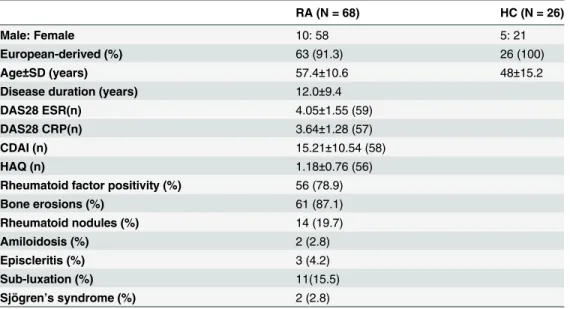
Table 1. Demographic profile of RA patients and healthy controls.
RA (N = 68) HC (N = 26)
Male: Female 10: 58 5: 21
European-derived (%) 63 (91.3) 26 (100)
Age±SD (years) 57.4±10.6 48±15.2
Disease duration (years) 12.0±9.4
DAS28 ESR(n) 4.05±1.55 (59)
DAS28 CRP(n) 3.64±1.28 (57)
CDAI (n) 15.21±10.54 (58)
HAQ (n) 1.18±0.76 (56)
Rheumatoid factor positivity (%) 56 (78.9)
Bone erosions (%) 61 (87.1)
Rheumatoid nodules (%) 14 (19.7)
Amiloidosis (%) 2 (2.8)
Episcleritis (%) 3 (4.2)
Sub-luxation (%) 11(15.5)
Sjögren’s syndrome (%) 2 (2.8)
DAS = Disease Activity Score, CDAI = Clinical Disease Activity Index HAQ = Health Assessment Questionaire
HC = healthy controls
Quantification of sHLA-G
EDTA plasma samples were obtained from peripheral blood. The determination of sHLA-G was performed as described previously [26]. For sHLA-G ELISA the specific capture reagent was the monoclonal antibody G233 (Exbio, Czech Republic). Bound molecules were detected by a polyclonal antiserum rabbit anti-humanβ2-microglobulin (B2M) (Dako, Hamburg,
Ger-many) followed by Envision goat anti-rabbit horseradish peroxidase (Dako, GerGer-many). Plasma samples were diluted 1:2 in PBS. Purified sHLA-G5 protein served as standard reagent [27] and 3,30,5,50-tetramethybenzidine as substrate solution. After stopping the enzyme reaction
with 1 M H2SO4, the optical density was measured at 450 nm (Biotek Instruments, Winooski,
VT). Determination of plasma sHLA-G levels was performed by four-parameter curve fitting. HLA-G5 was used as standard in a concentration ranging from 0.4375–112 ng/ml. PBS was used as a negative control. For the calculation of the ELISA detection limits, a standard curve starting from a concentration of 8 ng/ml was performed in equimolar dilution steps of 5 and 1 ng/ml, respectively. The results obtained were subjected to the software DINTEST (Institute für Rechts-und Verkehrsmedizin, Universitätsklinikum Heidelberg, Germany). According to this procedure, the detection limit of sHLA-G ELISA was 0.94 ng/ml.
Quantification of sHLA-G recognition by the LILRB1 receptor
For the measurement of LILRB1 receptor recognition to sHLA-G molecules in plasma the Luminex-x-MAP technology and instruments were used (Luminex). Microspheres with color code 36 were covalently coupled with the G233 mAb [28]. Recognition of the mAb to the micropheres was performed as recently described [29]: G233 coupled microspheres (1250 per sample) were incubated in a total volume of 50μl with plasma diluted 1:4 in Luminex buffer
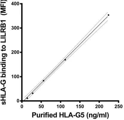
(Cayman). Thereafter the bound HLA-G molecules were exposed to recombinant human LILRB1 receptor protein fused to the Fc region of human IgG1 (R&D Systems). Then, the bound LILRB1 receptor was detected by the anti-human LILRB1 mAb (BD Biosciences), PE conjugated. Measurement of the microspheres was carried out by the Luminex 100 IS System (Luminex). In total, the median fluorescence intensity from 100 microspheres was calculated in each sample. For the determination of HLA-G molecules recognized by the LILRB1 receptor, purified HLA-G5 was used in concentrations ranging from 0–224 ng/ml (Fig 1). The LILRB1 recognition of sHLA-G is given in fluorescence units (FU)/ml. One unit corresponds to 1 ng purified HLA-G5. The detection limit was 11.9 units/ml. Luminex buffer was used as a negative control.
Polymerase chain reaction amplification of the 14bp polymorphism in
exon 8 (3
’
UTR) of the
HLA-G
gene and genotyping
DNA was isolated from peripheral blood of patients and controls using a salting out method [30]. The genotyping of the 14bp polymorphism of the HLA-G gene was performed as previ-ously described [31,32].
Statistical analysis
Results
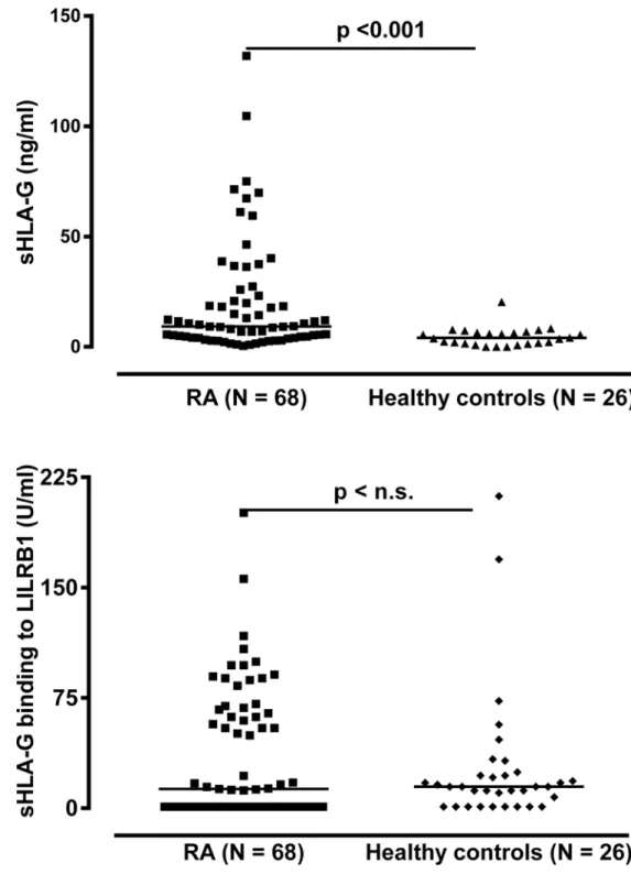
Circulating sHLA-G levels are increased in late RA patients
In order to investigate whether sHLA-G levels were altered in the periphery among late rheu-matoid arthritis patients, we analyzed sHLA-G in plasma samples in 68 RA patients with mean disease duration of about 12 years (Table 1). The sHLA-G plasma levels were significantly in-creased in RA patients as compared to healthy controls (p<0.001, Mann-Whitney test,Fig 2):
The median sHLA-G level of RA patients was 9.3 ng/ml (range: 0–131.9 ng/ml) and in healthy individuals 4.6 ng/ml (range: 0–20.4 ng/ml). Of note, 18 patients (26.5%) exhibited sHLA-G lev-els above the controls’highest concentration (Fig 2). With respect to sHLA-G levels and disease activity parameters, however, no significant correlations were observed (Table A inS1 File). In addition, no significant correlations could be observed between sHLA-G levels and anti-RA treatment regimen (Table B inS1 File).
Circulating sHLA-G molecules are not recognized by their cognate
LILRB1 receptor in a substantial number of late RA patients
In order to evaluate the potential recognition of sHLA-G molecules present in the RA patients plasma we analyzed the binding capacity of sHLA-G to its cognate LILRB1 receptor [28]. Im-portantly, in the recognition assay we used the same antibody (mAb G233) to capture sHLA-G as for the quantitative assessment, but this time bound molecules were exposed to recombinant human LILRB1 followed by human LILRB1 antibody instead of polyclonal rabbit anti-humanβ2-microglobulin antiserum. Results of these assay showed that, despite the high
sHLA-G levels in patients, no quantitative differences in recognition by LILRB1 were observed
Fig 1. sHLA-G recognition by LILRB1 receptor.Straight line indicates the linear regression and dotted line indicates the 95% confidence interval of regression. MFI = mean fluorescence intensity. One unit/ml sHLA-G5 corresponds to 1ng/ml of purified sHLA-G5.
between patients and controls (Fig 2). The median in LILRB1 recognition was 12.9 FU/ml in the patients’group and 15.3 FU/ml in the controls’group (p = 0.632, Mann-Whitney test). However, the proportion of patients presenting specific recognition of sHLA-G molecules by LILRB1 (above the calculated detection limit of the test) was significantly decreased as com-pared to healthy controls (56% vs. 81%, p = 0.027, Chi-square test). Of note, among individuals
Fig 2. Soluble HLA-G level in plasma and its recognition by LILRB1 in RA patients and healthy controls.RA = Rheumatoid arthritis
with no detectable LILRB1 recognition, significantly higher median values of sHLA-G were identified in patients as compared to controls (5.42 vs. 0.83 FU/mL, p<0.001, Mann-Whitney
test,Table 2). This suggests that lack of LILRB1 recognition is mainly due to low amounts (not detectable) of sHLA-G in healthy individuals, whereas in RA patients it is rather a consequence of a large amount of non-recognized sHLA-G molecules. This assumption is further supported by the correlation analysis between sHLA-G levels and LILRB1 recognition: Although a signifi-cant positive correlation between sHLA-G level and LILRB1 recognition was found in both healthy controls (r = 0.57, p = 0.003) and RA patients (r = 0.52, p<0.001), the correlation did
differ with respect to the slope of their linear regression line (Fig 3): For healthy controls the slope of regression line was with 4.43 ± 2.2 steeply rising, whereas for RA patients the slope of regression line was with 0.96 ± 0.18, clearly decreased. In addition to 30 RA patients without detectable LILRB1 recognition, despite substantial levels of circulating sHLA-G molecules, two RA patients revealed sHLA-G molecules in a concentration above 45 ng/ml with a very weak recognition by the LILRB1 receptor. Thus, in a substantial number of late RA patients the cir-culating sHLA-G molecules in the blood were not or only hardly recognized by the LILRB1 re-ceptor suggesting that these sHLA-G molecules are functionally inactive with regard to this receptor.
RF- negative patients were significantly overrepresented in the group of
patients positive for LILRB1 binding
LILRB1 recognition was also analyzed with respect to the presence and absence of rheumatoid factor (RF). The group of patients with detectable LILRB1 binding presented a higher propor-tion of RF-negative patients (31%) compared to the group of patients without LILRB1 recogni-tion (10%, p = 0.033, Chi-square test,Table 3). Also, from the 5 patients where ACCP was accessed, four presented positivity to LILRB1 recognition and were simultaneously negative to ACCP, whereas the only patient negative to LILRB1 recognition was positive to ACCP.
Methotrexate treated patients presented lower LILRB1 binding to
sHLA-G molecules
As the anti-rheumatic drug methotrexate (MTX) is reported to mediate the up-regulation of interleukin-10 and HLA-G [33], we additionally analyzed the LILRB1 recognition to sHLA-G molecules in treated (N = 58) and non-treated (N = 10) RA patients. Interestingly, patients treated with MTX revealed significantly lower sHLA-G recognition of LILRB1 than non-treat-ed patients (mnon-treat-edians: 12.2 vs. 67.7 FU/ml, p = 0.040, Mann Whitney test,Fig 4).
Table 2. Correlation of sHLA-G recognition by LILRB1 and sHLA-G levels.
LILRB1 recognition N (%) RA N (%) HC sHLA-G (range) RAa sHLA-G (range) HCa P-valueb
Positive 38 (56) 21 (81) 16.1 (1.1–131.9) 5.8 (1.4–131.9)
Negative 30 (44) 5 (19) 5.4 (0.5–61.1) 0.8 (0–2.4) <0.001
P-value 0.032c
amedian (range) in ng/ml
bComparison between patients and controls, Mann-Whitney test
cComparison between patients and controls of LILRB1 recognition frequencies by Fisher
’s exact test HC = healthy controls
Circulating sHLA-G levels are not associated to genotypes of the 14bp
polymorphism of the HLA-G gene
Given that polymorphisms at theHLA-Ggene are potentially involved in the susceptibility to autoimmune diseases such as arthritis and were suggested as prognostic factors enabling to stratify patients in groups of responders and non-responders [23], HLA-G variants for the 14 bp were additionally taken into account in the present study (Table 4). No significant differ-ences were observed in genotype frequencies between patients and controls. The already de-scribed difference in blood sHLA-G levels among 14 bpHLA-Ggenotypes was observed in healthy controls, with individuals homozygous for the insertion allele (ins/ins) presenting the lowest median values, as expected (0.7 vs. 5.9 in heterozygotes and 5.5 ng/mL in deletion ho-mozygotes, p = 0.018). However, in late RA patients a different pattern of expression was ob-served: All three genotypes expressed comparable levels of sHLA-G (medians: 5.5, 10.6 and 6.4 ng/mL, respectively, p = 0.534 Mann Whitney test).
Fig 3. Correlation of sHLA-G levels and its recognition by LILRB1 receptor in RA patients and healthy controls.Straight line indicates the linear regression and dotted line indicates the 95% confidence interval of regression. Closed cycles indicate plasma samples of RA patients with HLA-G molecules with an impaired LILRB1 recognition.
doi:10.1371/journal.pone.0123838.g003
Table 3. Correlation of sHLA-G recognition by LILRB1 and the presence of RF.
LILRB1 recognition N (%) RF-positive N (%) RF-negative P-valuea
Positive 27 (69) 12 (31) 0.033
Negative 27 (90) 3 (10)
aChi-square test
Discussion
HLA-G has been described as a molecule involved in tissue protection against inflammatory ag-gression and its expression has been described in several inflammatory conditions, including mul-tiple sclerosis, inflammatory bowel disease, RA and juvenile idiopathic arthritis [20,22,34,35]. Despite of the fact that previous studies have already analyzed soluble HLA-G levels in RA plasma samples, this is the first study to investigate sHLA-G in terms of its binding capacity to one of its receptors (LILRB1) in this disease. From that analysis it was possible to identify an impaired
Fig 4. Methotrexate treatment and sHLA-G recognition by LILRB1 in RA patients.
doi:10.1371/journal.pone.0123838.g004
Table 4. HLA-G 14 bp genotype in relation to sHLA-G levels in adult patient groups and healthy controls.
RA HC RA HC
HLA-G genotype
N (%) RA
N (%) HC
sHLA-Ga (rangeb)
sHLA-Ga (rangeb)
Pbonfc LILRB1 recognitiond (rangee)
LILRB1 recognitiond (rangee)
Pbonfc
del/del 22 (32) 11 (42) 6.4 (1.5–131.9) 5.5 (1.6–11.0) 0.178 0 (0–155.9) 17.3 (0–212.2) 0.074 del/ins 37 (55) 11 (42) 10.6 (0.5–104.6) 5.9 (0.8–20.4) 0.013 16.1 (0–117.1) 14.7 (0–169.2) 0.970 ins/ins 9 (13) 4 (15) 5.5 (1.0–59.5) 0.7 (0–2.1) 0.020 50.8 (0–201.0) 3.8 (0–24.6) 0.148
P-value 0.567f 0.534g
0.018g 0.200g 0.182g
amedian in ng/ml b(minimum
—maximum sHLA-G in ng/ml)
cComparison between patients and controls of the same genotype, Mann Whitney test dmedian in units/ml (measured by LUMINEX)
e(minimum
—maximumfluorescence intensity)
fComparison of genotype frequencies between patients and controls, Chi-square test gComparison among genotypes, Kruskall-Wallis test
HC = healthy controls
binding capacity of sHLA-G circulating molecules to LILRB1 in RA patients, suggesting an im-paired functionality of these molecules regarding this receptor.
The first important finding was that circulating sHLA-G levels were increased in late RA pa-tients. This is in agreement with the results from Rizzo et al., which showed that in early un-treated RA patients, detectable levels of sHLA-G in plasma could be observed in all subjects, as compared to a minority (23%) of healthy controls, and that those levels increased upon anti-RA treatment in those patients [23]. This increase could reflect an attempt of the immune sys-tem to counterbalance the autoimmune process. However, in our RA patient cohort, no notice-able correlation between plasma sHLA-G levels and disease activity parameters was observed (Table A inS1 File). Furthermore, in our study, sHLA-G levels did not differ with respect to anti-RA treatment. Our results are at variance to the first report on sHLA-G in RA describing lower plasma HLA-G levels in late RA patients [22] and to those from Rizzo et al. [23]. Never-theless in the latter study only 23% of the controls were positive for sHLA-G whereas substan-tial amounts of sHLA-G molecules could be detected in all blood samples of RA patients (100%). The differences in mean sHLA-G levels might be partially explained by different proto-cols of the HLA-G measurement: while the study from Verbruggen et al. [22] used a two-step ELISA that included the depletion of classic HLA-I and HLA-E and detection by a pan-HLA-I antibody (W6/32), our assay and also that one used in the work from Rizzo et al. skipped the depletion step and used a more direct detection strategy by an anti-HLA-G antibody. Impor-tantly, the study of Rizzo et al. [23] used the antibody MEM-G/09 as capture antibody where we were using the HLA-G specific antibody G233 and for these two antibodies discrepancies in sHLA-G concentration readouts have been previously reported [36]. Other explanations for those discrepancies might include sample composition and differences in treatment regimens.
of HLA-G, which could not be identified by our detection systems. However, these HLA-G structures might additionally play a functional role in the immune pathology of RA.
Our results also showed an overrepresentation of RF- patients in the group of patients with detectable binding of HLA-G to LILRB1. Recently, Naji et al. demonstrated that HLA-G recog-nition to LILRB1 suppresses B cell responses, including B cell proliferation, differentiation, and Ig secretion [40]. Therefore, we could suggest that in rheumatoid arthritis patients, high levels of sHLA-G molecules with capability to bind to LILRB1 could also impair the production of auto-antibodies. In this sense, and also considering the existence in the literature of controver-sial results in sHLA-G levels and severity in different autoimmune diseases (such as in Systemic Lupus Erythematosus [41,42]), it would be important not only to access the presence and lev-els of sHLA-G in autoimmune patients but also to perform tests evaluating the its recognition by its cognate receptors.
Considering anti-RA therapy, patients treated with MTX revealed lower sHLA-G recogni-tion of LILRB1. This suggests that MTX does not facilitate the up-regularecogni-tion of HLA-G dimers, at least in late RA patients after long lasting treatment. Further studies need to be performed in order to clarify how HLA-G dimerization will behave in late RA patient treatment.
Previous studies have associated the 14 bp insertion allele and someHLA-Galleles linked to it as low sHLA-G producers, [4,43,44]. This finding was confirmed in our control sample but not among RA patients. This suggests that, despite being associated to lower levels of sHLA-G expression in plasma of healthy individuals, the 14 bp insertion allele (and consequently the ins/ins genotype) is responsive in situations in which sHLA-G mediated regulation of inflam-mation is required (i.e. immunologic stress). Furthermore, the lack of association between ge-notype and sHLA-G plasma levels in RA patients suggests that post-transcriptional
mechanisms affecting both the level and function of sHLA-G might be operative in late RA.
Conclusions
In this study we present for the first time that in a substantial number of chronically inflamed RA patients, the circulating sHLA-G molecules in the blood are impaired with respect to the LILRB1 receptor recognition. Thus, our findings offer new insights into the immune patholo-gy of chronically inflamed RA patients after long-lasting anti-RA treatment. In this scenario, sHLA-G levels are indeed increased but these molecules are not qualified to exert its immune suppressive and protective functions against inflammation via LILRB1 receptor. Giving con-sideration to the inherent complexity of the HLA-G molecule, these observations call atten-tion to the importance of receptor binding assays as a complementaatten-tion to the quantificaatten-tion of HLA-G, in order for a better understanding the physiological phenomena involving this molecule.
Supporting Information
S1 File.Table A, Correlations of RA disease parameters and levels of sHLA-G molecules with
their LILRB1 recognition. Table B, Relationship of RA treatment and levels of sHLA-G mole-cules with their LILRB1 recognition.
(DOCX)
Acknowledgments
Author Contributions
Conceived and designed the experiments: TDV JABC MS BW PAH MB CVB JCTB RMX VR. Performed the experiments: TDV JABC MS BW PAH MB CVB JCTB RMX VR. Analyzed the data: TDV JABC MS BW PAH MB CVB JCTB RMX VR. Contributed reagents/materials/ analysis tools: TDV JABC MB CVB JCTB RMX VR. Wrote the paper: TDV JABC BW PAH VR.
References
1. Paul P, Cabestre FA, Ibrahim EC, Lefebvre S, Khalil-Daher I, Vazeux G, et al. Identification of HLA-G7 as a new splice variant of the HLA-G mRNA and expression of soluble HLA-G5, -G6, and -G7 tran-scripts in human transfected cells. Hum Immunol. 2000; 61: 1138–49. PMID:11137219
2. Fujii T, Ishitani A, Geraghty DE. A soluble form of the HLA-G antigen is encoded by a messenger ribo-nucleic acid containing intron 4. J Immunol. 1994; 153: 5516–24. PMID:7989753
3. Kirszenbaum M, Moreau P, Gluckman E, Dausset J, Carosella E. An alternatively spliced form of HLA-G mRNA in human trophoblasts and evidence for the presence of HLA-HLA-G transcript in adult lympho-cytes. Proc Natl Acad Sci U S A. 1994; 91: 4209–13. PMID:8183892
4. Hviid TV, Hylenius S, Rorbye C, Nielsen LG. HLA-G allelic variants are associated with differences in the HLA-G mRNA isoform profile and HLA-G mRNA levels. Immunogenetics. 2003; 55: 63–79. PMID:
12712263
5. Paul P, Cabestre FA, Ibrahim EC, Lefebvre S, Khalil-Daher I, Vazeux G, et al. Identification of HLA-G7 as a new splice variant of the HLA-G mRNA and expression of soluble HLA-G5, -G6, and -G7 tran-scripts in human transfected cells. Hum Immunol. 2000; 61: 1138–49. PMID:11137219
6. Carosella ED, Gregori S, LeMaoult J. The tolerogenic interplay(s) among HLA-G, myeloid APCs, and regulatory cells. Blood. 2011; 118: 6499–505. doi:10.1182/blood-2011-07-370742PMID:21960588
7. Veit TD, Vianna P, Chies JAB. HLA-G—From Fetal Tolerance to a Regulatory Molecule in Inflammatory Diseases. Curr Immunol Rev. 2010; 6: 1–15.
8. Gonzalez A, Rebmann V, LeMaoult J, Horn PA, Carosella ED, Alegre E. The immunosuppressive mol-ecule HLA-G and its clinical implications. Crit Rev Clin Lab Sci. 2012; 49: 63–84. doi:10.3109/ 10408363.2012.677947PMID:22537084
9. Shiroishi M, Kuroki K, Ose T, Rasubala L, Shiratori I, Arase H, et al. Efficient leukocyte Ig-like receptor signaling and crystal structure of disulfide-linked HLA-G dimer. J Biol Chem. 2006; 281: 10439–47. PMID:16455647
10. Apps R, Gardner L, Sharkey AM, Holmes N, Moffett A. A homodimeric complex of HLA-G on normal tro-phoblast cells modulates antigen-presenting cells via LILRB1. Eur J Immunol. 2007; 37: 1924–37. PMID:17549736
11. Zhong M, Weng X, Liang Z, Lu S, Li J, Chen X, et al. Dimerization of soluble HLA-G by IgG-Fc fragment augments ILT2-mediated inhibition of T-cell alloresponse. Transplantation. 2009; 87: 8–15. doi:10. 1097/TP.0b013e31818b6141PMID:19136885
12. Baricordi OR, Stignani M, Melchiorri L, Rizzo R. HLA-G and inflammatory diseases. Inflamm Allergy Drug Targets. 2008; 7: 67–74. PMID:18691135
13. Fainardi E, Castellazzi M, Stignani M, Morandi F, Sana G, Gonzalez R, et al. Emerging topics and new perspectives on HLA-G. Cell Mol Life Sci. 2011; 68: 433–51. doi:10.1007/s00018-010-0584-3PMID:
21080027
14. Khosrotehrani K, Le Danff C, Reynaud-Mendel B, Dubertret L, Carosella ED, Aractingi S. HLA-G ex-pression in atopic dermatitis. J Invest Dermatol. 2001; 117: 750–2. PMID:11564188
15. Wiendl H, Behrens L, Maier S, Johnson MA, Weiss EH, Hohlfeld R. Muscle fibers in inflammatory my-opathies and cultured myoblasts express the nonclassical major histocompatibility antigen HLA-G. Ann Neurol. 2000; 48: 679–84. PMID:11026456
16. Aractingi S, Briand N, Le Danff C, Viguier M, Bachelez H, Michel L, et al. HLA-G and NK receptor are expressed in psoriatic skin: a possible pathway for regulating infiltrating T cells? Am J Pathol. 2001; 159: 71–7. PMID:11438456
17. Kapasi K, Albert SE, Yie S, Zavazava N, Librach CL. HLA-G has a concentration-dependent effect on the generation of an allo-CTL response. Immunology. 2000; 101: 191–200. PMID:11012772
19. Kanai T, Fujii T, Kozuma S, Yamashita T, Miki A, Kikuchi A, et al. Soluble HLA-G influences the release of cytokines from allogeneic peripheral blood mononuclear cells in culture. Mol Hum Reprod. 2001; 7: 195–200. PMID:11160846
20. Torres MI, Le Discorde M, Lorite P, Rios A, Gassull MA, Gil A, et al. Expression of HLA-G in inflammato-ry bowel disease provides a potential way to distinguish between ulcerative colitis and Crohn's disease. Int Immunol. 2004; 16: 579–83. PMID:15039388
21. Mitsdoerffer M, Schreiner B, Kieseier BC, Neuhaus O, Dichgans J, Hartung HP, et al. Monocyte-derived HLA-G acts as a strong inhibitor of autologous CD4 T cell activation and is upregulated by interferon-beta in vitro and in vivo: rationale for the therapy of multiple sclerosis. J Neuroimmunol. 2005; 159: 155–64. PMID:15652415
22. Verbruggen LA, Rebmann V, Demanet C, De Cock S, Grosse-Wilde H. Soluble HLA-G in rheumatoid arthritis. Hum Immunol. 2006; 67: 561–7. PMID:16916651
23. Rizzo R, Farina I, Bortolotti D, Galuppi E, Rotola A, Melchiorri L, et al. HLA-G may predict the disease course in patients with early rheumatoid arthritis. Hum Immunol. 2013.
24. Rizzo R, Rubini M, Govoni M, Padovan M, Melchiorri L, Stignani M, et al. HLA-G 14-bp polymorphism regulates the methotrexate response in rheumatoid arthritis. Pharmacogenet Genomics. 2006; 16: 615–23. PMID:16906016
25. Ferraz MB, Oliveira LM, Araujo PM, Atra E, Tugwell P. Crosscultural reliability of the physical ability di-mension of the health assessment questionnaire. J Rheumatol. 1990; 17: 813–7. PMID:2388204
26. Schutt P, Schutt B, Switala M, Bauer S, Stamatis G, Opalka B, et al. Prognostic relevance of soluble human leukocyte antigen-G and total human leukocyte antigen class I molecules in lung cancer pa-tients. Hum Immunol. 2010; 71: 489–95. doi:10.1016/j.humimm.2010.02.015PMID:20156510
27. Rebmann V, Lemaoult J, Rouas-Freiss N, Carosella ED, Grosse-Wilde H. Report of the Wet Workshop for Quantification of Soluble HLA-G in Essen, 2004. Hum Immunol. 2005; 66: 853–63. PMID:16216668
28. Verloes A, Van de Velde H, LeMaoult J, Mateizel I, Cauffman G, Horn PA, et al. HLA-G expression in human embryonic stem cells and preimplantation embryos. J Immunol. 2011; 186: 2663–71. doi:10. 4049/jimmunol.1001081PMID:21248264
29. Rebmann V, Switala M, Eue I, Schwahn E, Merzenich M, Grosse-Wilde H. Rapid evaluation of soluble HLA-G levels in supernatants of in vitro fertilized embryos. Hum Immunol. 2007; 68: 251–8. PMID:
17400060
30. Lahiri DK, Nurnberger JI Jr. A rapid non-enzymatic method for the preparation of HMW DNA from blood for RFLP studies. Nucleic Acids Res. 1991; 19: 5444. PMID:1681511
31. Hviid TV, Hylenius S, Hoegh aM, Kruse C, Christiansen OB. HLA-G polymorphisms in couples with re-current spontaneous abortions. Tissue Antigens. 2002; 60: 122–32. PMID:12392506
32. Cordero EAA, Veit TD, Silva MAL, Jacques SMC, Silla LMDR, Chies JAB. HLA-G polymorphism influ-ences the susceptibility to HCV infection in sickle cell disease patients. Tissue Antigens. 2009: 308–13. doi:10.1111/j.1399-0039.2009.01331.xPMID:19775370
33. Rizzo R, Rubini M, Govoni M, Padovan M, Melchiorri L, Stignani M, et al. HLA-G 14-bp polymorphism regulates the methotrexate response in rheumatoid arthritis. Pharmacogenetics and genomics. 2006; 16: 615–23. PMID:16906016
34. Fainardi E, Rizzo R, Melchiorri L, Vaghi L, Castellazzi M, Marzola A, et al. Presence of detectable levels of soluble HLA-G molecules in CSF of relapsing-remitting multiple sclerosis: relationship with CSF solu-ble HLA-I and IL-10 concentrations and MRI findings. J Neuroimmunol. 2003; 142: 149–58. PMID:
14512174
35. Prigione I, Penco F, Martini A, Gattorno M, Pistoia V, Morandi F. HLA-G and HLA-E in patients with ju-venile idiopathic arthritis. Rheumatology (Oxford). 2011; 50: 966–72. doi:10.1093/rheumatology/ keq418PMID:21186170
36. Gonzalez A, Alegre E, Arroyo A, LeMaoult J, Echeveste JI. Identification of circulating nonclassic human leukocyte antigen G (HLA-G)-like molecules in exudates. Clin Chem. 2011; 57: 1013–22. doi:
10.1373/clinchem.2010.159673PMID:21527645
37. Apps R, Sharkey A, Gardner L, Male V, Kennedy P, Masters L, et al. Ex vivo functional responses to HLA-G differ between blood and decidual NK cells. Molecular human reproduction. 2011; 17: 577–86. doi:10.1093/molehr/gar022PMID:21471023
38. Zilberman S, Schenowitz C, Agaugue S, Benoit F, Riteau B, Rouzier R, et al. HLA-G1 and HLA-G5 ac-tive dimers are present in malignant cells and effusions: the influence of the tumor microenvironment. European journal of immunology. 2012; 42: 1599–608. doi:10.1002/eji.201141761PMID:22678912
40. Naji A, Menier C, Maki G, Carosella ED, Rouas-Freiss N. Neoplastic B-cell growth is impaired by HLA-G/ILT2 interaction. Leukemia. 2012; 26: 1889–92. doi:10.1038/leu.2012.62PMID:22441169
41. Rosado S, Perez-Chacon G, Mellor-Pita S, Sanchez-Vegazo I, Bellas-Menendez C, Citores MJ, et al. Expression of human leukocyte antigen-G in systemic lupus erythematosus. Hum Immunol. 2008; 69: 9–15. doi:10.1016/j.humimm.2007.11.001PMID:18295670
42. Rizzo R, Hviid TV, Govoni M, Padovan M, Rubini M, Melchiorri L, et al. HLA-G genotype and HLA-G ex-pression in systemic lupus erythematosus: HLA-G as a putative susceptibility gene in systemic lupus erythematosus. Tissue Antigens. 2008; 71: 520–9. doi:10.1111/j.1399-0039.2008.01037.xPMID:
18380776
43. Rebmann V, van der Ven K, Pässler M, Pfeiffer K, Krebs D, Grosse-Wilde H. Association of soluble HLA-G plasma levels with HLA-G alleles. Tissue antigens. 2001; 57: 15–21. PMID:11169254