Replication
Andrea Marzi1,2, Thomas Reinheckel3,4, Heinz Feldmann1,2,5*
1Laboratory of Virology, Division of Intramural Research, National Institute of Allergy and Infectious Diseases, National Institutes of Health, Hamilton, Montana, United States of America,2Special Pathogens Program, National Microbiology Laboratory, Public Health Agency of Canada, Winnipeg, Manitoba, Canada,3Institute of Molecular Medicine and Cell Research, Albert-Ludwigs-University, Freiburg, Germany,4BIOSS Centre for Biological Signaling Studies, Albert-Ludwigs-University, Freiburg, Germany, 5Department of Medical Microbiology, University of Manitoba, Winnipeg, Manitoba, Canada
Abstract
Ebola virus (EBOV), familyFiloviridae, emerged in 1976 on the African continent. Since then it caused several outbreaks of viral hemorrhagic fever in humans with case fatality rates up to 90% and remains a serious Public Health concern and biothreat pathogen. The most pathogenic and best-studied species isZaire ebolavirus (ZEBOV). EBOV encodes one viral surface glycoprotein (GP), which is essential for replication, a determinant of pathogenicity and an important immunogen. GP mediates viral entry through interaction with cellular surface molecules, which results in the uptake of virus particles via macropinocytosis. Later in this pathway endosomal acidification activates the cysteine proteases Cathepsin B and L (CatB, CatL), which have been shown to cleave ZEBOV-GP leading to subsequent exposure of the putative receptor-binding and fusion domain and productive infection. We studied the effect of CatB and CatL onin vitroandin vivoreplication of EBOV. Similar to previous findings, our results show an effect of CatB, but not CatL, on ZEBOV entry into cultured cells. Interestingly, cell entry by other EBOV species (Bundibugyo,Coˆte d’Ivoire,RestonandSudan ebolavirus) was independent of CatB or CatL as was EBOV replication in general. To investigate whether CatB and CatL have a rolein vivoduring infection, we utilized the mouse model for ZEBOV. Wild-type (control),catB2/2andcatL2/2mice were equally susceptible to lethal
challenge with mouse-adapted ZEBOV with no difference in virus replication and time to death. In conclusion, our results show that CatB and CatL activity is not required for EBOV replication. Furthermore, EBOV glycoprotein cleavage seems to be mediated by an array of proteases making targeted therapeutic approaches difficult.
Citation:Marzi A, Reinheckel T, Feldmann H (2012) Cathepsin B & L Are Not Required for Ebola Virus Replication. PLoS Negl Trop Dis 6(12): e1923. doi:10.1371/ journal.pntd.0001923
Editor:Thomas Geisbert, University of Texas Medical Branch, United States of America ReceivedJuly 13, 2012;AcceptedOctober 12, 2012;PublishedDecember 6, 2012
This is an open-access article, free of all copyright, and may be freely reproduced, distributed, transmitted, modified, built upon, or otherwise used by anyone for any lawful purpose. The work is made available under the Creative Commons CC0 public domain dedication.
Funding:This research was supported in part by the Intramural Research Program of the National Institute of Allergy and Infectious Diseases (NIAID), National Institutes of Health (NIH) and the Public Health Agency of Canada (PHAC). A.M. was in part funded by the Natural Science and Engineering Council of Canada (NSERC). The funders had no role in study design, data collection and analysis,decision to publish, or preparation of the manuscript.
Competing Interests:The authors have declared that no competing interests exist. * E-mail: feldmannh@niaid.nih.gov
Introduction
Members of the family Filoviridae, Ebola virus (EBOV) and Marburg virus (MARV), are the causative agents of viral hemorrhagic fever in central Africa and a major public health threat in their endemic areas. Worldwide concern relates to the importation of infected individuals and the potential use of filoviruses as biothreat pathogens [1]. All current MARV strains belong to the Lake Victoria marburgvirus species, while Ebola virus (EBOV) strains are attributed to five different species:Zaire ebolavirus (ZEBOV), Sudan ebolavirus (SEBOV), Coˆte d’Ivoire ebolavirus (CIE-BOV),Reston ebolavirus(REBOV) andBundibugyo ebolavirus(BEBOV) [1,2]. The species vary in their pathogenicity for humans with ZEBOV being most pathogenic (up to 90% case fatality rate), followed by SEBOV and BEBOV with about 50% and.25% case fatality rates, respectively. CIEBOV and REBOV cause lethal infections in nonhuman primates, but have not yet been associated with fatal human cases [1,2]. Although EBOV (mainly ZEBOV) and MARV have been extensively studiedin vitroandin vivo, today there is neither a licensed vaccine nor treatment available.
EBOV entry into target cells is still not fully understood and remains a focus of ongoing research. While a number of
attachment factors seem to facilitate EBOV entry [3], so far no specific cell surface receptor molecule has been identified. Recently, it was shown that the cholesterol transporter Nie-mann-Pick C1 (NPC1) is required for ZEBOV infection [4,5]. NPC1 is a ubiquitously expressed endosomal membrane protein involved in the fusion and fission of endosomes and lysosomes [6], and its deficiency has been shown to impact on HIV-1 particle release [7]. Its presence in the endosome fits the current EBOV entry model based on macropinocytosis, a cellular pathway proposed to be the main uptake mechanism of EBOV particles into cells [8–10]. Previously, Chandranet al.identified the cysteine proteases cathepsin B (CatB) and cathepsin L (CatL), which are also present in endosomes, as important factors for ZEBOV entry [11]. According to the current model, cleavage of the ZEBOV glycoprotein (GP) by CatB is necessary for exposure of the core receptor-binding domain and fusion machinery, otherwise buried in the GP structure, to initiate fusion of the viral and the endosomal membrane [12–14]. Subsequently, the viral genome along with the replication complex is released into the cytoplasm where replication and virus progeny production occur.
and exhibit nonspecific proteolytic activity within lysosomes [15,16]. CatB has been associated with TNF-a induced liver damage and seems to play a critical role for the development of pancreatitis [17,18]. Despite these facts, catB2/2 mice are phenotypically similar to wild-type control mice and fully immunocompetent [18]. CatL is important for epidermal homeo-stasis and the regulation of the hair cycle and as suchcatL2/2mice are hairless [19]. Furthermore, CatL is involved in MHC II-mediated antigen presentation in epithelial cells of the thymus [20]. Consequently,catL2/2mice have reduced numbers of CD4+ T helper cells, which are however fully functional. A double knockout mouse lacking CatB and CatL has been generated but is not viable long enough for experimental use [21,22]. To determine the importance of cathepsins in viral infections, various inhibitors of endosomal acidification, cathepsin or specifically CatB and CatL activity have been usedin vitro. In these studies the role of cathepsins have been demonstrated to be important for the entry of reovirus, SARS-CoV, henipaviruses and ZEBOV [11,14,23–27].
Here we show that while CatB mediates ZEBOV entryin vitro, it is not important for entry and replication of any other EBOV species. We further show thatin vivoreplication of mouse-adapted ZEBOV (MA-ZEBOV) is independent of CatB and CatL, indicating that both of these cathepsins are not required for ZEBOV replicationin vivo.
Methods
Animal ethics statement
Animals were handled in the Biosafety Level 4 (BSL4) containment space of the Integrated Research Facility (IRF) at the Rocky Mountain Laboratories (RML), Division of Intramural Research (DIR), National Institute of Allergy and Infectious Diseases (NIAID), National Institutes of Health (NIH). Research was conducted in compliance with the guidelines of the NIAID/ RML Institutional Animal Care and Use Committee (IACUC). The facility, where this research was conducted, is fully accredited by the Association for the Assessment and Accreditation of Laboratory Animal Care International (AAALAC) with approved Office of Laboratory Animal Welfare (OLAW) assurance (#A4149-01). Research was conducted under a protocol approved by the IACUC. All procedures were conducted by trained personnel under veterinary supervision, and all invasive clinical
procedures were performed while animals were anesthetized. Endpoint criteria, as specified by the IACUC approved scoring parameters, were used to determine when animals should be humanely euthanized.
Cell culture and virus propagation
Vero E6 cells and Mouse Embryonic Fibroblast (MEF) cell lines lacking cathepsin B, cathepsin L or both cathepsins were cultured in Dulbecco’s Modified Eagle Medium (DMEM) (Sigma, St. Louis, MO) supplemented with 10% FBS, penicillin/streptomycin and L-glutamine in a 37uC incubator, 5% CO2. VSV (serotype
Indiana), SARS (strain Tor 2), BEBOV [2], CIEBOV (strain Tai Forest), REBOV (strain Pennsylvania), SEBOV (strain Boniface), ZEBOV (strain Mayinga) and the furin knockout and mouse-adapted variants of ZEBOV (ZEBOV-Fko and MA-ZEBOV, respectively) [28–30] were propagated in Vero E6 cells. The supernatants were cleared of cell debris by centrifugation at 1,5006g for 10 min, aliquoted and stored in liquid nitrogen. Viral
titers were determined conducting conventional plaque assay or immunoplaque assay. All infectious work with EBOV was performed in the Biosafety Level 4 (BSL4) laboratories at the National Microbiology Laboratory (NML) of the Public Health Agency of Canada (PHAC) or the IRF, RML, DIR, NIAID, NIH.
Foci reduction experiments
The endosomal acidification inhibitor Bafilomycin A1 (BafA1) and the inhibitor CA074 (specific for CatB) were obtained from Sigma (St. Louis, MO); CatL-inhibitor V was purchased from Calbiochem (via EMD Chemicals Inc., Gibbstown, NJ). All inhibitors were dissolved in DMSO and stored at 220uC. Dilutions were prepared in plain DMEM (no supplements) prior to every experiment. For foci reduction experiments Vero E6 cells were seeded in 48-well plates the day before infection. Media was removed and cells were incubated for one hour with 50ml inhibitor in the following concentrations as described previously [11]: BafA1 200, 100, 50, 20 nM; CA074 (CatB) 200, 100, 50, 20mM; CatL-inhibitor V 20, 10, 5, 2mM. Then 50ml plain DMEM with 100 focus forming units (ffu) virus were added and kept for one hour at 37uC. After three washes with plain DMEM a 1:1 mixture of 2.4% carboxymethyl cellulose (CMC) and 26
Minimal Essential Medium (MEM) (Life Technologies, Carlsbad, CA) supplemented with 4% FBS, penicillin/streptomycin and L-glutamine was added containing the appropriate concentration of inhibitor (BafA1 100, 50, 25, 10 nM; CA074 (CatB) 100, 50, 25, 10mM; CatL-inhibitor V 10, 5, 2.5, 1mM). SARS infected cells were fixed and stained with crystal violet and plaques were counted three days after infection. Four days after infection EBOV inoculated cells were fixed with 10% neutral buffered formalin and removed from BSL4 following standard operating procedures. Subsequently, the cells were permeabilized and foci were stained with a rabbit anti-VP40 antibody (kindly provided by Y. Kawaoka, University of Wisconsin, Madison, WI) or a rabbit serum directed against REBOV-NP (kindly provided by A. Takada, Hokkaido University, Sapporo, Japan) followed by a FITC-labeled secondary antibody (Sigma, ST. Louis, MO). Foci were counted using a fluorescent microscope (Carl Zeiss Micro-imaging LLC, Thornwood, NY).
Viral growth kinetics
Vero E6 cells were seeded in a 24-well plate the day before the experiment. Pretreatment occurred with 200ml of 100 nM BafA1, 100mM CA074, 10mM CatL-inhibitor V or no inhibitor for 1 hour. Thereafter, 200ml ZEBOVwt (MOI = 1) were added and cells were incubated for another hour. Following three washes with
Author Summary
It is currently believed that Ebola virus (EBOV) enters cells via macropinocytosis following which, the cysteine prote-ases cathepsin B and L (CatB, CatL) cleave the viral glycoprotein (GP) allowing exposure of its core receptor-binding and fusion domain thus facilitating subsequent infection. We studied the effect of CatB and CatL onin vitro and in vivoEBOV replication. Our results demonstrate a reduction ofZaire ebolavirus(ZEBOV) entry upon selective inhibition of CatB, but not CatL in cell culture. Interestingly, all other EBOV species enter the cells efficiently when CatB and/or CatL activity is blocked. Moreover, when wild-type (control),catB2/2 andcatL2/2mice were infected with a
plain DMEM, cells in each well were covered with 1 ml DMEM (supplemented with 2% FBS) containing 50 nM BafA1, 50mM CA074, 5mM CatL-inhibitor V or no inhibitor. MEF cell lines were seeded in a 24-well plate the day before the experiment. For infection, 200ml ZEBOVwt or MA-ZEBOV (MOI = 1) were added and incubated for one hour. Following three washes with plain DMEM, cells in each well were covered with 1 ml DMEM (supplemented with 2% FBS). At time points 0, 12, 24, 48, 72 and 96 hours post infection 200ml supernatant were collected from all infected cells, and 200ml DMEM with 2% FBS containing the appropriate concentration of inhibitor were added back into each well. Samples were stored at280uC before titration on Vero E6 cells.
Mouse infections
Groups of C57BL/6catB2/2, C57BL/6 catL2/2or C57BL/6 (control) mice were infected intraperitoneally (i.p.) with 10 ffu MA-ZEBOV (1,000 LD50) or 16105pfu VSVwt (serotype Indiana)
and monitored daily for weight loss and signs of disease. On day 3 and 7 post infection 3 mice of each group were euthanized and blood, liver and spleen samples were collected and stored at
280uC for virus titration. Surviving animals were euthanized at day 28 (study endpoint) and final serum was collected to determine antibody titers.
Virus load determination
Vero E6 cells were seeded in 96-well plates the day before titration. Liver and spleen samples were thawed, weighed, homogenized in 10-fold weight/volume of plain DMEM and serial 10-fold dilutions were prepared. Blood samples and supernatant collected from infected cells to determine viral growth for ZEBOVwt and MA-ZEBOV were thawed and serial 10-fold dilutions were prepared. Media was removed from cells and triplicates were inoculated with each dilution. After one hour DMEM supplemented with 2% FBS, penicillin/streptomycin and L-glutamine was added and incubated at 37uC. Cells were monitored for cytopathic effect (CPE) and 50% tissue culture infectious dose (TCID50) was calculated for each sample
employ-ing the Reed and Muench method [31].
Humoral immune responses in mice
For the detection of ZEBOV-GP specific antibodies in mouse sera, soluble ZEBOV-GP antigen (ZEBOV-GPDTM) was pro-duced and used in an enzyme-linked immunosorbent assay (ELISA) as described before [32,33]. For the detection of VSV-specific antibodies VSV was propagated in Vero E6 cells. VSV particles were harvested after 48 hrs, purified by centrifugation through a 20% sucrose cushion and treated with 0.05% Triton-X100 in PBS prior to storage at280uC. The ELISA used VSV antigen in a 1:100 dilution. Sera from mice infected with MA-ZEBOV were inactivated by c-irradiation as per standard operating protocol.
Statistical analyses
One-way ANOVA was performed using Prism 5 (Graph Pad Software Inc.).
Results
ZEBOV entryin vitrois CatB-mediated
The observation that entry of ZEBOV into Vero E6 cells is CatB- and CatL-dependent was initially described in 2005 by Chandran and colleagues [11]. For further analysis of ZEBOV uptake into Vero E6 cells we performed a differentin vitroassay
based on foci reduction using the same inhibitors. Cells were seeded and treated with various lysosome and cathepsin inhibitors for one hour, infected with ZEBOV wild-type (wt) for one hour, covered with carboxymethyl cellulose containing inhibitors and fixed after incubation for 4 days. The number of foci was determined by immunostaining. Only the inhibition of CatB resulted in significantly reduced virus entry, whereas CatL proteolytic activity did not appear to be obligatory for ZEBOV uptake into Vero E6 cells, although CatL is highly expressed in these cells [27]. SARS-CoV was used as a CatL-dependent control virus [26] to verify the activity of the CatL inhibitor (Fig. 1). In addition to ZEBOVwt, we also tested a furin cleavage site knockout ZEBOV (ZEBOV-Fko) [28,29] and the MA-ZEBOV in this assay to determine whether furin cleavage of the glycoprotein or mutations caused by the adaptation process of the virus to mice would influence the requirement for cathepsin cleavage. Both viruses entered Vero E6 cells in a CatB-dependent manner similar to ZEBOVwt and were not affected by the presence of the CatL inhibitor (Fig. 1). These results show that the uptake of all tested viruses occurred by utilizing the cellular endocytotis machinery as indicated by the inhibitory effect of Bafilomycin A1 (BafA1), an endosomal acidification inhibitor. Furthermore, proteolytic pro-cessing of the ZEBOV-GP by furin is not a prerequisite for cathepsin cleavage in the virus maturation or entry process. In addition, mutations throughout the viral genome acquired during the adaptation process of ZEBOV to the mouse have no influence on ZEBOV uptake.
The role of CatB in cellular uptake differs among Ebola virus species
Our data on ZEBOV only partially support previously published observations [11], but confirm more recent studies claiming that CatB, rather than CatL, plays a role in ZEBOV entry [34,35]. Therefore, we studied the role of cathepsin cleavage on virus entry for BEBOV, CIEBOV, REBOV and SEBOV using a representative strain for each EBOV species. Foci reduction assays were performed and evaluated as described above. Infection of all viruses was reduced in the presence of BafA1 (Fig. 2), confirming that endosomal acidification plays a critical role during the entry process of all these EBOVs. Interestingly, only the maximum concentration of the CatB inhibitor had an effect on BEBOV uptake into Vero E6 cells, whereas CIEBOV, REBOV and SEBOV produced foci independent of the presence of the inhibitor (Fig. 2). The entry of none of the EBOVs was affected by the CatL inhibitor. This data suggests that CatB is not equally important for the entry of other EBOVs and seems to have a selective effect on ZEBOV and a limited effect on BEBOV entry.
ZEBOV replication is largely independent on cathepsin B cleavage
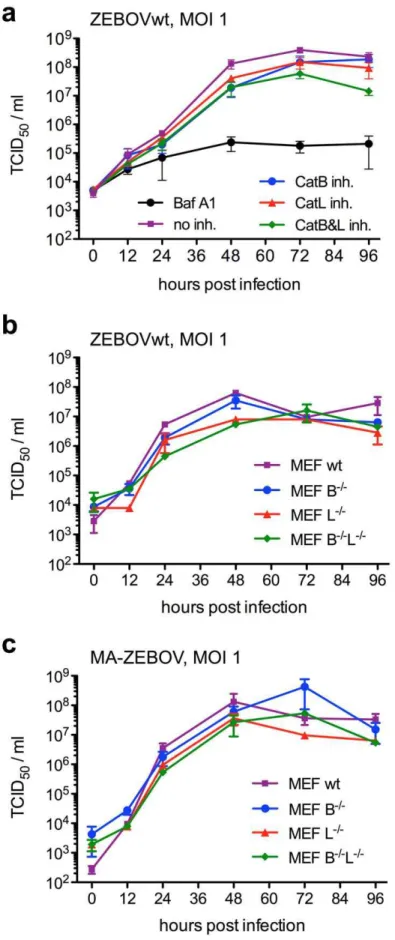
After confirmation that CatB has a role in ZEBOV entry we analyzed virus replication in the presence of cathepsin inhibitors. Vero E6 cells were treated with inhibitors (BafA1 100 nM; CatB 100mM; CatL 10mM) for one hour prior to ZEBOVwt infection (MOI = 1). After unbound virus was washed away the cells were covered with medium containing inhibitor (BafA1 50 nM; CatB 50mM; CatL 5mM), samples were taken at the indicated time points and stored at280uC for virus titration. Virus titers were determined on Vero E6 cells using a 50% tissue culture infectious dose (TCID50) assay and calculated using the Reed and Muench
ZEBOV replication by about 1 log. This indicates that the CatB inhibitory effect on entry is likely compensated for by other cellular proteases. However, BafA1 reduces virus growth by more than 2 logs, showing that endosomal acidification is important for efficient ZEBOV entry and replication (Fig. 1, 3A). In order to further confirm the data we performed ZEBOVwt and MA-ZEBOV infections in MEF cell lines deficient in the expression of CatB, CatL or both cathepsins. The cells were infected for one hour with ZEBOVwt or MA-ZEBOV (MOI = 1) and samples were taken at the indicated time points. Virus titers were determined on Vero E6 cells using a TCID50assay and calculated
using the Reed and Muench formula (Fig. 3B,C) [31]. ZEBOVwt and MA-ZEBOV replicated similarly well in the absence of CatB or CatL or both proteases. Although small differences in titer were
observed at certain time points, one-way ANOVA did not find statistically significantpvalues (Fig. 3).
CatB2/2 andcatL2/2mice succumb to ZEBOV infection Finally, the in vitro data were verified in vivo using the well-established lethal mouse model for ZEBOV [36]. ThecatB2/2and catL2/2 mice are well characterized [37] and therefore ideal to determine the importance of CatB and CatL for ZEBOV replicationin vivo. MEF cell lines obtained from these mice have been used here and in previous studies to demonstrate that mouse cathepsins are functionally similar to human cathepsins and important for ZEBOV-GP-mediated entry [11,38]. Groups of catB2/2,catL2/2and control mice were infected with 1,000 LD
50
of MA-ZEBOV and monitored daily for signs of disease including
Figure 1.Zaire ebolavirusentry is CatB mediated.Vero E6 cells were treated prior to infection for one hour with inhibitors directed against the indicated cathepsin(s) or endosomal acidification (BafA1). Following infection the cells were washed and a carboxymethyl cellulose overlay was added containing inhibitor (BafA1 100, 50, 25, 10 nM; CA-074 (CatB) 100, 50, 25, 10mM; CatL inhibitor V 10, 5, 2.5, 1mM). SARS-CoV infected cells were
fixed on day 3, stained with crystal violet and plaques were counted. ZEBOV infected cells were fixed on day 4, foci were stained with an antibody against ZEBOV-VP40 and counted. The number of foci and plaques without inhibitor was set as 100%. A representative experiment performed in triplicates is shown. Error bars indicate the standard error of the mean. ZEBOVwt =Zaire ebolavirus, strain Mayinga; ZEBOV-Fko =Zaire ebolavirusfurin cleavage site knockout mutant; MA-ZEBOV = mouse-adaptedZaire ebolavirus.
weight loss (Fig. 4A,B). On day 3 and 7 post-infection 3 mice in each group were euthanized and blood, liver and spleen samples were taken to determine the viral load (Fig. 5). All animals in the catL2/2 group succumbed to MA-ZEBOV infection between days 7 and 9, similar to most control mice. All but one of the 22 catB2/2 mice challenged with MA-ZEBOV succumbed to infection; the one surviving mouse showed signs of disease and recovered (Fig. 4A,B). As previously observed, the infection of control mice is not always uniformly lethal [33]. Here 3 out of 22 control mice developed disease but survived the challenge (Fig. 4A,B). There were no significant differences between viral titers in liver, spleen and blood samples taken on day 3 post MA-ZEBOV infection from the three different mouse strains (Fig. 5A). At day 7 post Ma-ZEBOV infection liver and spleen titers were
similar among all three mouse strains but higher in the blood of catB2/2andcatL2/2mice compared with wt mice (Fig. 5B). This indicates that disease progression and outcome were similar in all the animals (Fig. 4, 5). In order to exclude the occurrence of mutations in the glycoprotein (GP) gene of MA-ZEBOV duringin vivoreplication, we determined the full GP sequence and found no mutations (data not shown).
To exclude thatcatB2/2andcatL2/2mice are in general more susceptible to viral infections, groups of mice were infected with recombinant vesicular stomatitis virus (VSV), strain Indiana, and monitored daily for weight loss and signs of illness (Fig. 4C). VSV did not cause disease in any of the mice demonstrating that neither the catB2/2 nor the catL2/2 mice do possess an increased susceptibility to virus infection. In blood, liver or spleen samples
Figure 2. Entry of other Ebola viruses is CatB-independent.Vero E6 cells were treated prior to infection for one hour with inhibitors directed against the indicated cathepsin(s) or endosomal acidification (BafA1). Following infection the cells were washed and a carboxymethyl cellulose overlay containing inhibitor (BafA1 100, 50, 25, 10 nM; CA-074 (CatB) 100, 50, 25, 10mM; CatL inhibitor V 10, 5, 2.5, 1mM) was added. After fixation on
day 4, foci were stained with antibodies against VP40 (BEBOV =Bundibugyo ebolavirus, CIEBOV =Coˆte d’Ivoire ebolavirus, SEBOV =Sudan ebolavirus, strain Boniface) or NP (REBOV =Reston ebolavirus, strain Pennsylvania) and counted. The number of foci without inhibitor was set as 100%. A representative experiment performed in triplicates is shown. Error bars indicate the standard error of the mean.
taken on day 3 and 7 post VSV infection no viral RNA was detected (data not shown), indicating thatcatB2/2andcatL2/2as well as control mice were able to efficiently clear the virus. ELISA performed with serum samples of these mice showed that all animals were infected as indicated by the detection of VSV-specific antibodies (Fig. 4D). This data demonstrates that there is no obvious difference betweencatB2/2,catL2/2and control mice in susceptibility to viral infections and the development of immune responses.
Discussion
The present study demonstrates that CatB, but not CatL, mediates ZEBOV uptake into Vero E6 cells. This observation is only partially in line with the initialin vitrostudies demonstrating both CatB and CatL dependent uptake of ZEBOV [11,14] and
supports more recent studies showing that only CatB mediates ZEBOV entry into target cells [34,35]. In addition, we could demonstrate that post-translational furin cleavage of ZEBOV-GP into the fragments GP1and GP2is not a prerequisite for cathepsin
processing. This result does not come as a big surprise considering earlier studies reporting that ZEBOV replication and pathogenic-ity were independent of furin cleavage [28,29].
Interestingly, all other EBOV species tested in our study seem to enter Vero E6 cells in a CatB- and CatL-independent manner suggesting that other endosomal proteases might functionally replace CatB in virus entry. This finding is in disagreement with recently published data showing that cell entry of VSV- and HIV-1-based pseudotype particles expressing different EBOV-GPs is CatB-dependent [38,39]. Interestingly, one of these studies also showed that cell entry of infectious SEBOV (strain Gulu) was CatB- and CatL-independent as we could demonstrate here for a
infection in a 24-well-plate. Cells were infected for 1 hour with 0.2 ml ZEBOVwt (B) or MA-ZEBOV (C) at a MOI of 1. After three washes the cells were covered with 1 ml medium and incubated for 4 days. For all experiments, samples were collected at 0, 12, 24, 48, 72 and 96 hours post infection and infectious titers were determined. A representative experiment performed in triplicates is shown. Error bars indicate the standard error of the mean. doi:10.1371/journal.pntd.0001923.g003
Figure 4.CatB2/2andcatL2/2mice succumb to Ebola virus but not to VSV infection.Groups of mice were i.p. infected with 10 ffu
MA-ZEBOV (1,000 LD50) or 16105pfu VSV (serotype Indiana) and monitored daily for weight loss and other signs of illness. Survival (A) and weight curves
(B) for MA-ZEBOV infection are shown. Body weights of VSV-infected mice are shown in (C). VSV antibodies were detected using ELISA to confirm infection (D).
different SEBOV strain (strain Boniface) [38]. The discrepancies among our data and some of the previously published reports might be explained by the differences in size and shape of particles
as well as the mechanism of particle uptake. HIV-1 particles are largely spherical and the mechanism of uptake is receptor mediated through the interaction of its surface glycoprotein
Figure 5.Zaire ebolavirusreplicates to similar titers in knockout and control mice.CatB2/2,catL2/2or control mice (n = 3) were i.p. infected
with 1,000 LD50of MA-ZEBOV and euthanized at the indicated time point. Liver, spleen and blood samples were taken on day 3 (A) and day 7 (B) and
viral titers were determined. A single 50% tissue culture infectious dose (TCID50) value is depicted for each mouse. Bars indicate the mean value.
gp160 with CD4 and CCR5 or CXCR4 [40]. VSV particles are short and bullet-shaped and cell uptake occurs via the endocytic pathway [41]. In the infectious ZEBOV context the interactions of GP with VP24 and VP40 (missing in pseudotype particles) may further influence the cellular uptake mechanism [14], altogether suggesting that HIV-1- and VSV-based pseudotype particle entry could be different from those of filovirus particles, which are extremely long and filamentous in shape and mainly utilize macropinocytosis for particle uptake [8–10]. In addition, the CIEBOV-GP used to produce VSV-based pseudotype particles in one study lacked the mucin-like domain, which could have had impact on the GP structure and thus might have affected cleavage and entry [38]. In our view this highlights the need for confirmation of data obtained from pseudotype particle systems by live EBOV infections or at least by the use of EBOV-like particles. Finally, cell type and origin may also influence CatB-and CatL-mediated cleavage as studies were performed in different cell lines.
For SARS-CoV entry, which is reported to be highly CatL-dependent, it has been shown that expression of the cellular transmembrane protease serine 2 (TMPRSS2) can overcome the block in SARS-CoV infection and replication caused by CatL inhibitors [42–45]. Moreover, signaling of toll-like receptor 9 (TLR9) has initially been associated with CatB, CatL and CatK activities [46,47]. However, studies using bone marrow derived macrophages and dendritic cells derived from cathepsin knockout mice did not identify a single cathepsin as an essential factor for TLR9 signaling [48,49] and rather point towards a role of other endolysosomal proteases, such as asparagine endopeptidase (AEP) [50] for activation. Thus, it seems reasonable to speculate that in the absence of CatB and CatL, such as in corresponding knockout mice, other endosomal proteases will mediate EBOV-GP cleavage enabling cathepsin-independent EBOV entry into target cells.
Previous reports have shown that EBOV-GPs were also processed by cathepsins in MEFs indicating that mouse CatB and CatL are functionally active [11,38]. Here we have demonstrated that, similarly to ZEBOVwt, cell entry by
MA-ZEBOV into Vero E6 cells was CatB-dependent but CatL-independent (Fig. 1). Furthermore, both EBOVs replicated to high titers in MEF cell lines independent of CatB and/or CatL (Fig. 3B,C). Therefore, we used the mouse disease model to investigate the effect of these cathepsins on ZEBOV replicationin vivo. C57BL/6 mice (genetic background), CatB or CatL knockout mice (catB2/2orcatL2/2) did not show an increased susceptibility to viral infection in general as determined here with VSV (Fig. 4C,D). In contrast, all knockout and C57BL/6 mice infected with MA-ZEBOV succumbed to infection with no difference in disease progression and time to death (Fig. 4A) or viral loads in liver, spleen and blood (Fig. 5) demonstrating that MA-ZEBOV replicationin vivois CatB- and CatL-independent.
In conclusion, our studies indicate that CatB and CatL are not absolutely required for EBOV replication. For yet unknown reasons, CatB seems to play a more considerable role in ZEBOV uptake than it does for any other EBOV species. EBOV seems to have evolved to use a broader spectrum of endosomal proteases to ensure GP cleavage and thus facilitates successful infection of target cells. Therefore, therapeutic approaches targeting single proteases are unlikely to be beneficial to combat EBOV infections.
Acknowledgments
We thank Hideki Ebihara (DIR, NIAID, NIH) for critical discussion throughout this study and Darryl Falzarano (DIR, NIAID, NIH) for reviewing the manuscript. The authors thank Julie Callison (DIR, NIAID, NIH) for excellent technical assistance and are grateful to Laura Tally (RMVB, NIAID, NIH) for assistance with the knockout mice. We thank Kathleen Meuchel, Jayne Faris, and Sandy Skorupa (RMVB, NIAID, NIH) for assistance with animal care in BSL4. Opinions, interpretations, conclusions, and recommendations are those of the authors and are not necessarily endorsed by NIH or PHAC.
Author Contributions
Conceived and designed the experiments: AM HF. Performed the experiments: AM HF. Analyzed the data: AM HF. Contributed reagents/materials/analysis tools: TR. Wrote the paper: AM TR HF.
References
1. Sanchez A, Geisbert TW, Feldmann H (2006) Filoviridae: Marburg and Ebola viruses Fields Virology: 1409–1448.
2. Towner JS, Sealy TK, Khristova ML, Albarino CG, Conlan S, et al. (2008) Newly discovered ebola virus associated with hemorrhagic fever outbreak in Uganda. PLoS Pathog 4: e1000212.
3. Takada A (2012) Filovirus tropism: cellular molecules for viral entry. Frontiers in Microbiology 3/00034.
4. Carette JE, Raaben M, Wong AC, Herbert AS, Obernosterer G, et al. (2011) Ebola virus entry requires the cholesterol transporter Niemann-Pick C1. Nature 477: 340–343.
5. Cote M, Misasi J, Ren T, Bruchez A, Lee K, et al. (2011) Small molecule inhibitors reveal Niemann-Pick C1 is essential for Ebola virus infection. Nature 477: 344–348.
6. Goldman SD, Krise JP (2010) Niemann-Pick C1 functions independently of Niemann-Pick C2 in the initial stage of retrograde transport of membrane-impermeable lysosomal cargo. J Biol Chem 285: 4983–4994.
7. Tang Y, Leao IC, Coleman EM, Broughton RS, Hildreth JE (2009) Deficiency of niemann-pick type C-1 protein impairs release of human immunodeficiency virus type 1 and results in Gag accumulation in late endosomal/lysosomal compartments. J Virol 83: 7982–7995.
8. Saeed MF, Kolokoltsov AA, Albrecht T, Davey RA (2010) Cellular entry of ebola virus involves uptake by a macropinocytosis-like mechanism and subsequent trafficking through early and late endosomes. PLoS Pathog 6: e1001110. 9. Nanbo A, Imai M, Watanabe S, Noda T, Takahashi K, et al. (2010) Ebolavirus
is internalized into host cells via macropinocytosis in a viral glycoprotein-dependent manner. PLoS Pathog 6: e1001121.
10. Aleksandrowicz P, Marzi A, Biedenkopf N, Beimforde N, Becker S, et al. (2011) Ebola virus enters host cells by macropinocytosis and clathrin-mediated endocytosis. J Infect Dis 204 Suppl 3: S957–967.
11. Chandran K, Sullivan NJ, Felbor U, Whelan SP, Cunningham JM (2005) Endosomal proteolysis of the Ebola virus glycoprotein is necessary for infection. Science 308: 1643–1645.
12. Schornberg K, Matsuyama S, Kabsch K, Delos S, Bouton A, et al. (2006) Role of endosomal cathepsins in entry mediated by the Ebola virus glycoprotein. J Virol 80: 4174–4178.
13. Kaletsky RL, Simmons G, Bates P (2007) Proteolysis of the Ebola virus glycoproteins enhances virus binding and infectivity. J Virol 81: 13378–13384. 14. Sanchez A (2007) Analysis of filovirus entry into vero e6 cells, using inhibitors of endocytosis, endosomal acidification, structural integrity, and cathepsin (B and L) activity. J Infect Dis 196 Suppl 2: S251–258.
15. Rossi A, Deveraux Q, Turk B, Sali A (2004) Comprehensive search for cysteine cathepsins in the human genome. Bio Chem 385: 363–372.
16. Barrett AJ (1992) Cellular proteolysis. An overview. Annals of the New York Academy of Sciences 674: 1–15.
17. Guicciardi ME, Deussing J, Miyoshi H, Bronk SF, Svingen PA, et al. (2000) Cathepsin B contributes to TNF-alpha-mediated hepatocyte apoptosis by promoting mitochondrial release of cytochrome c. J Clin Invest 106: 1127–1137. 18. Halangk W, Lerch MM, Brandt-Nedelev B, Roth W, Ruthenbuerger M, et al. (2000) Role of cathepsin B in intracellular trypsinogen activation and the onset of acute pancreatitis. J Clin Invest 106: 773–781.
19. Roth W, Deussing J, Botchkarev VA, Pauly-Evers M, Saftig P, et al. (2000) Cathepsin L deficiency as molecular defect of furless: hyperproliferation of keratinocytes and pertubation of hair follicle cycling. The FASEB journal : official publication of the Federation of American Societies for Experimental Biology 14: 2075–2086.
20. Nakagawa T, Roth W, Wong P, Nelson A, Farr A, et al. (1998) Cathepsin L: critical role in Ii degradation and CD4 T cell selection in the thymus. Science 280: 450–453.
21. Sevenich L, Pennacchio LA, Peters C, Reinheckel T (2006) Human cathepsin L rescues the neurodegeneration and lethality in cathepsin B/L double-deficient mice. Biol Chem 387: 885–891.
23. Ebert DH, Deussing J, Peters C, Dermody TS (2002) Cathepsin L and cathepsin B mediate reovirus disassembly in murine fibroblast cells. J Biol Chem 277: 24609–24617.
24. Chandran K, Nibert ML (2003) Animal cell invasion by a large nonenveloped virus: reovirus delivers the goods. Trends Micro 11: 374–382.
25. Pager CT, Dutch RE (2005) Cathepsin L is involved in proteolytic processing of the Hendra virus fusion protein. J Virol 79: 12714–12720.
26. Simmons G, Gosalia DN, Rennekamp AJ, Reeves JD, Diamond SL, et al. (2005) Inhibitors of cathepsin L prevent severe acute respiratory syndrome coronavirus entry. Proc Natl Acad Sci USA 102: 11876–11881.
27. Diederich S, Sauerhering L, Weis M, Altmeppen H, Schaschke N, et al. (2012) Activation of the Nipah virus fusion protein in MDCK cells is mediated by cathepsin B within the endosomal-recycling compartment. J Virol.
28. Neumann G, Feldmann H, Watanabe S, Lukashevich I, Kawaoka Y (2002) Reverse genetics demonstrates that proteolytic processing of the Ebola virus glycoprotein is not essential for replication in cell culture. Journal of virology 76: 406–410.
29. Neumann G, Geisbert TW, Ebihara H, Geisbert JB, Daddario-DiCaprio KM, et al. (2007) Proteolytic processing of the Ebola virus glycoprotein is not critical for Ebola virus replication in nonhuman primates. Journal of virology 81: 2995– 2998.
30. Bray M (2001) The role of the Type I interferon response in the resistance of mice to filovirus infection. J Gen Virol 82: 1365–1373.
31. Reed LJ, Muench H (1938) A simple method of estimating fifty percent endpoints. The AMerican Journal of Hygiene 27: 493–497.
32. Nakayama E, Yokoyama A, Miyamoto H, Igarashi M, Kishida N, et al. (2010) Enzyme-linked immunosorbent assay for the detection of filovirus species-specific antibodies. Clin Vaccine Immunol.
33. Marzi A, Ebihara H, Callison J, Groseth A, Williams KJ, et al. (2011) Vesicular stomatitis virus-based Ebola vaccines with improved cross-protective efficacy. J Infect Dis 204 Suppl 3: S1066–1074.
34. Wong AC, Sandesara RG, Mulherkar N, Whelan SP, Chandran K (2010) A forward genetic strategy reveals destabilizing mutations in the Ebolavirus glycoprotein that alter its protease dependence during cell entry. J Virol 84: 163– 175.
35. Martinez O, Johnson J, Manicassamy B, Rong L, Olinger GG, et al. (2010) Zaire Ebola virus entry into human dendritic cells is insensitive to cathepsin L inhibition. Cellular microbiology 12: 148–157.
36. Bray M, Davis K, Geisbert T, Schmaljohn C, Huggins J (1999) A mouse model for evaluation of prophylaxis and therapy of Ebola hemorrhagic fever. J Infect Dis 179 Suppl 1: S248–258.
37. Reiser J, Adair B, Reinheckel T (2010) Specialized roles for cysteine cathepsins in health and disease. J Clin Invest 120: 3421–3431.
38. Misasi J, Chandran K, Yang JY, Considine B, Filone CM, et al. (2012) Filoviruses require endosomal cysteine proteases for entry but exhibit distinct protease preferences. J Virol 86: 3284–3292.
39. Gnirss K, Kuhl A, Karsten C, Glowacka I, Bertram S, et al. (2012) Cathepsins B and L activate Ebola but not Marburg virus glycoproteins for efficient entry into cell lines and macrophages independent of TMPRSS2 expression. Virology 424: 3–10.
40. Wilen CB, Tilton JC, Doms RW (2012) Molecular Mechanisms of HIV entry. Adv Exp Med Biol 726: 223–242.
41. Albertini AA, Baquero E, Ferlin A, Gaudin Y (2012) Molecular and cellular aspects of rhabdovirus entry. Viruses 4: 117–139.
42. Bertram S, Glowacka I, Muller MA, Lavender H, Gnirss K, et al. (2011) Cleavage and activation of the severe acute respiratory syndrome coronavirus spike protein by human airway trypsin-like protease. J Virol 85: 13363–13372. 43. Glowacka I, Bertram S, Muller MA, Allen P, Soilleux E, et al. (2011) Evidence that TMPRSS2 activates the severe acute respiratory syndrome coronavirus spike protein for membrane fusion and reduces viral control by the humoral immune response. J Virol 85: 4122–4134.
44. Matsuyama S, Nagata N, Shirato K, Kawase M, Takeda M, et al. (2010) Efficient activation of the severe acute respiratory syndrome coronavirus spike protein by the transmembrane protease TMPRSS2. J Virol 84: 12658–12664. 45. Shulla A, Heald-Sargent T, Subramanya G, Zhao J, Perlman S, et al. (2011) A
transmembrane serine protease is linked to the severe acute respiratory syndrome coronavirus receptor and activates virus entry. J Virol 85: 873–882. 46. Asagiri M, Hirai T, Kunigami T, Kamano S, Gober HJ, et al. (2008) Cathepsin
K-dependent toll-like receptor 9 signaling revealed in experimental arthritis. Science 319: 624–627.
47. Matsumoto F, Saitoh S, Fukui R, Kobayashi T, Tanimura N, et al. (2008) Cathepsins are required for Toll-like receptor 9 responses. Biochem Biophys Res Commun 367: 693–699.
48. Ewald SE, Lee BL, Lau L, Wickliffe KE, Shi GP, et al. (2008) The ectodomain of Toll-like receptor 9 is cleaved to generate a functional receptor. Nature 456: 658–662.
49. Park B, Brinkmann MM, Spooner E, Lee CC, Kim YM, et al. (2008) Proteolytic cleavage in an endolysosomal compartment is required for activation of Toll-like receptor 9. Nature immunology 9: 1407–1414.