Digitalized Design of Extraforaminal Lumbar Interbody
Fusion: A Computer-Based Simulation and Cadaveric
Study
Mingjie Yang", Cheng Zeng", Song Guo", Jie Pan, Yingchao Han, Zeqing Li, Lijun Li*, Jun Tan* Department of Spine Surgery, Shanghai East Hospital, Tongji University School of Medicine, Shanghai, People’s Republic of China
Abstract
Purpose:This study aims to investigate the feasibility of a novel lumbar approach named extraforaminal lumbar interbody fusion (ELIF), a newly emerging minimally invasive technique for treating degenerative lumbar disorders, using a digitalized simulation and a cadaveric study.
Methods:The ELIF surgical procedure was simulated using the Mimics surgical simulator and included dissection of the superior articular process, dilation of the vertebral foramen, and placement of pedicle screws and a cage. ELIF anatomical measures were documented using a digitalized technique and subsequently validated on fresh cadavers.
Results:The use of the Mimics allowed for the vivid simulation of ELIF surgical procedures, while the cadaveric study proved the feasibility of this novel approach. ELIF had a relatively lateral access approach that was located 8–9 cm lateral to the median line with an access depth of approximately 9 cm through the intermuscular space. Dissection of the superior articular processes could fully expose the target intervertebral discs and facilitate a more inclined placement of the pedicle screws and cage with robust enhancement.
Conclusions: According to the computer-based simulation and cadaveric study, it is feasible to perform ELIF. Further research including biomechanical study is needed to prove ELIF has a superior ability to preserve the posterior tension bands of the spinal column, with similar effects on spinal decompression, fixation, and fusion, and if it can enhance post-fusion spinal stability and expedites postoperative recovery.
Citation:Yang M, Zeng C, Guo S, Pan J, Han Y, et al. (2014) Digitalized Design of Extraforaminal Lumbar Interbody Fusion: A Computer-Based Simulation and Cadaveric Study. PLoS ONE 9(8): e105646. doi:10.1371/journal.pone.0105646
Editor:Paul Park, University of Michigan, United States of America
ReceivedMay 18, 2014;AcceptedJuly 22, 2014;PublishedAugust 26, 2014
Copyright:ß2014 Yang et al. This is an open-access article distributed under the terms of the Creative Commons Attribution License, which permits unrestricted use, distribution, and reproduction in any medium, provided the original author and source are credited.
Funding:This study was supported by grants from Discipline leader plan of the health ministry of pudong area, Shanghai, China. Program number PWRD 2011-2. The funders had no role in study design, data collection and analysis, decision to publish, or preparation of the manuscript.
Competing Interests:The authors have declared that no competing interests exist. * Email: [email protected] (LL); [email protected] (JT)
"These authors are co-first authors on this work.
Introduction
Lumbar interbody fusion (LIF), such as posterior lumbar interbody fusion (PLIF) and transforaminal lumbar interbody fusion (TLIF), is the mainstay surgical treatment for degenerative lumbar disease, lumbar instability, and intervertebral disc disor-ders. Spinal surgeons are always attempting to modify the surgical approaches to LIF in more minimal invasive ways. These newly emerging modifications include anterior LIF, with a trans- or extraperitoneal approach anterior to the lumbar vertebrae [1], extreme lateral LIF, with a trans-psoas-major-muscle approach that is lateral to the lumbar vertebrae [2], and axial LIF, with a presacral approach [3]. However, these modified techniques are subject to some limitations including a steep learning curve, technical difficulty of manipulation, and high risk of procedural complications such as retrograde ejaculation, vascular or ureteral injury, and compromised lumbosacral plexus or genitofemoral nerve function [4,5]. Therefore, conventional PLIF and TLIF are still preferred in current practice [6].
Inspired by the conception of transforaminal endoscopy (the Tessys technique), we developed a modified TLIF technique, namely extreme lateral TLIF (ELIF) [7]. ELIF has a more lateral access approach than TLIF, avoiding the inferior articular process and allowing for full exposure of the superior counterpart, which causes the nerve compression-associated symptoms. ELIF also facilitates the decompression of the lateral vertebral canal and the fusion of vertebral bodies in a ‘‘safety triangle.’’
The primary objective of this study was to assess the feasibility of ELIF using a computer-based simulation and to validate the digitalized anatomic measurements using a cadaveric model.
Materials and Methods
Ethics Statement
This study has been reviewed and approved by ethics committee of Shanghai East Hospital, Tongji University school of medcine before the study began. And the ethic statement form has been upload in the attach files.
Siemens Medical Solutions Inc., Forchheim, Germany). The scanning parameters were as follows: tube current = 250 mA, tube voltage = 120 kV, scanning slice thickness = 1.0 mm, and reconstruction slice thickness = 1.0 mm. Digital imaging and communications in medicine (DICOM) data obtained from the scanning were imported into the Mimics V14.0 system (Materi-alise NV, Leuven, Belgium) for image post-processing. The region of interest was selected and reconstructed using the automatic reconstruction module. The bone tissue threshold was also automatically set to clearly visualize the anatomy in the region of interest and produce the mask of the target tissue. The region growing technique was used to acquire the masks for the femoral head, pelvis, sacrum, and five lumbar vertebrae, each of which was independently processed and individually colored. The masks were selected for further three-dimensional (3D) reconstruction and smoothing to generate the 3D solid model for the bony pelvis, including the lumbar and sacral vertebrae.
The L5 vertebral body was separately extracted, and the
superior articular processes were partially (approximately 3/4) dissected using the simulated cutter with an appropriate angle. The processed L5vertebral body was restored to the original spinal
column. The bases of the superior articular process residuals were shown to partially articulate with the inferior articular processes; however, the intervertebral foramen was dilated roughly 30% compared to prior to the operation. This enlarged foramen allowed the surgeon to perform lateral recess decompression, intervertebral space dissection, and cage placement. A sterolitho-graphy (STL) file was imported to this model to simulate the instrumentation of the body fusion cage and pedicle screws into an appropriate location.
Digitalized measurement of the ELIF anatomical indices Lumbar CT scanning data were obtained from 60 adult outpatients, including 30 males and 30 females aged 18–55 years, none of whom had a known lumbar tumor, inflammation, scoliosis, spondylolisthesis, or congenital spinal malformation. All patients were placed in the supine position, and a 64-detector spiral CT scanner equipped with a SYNGO workstation (SOMATOM Sensation) was used for the lumbar CT scan. The scanning parameters were as follows: tube current = 250 mA; tube voltage = 120 kV; scanning slice thickness = 1.0 mm; and reconstruction slice thickness = 1.0 mm. The scanning levels included the vertebrae from the T12level to the S3level and the
neighboring structures. The DICOM data were imported to Mimics 14.0 equipped with a multifunctional 3D image measure-ment module to produce axial, sagittal, and coronal two-dimensional (2D) multi-planar rendering (MPR) images. These MPR images were combined with the 3D reconstructed solid
of the L5vertebra was palpable though an approximate 45uangle.
The L4-5 facet joint was dissected along the transverse process
toward the median line. The vertebral body was exposed along the convergence of the superior process and the transverse process base and slightly toward the ventral side. Upon palpation of the infrapedicular notch, the vertebral body was further dissected toward the ventral side to expose the L4-5disc. The superior part
of the intervertebral space was slightly dissected along the orientation of the nerve root to expose the L4 nerve root. A W2.0-mm orthopedic grinder or boning knife was used to resectL of the superior articular process to fully expose the entire intervertebral foramen, nerve root orientation and exit, and lateral spinal dural sac. The nerve root was well preserved and the intervertebral disc was dissected through the dilated foramen. Following the dissection of the intervertebral space, the interbody fusion cage was instrumented. The convergence of the L4-5
superior and transverse processes was drilled and threaded for the placement of the pedicle screws. In the placement of the pedicle screw, the inner wall of the pedicle was explored using a nerve hook, while the nerve root was retracted and well preserved. The connect rod was placed and the screws were tightened. A repeated exploration was performed to ensure good preservation of the nerve root and stability of the internal fixation system prior to ELIF completion.
Statistical analysis
All data were processed using the SPSS statistical software package (version 20.0; SPSS Inc., Chicago, IL, USA) and expressed as mean 6 SD. The means were compared using one-way analysis of variance and the independent samples were compared using Student’sttest.Pvalues,0.05 were considered statistically significantly.
Results
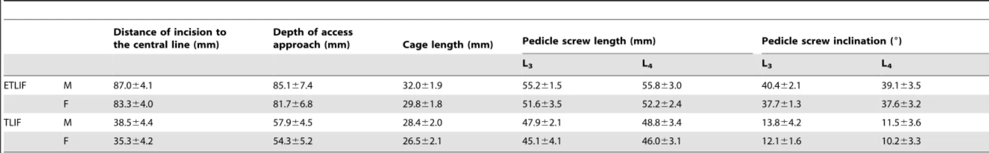
in TLIF; the angle of the screw placement was more inclined, at approximately 40u; and the cage length was approximately 30 mm. Compared to TLIF, ELIF had a more lateral incision that avoided the inferior articular process and allowed for direct access to the hypertrophic, compressed superior articular process through a lateroposterior approach, and the pedicle screw and cage were implanted in a more inclined angle and with a significantly greater length (P,0.01).
It was feasible to achieve a digitally designed surgical approach, procedure, and decompression on the cadaveric specimens with good consistency with the preoperative planning (Figure 3). Digitalized operative planning showed good accuracy, safety, and stability. The present digitalization technology could not accurately reconstruct images of the minute vascular vessels and nerves, and the mock cadaveric operation was not identical to the actual operation. For example, it was highly risky to access the para- and intraspinal venous plexuses.
Discussion
Digital medicine is a newly emerging technique which incorporates information technology into clinical medicine. This technique uses a laser scanner, CT, magnetic resonance (MR), and other digital equipment to acquire human body tomographic data. After further integrating these data, spinal digitalized 3D reconstruction can demonstrate the morphology of the spinal column from multiple angles and directions, which allows a clear and full visualization of the disease in whole and in detail. Furthermore, this technique can be used for the selection of surgical approach, the assessment of surgical risk and outcome, and preoperative planning on the digitalized model, for instance, simulating implantation of the cage [8–9].
In this study, we used Mimics 14.0 software, which is highly integrated and user-friendly for 3D image production and editing. The Mimics surgical simulation module is a surgical simulation platform for the data analysis of the human regional anatomy and the planning of the skin incision, dissection, and implantation.
Figure 1. Mimics measurement of anatomical indices on two-dimensional extraforaminal lumbar interbody fusion (ELIF) and TLIF multi-planar rendered images.(A) transverse view of a three-dimensional image; (B) measurement of the distance of the incision to the median line, the depth of the access approach, and the length of the pedicle screw; and (C) measurement of the length of the interbody fusion cage. doi:10.1371/journal.pone.0105646.g001
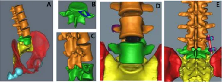
Figure 2. Mimics simulation of extraforaminal lumbar interbody fusion.(A) Three-dimensional reconstruction of the spinal column; (B) extraction of the L5vertebral body and resection of the superior articular process; (C) exposure of the intervertebral space following resection of the
superior articular process; (D) placement of the cage through the intervertebral foramen; (E) implantation of the pedicle screws. doi:10.1371/journal.pone.0105646.g002
Digitalized Design of ELIF
Table 2.L4-5ELIF and TLIF anatomical measures.
Distance of incision to the central line (mm)
Depth of access
approach (mm) Cage length (mm) Pedicle screw length (mm) Pedicle screw inclination (6)
L4 L5 L4 L5
ETLIF M 87.663.8 86.964.5 32.262.2 55.863.0 57.261.3 39.163.5 39.163.5
F 81.363.4 83.064.2 30.362.0 52.262.4 54.462.2 37.663.2 37.663.2
TLIF M 37.064.1 56.465.2 28.562.2 48.863.4 51.365.1 11.563.6 11.563.6
F 32.962.7 54.064.9 26.862.1 46.063.1 48.761.2 10.263.3 10.263.3
doi:10.1371/journal.pone.0105646.t002
Digitalized
Design
of
ELIF
4
August
2014
|
Volume
9
|
Issue
8
|
Compared to conventional gross anatomy measurements and solid operative presentations, 3D reconstruction technology can accu-rately reproduce the anatomical features of the target structure. The digitalized anatomical model can allow for a 3D dynamic and visualized manipulation in the computer system, which saves materials and time and improves the accuracy of the examination result. In this study, we used multiple digital techniques to simulate key ELIF procedures such as resection of the superior articular process, dissection of the intervertebral space, placement of the cage and pedicle screws, and acquisition of the relevant anatomical measures. All of these results were further validated by the results of the mock operation performed on the cadaveric specimens.
Harms et al. [10] reported a modified TLIF in 1982, which had a more lateral access approach and allowed for lumbar interbody fusion from the posterolateral foramen and the preservation of posterior tension structures such as the supraspinous and interspinous ligaments and laminal attachment of the sacrospinous muscle. Therefore, this modified TLIF has a minimal effect on the mechanical load distribution of the spinal column. Additionally, this modified procedure avoids excessive retraction of the dural sac and nerve roots and reduces the risk of intraspinal venous plexus bleeding and nerve root injury. As a major development in the
concept of minimally invasive spinal surgery, Holly et al. [11] innovated a minimally invasive TLIF based on conventional TLIF, which had an access approach through the intermuscular space between the multifidus muscle and the longissimus dorsi muscle. TLIF procedures could be completed inside the catheter and minimize operative injuries. Compared to PLIF, TLIF preserves the spinous process and lamina that are not causative of neurological symptoms, while only the symptomatic lateral spinal canal is specifically decompressed with a great emphasis on the maximized preservation of the lumbar muscles and bony structure [12]. As the inferior articular process overlies the dorsal side of the superior articular process, TLIF requires the preceding resection of the inferior and superior articular process for nerve root decompression and intervertebral space dissection. However, the inferior articular process is not the etiological cause in most cases, but it is sacrificed for the establishment of the access approach and the surgical field [13]. As inspired by the development of Joimax foraminal endoscopy, we designed a more lateral approach than TLIF, namely ELIF. This approach passes through the intermuscular space between the multifidus muscle and the longissimus dorsi muscle and avoids the inferior articular process. It also allows for the direct exposure and resection of the
Figure 3. Mock L4-5extraforaminal lumbar interbody fusion on cadaveric specimen.(A) palpation of the L5transverse process and
exposure of the superior articular process toward the medial side with preservation of the L4nerve root; (B) resection of the superior articular process,
dissection of the intervertebral space, and implantation of the cage and pedicle screws; (C) assembly and fixation of the connect rod; and (D) closure of the incision.
doi:10.1371/journal.pone.0105646.g003
Digitalized Design of ELIF
that ELIF had a more inclined placement of pedicle screws using longer pedicle screws and a longer cage compared to TLIF. Furthermore, ELIF can make direct decompression in lateral and even part of central canal which is totally different in ALIF and X/DLIF. But ELIF also has some potential shortcomings compared to TLIF. The more lateral incision of ELIF make the approach much deeper than TLIF, so this places a greater burden on lighting in operation site and surgical skills. And we can’t make reduction because the inferior facet joint is left intact, so it’s difficult to treat spondylolisthesis with ELIF.
We used the simulated ELIF to determine the anatomical indices in the mock ELIF on the cadaveric specimens. The incision was made 9 cm distal to the median line, and the approach passes the intermuscular space and reaches the transverse process. Further dissection along the transverse process toward the mediosuperior side was made to fully expose the superior articular process and reach the intervertebral foramen. The exit nerve is located on the ventral side of the inter-transverse-process ligament
dissection and cage placement. Further research including biomechanical study is needed to prove ELIF has a superior ability to preserve the posterior tension bands of the spinal column, with similar effects on spinal decompression, fixation, and fusion, and it can enhance post-fusion spinal stability and expedites postoperative recovery. However, this approach also has some disadvantages such as the intervertebral foramen area has abundant venous plexuses and is at high risk of bleeding and nerve injury, it is also difficult to decompress centrally and need a steep learning curve to perform ELIF.
Author Contributions
Conceived and designed the experiments: JT LJL MJY. Performed the experiments: CZ YCH SG ZQL. Analyzed the data: MJY CZ JP. Contributed reagents/materials/analysis tools: CZ YCH SG. Wrote the paper: MJY LJL CZ.
References
1. Hsieh PC, Koski TR, O’Shaughnessy BA, Sugrue P, Salehi S, et al. (2007) Anterior lumbar interbody fusion in comparison with transforaminal lumbar interbody fusion: implications for the restoration of foraminal height, local disc angle, lumbar lordosis, and sagittal balance. J Neurosurg Spine, 7:379–86. 2. Rodgers WB, Gerber EJ, Patterson J (2011) Intraoperative and early
postoperative complications in extreme lateral interbody fusion: an analysis of 600 cases. Spine, 36:26–32.
3. Lindley EM, McCullough MA, Burger EL, Brown CW, Patel VV (2011) Complications of axial lumbar interbody fusion. J Neurosurg Spine, 15:273–9. 4. Tobler WD, Ferrara LA (2011) The presacral retroperitoneal approach for axial lumbar interbody fusion: a prospective study of clinical outcomes, complications and fusion rates at a follow-up of two years in 26 patients. J Bone Joint Surg Br, 93:955–60.
5. Cahill KS, Martinez JL, Wang MY, Vanni S, Levi AD (2012) Motor nerve injuries following the minimally invasive lateral transpsoas approach. J Neur-osurg Spine, 17:227–31.
6. Hey HW, Hee HT (2010) Lumbar degenerative spinal deformity: Surgical options of PLIF, TLIF and MI-TLIF. Indian J Orthop, 44:159–62. 7. Di Paola CP, Molinari RW (2008) Posterior lumbar interbody fusion. J Am
Acad Orthop Surg, 16:130–9.
8. Kadoury S, Labelle H (2012) Classification of three-dimensional thoracic deformities in adolescent idiopathic scoliosis from a multivariate analysis. Eur Spine J, 21:40–9.
9. Hong JY, Suh SW, Easwar TR, Modi HN, Yang JH, et al. (2011) Evaluation of the three-dimensional deformities in scoliosis surgery with computed tomogra-phy: efficacy and relationship with clinical outcomes. Spine, 36:1259–65.
10. Rosenberg WS, Mummaneni PV (2001) Transforaminal lumbar interbody fusion: technique, complications, and early results. Neurosurgery, 48:569–74. 11. Holly LT, Schwender JD, Rouben DP, Foley KT (2006) Minimally invasive
transforaminal lumbar interbody fusion: indications, technique, and complica-tions. Neurosurg Focus, 20:E6.
12. Lee KH, Yue WM, Yeo W, Soeharno H, Tan SB (2012) Clinical and radiological outcomes of open versus minimally invasive transforaminal lumbar interbody fusion. Eur Spine J, 21:2265–70.
13. Mura PP, Costaglioli M, Piredda M, Caboni S, Casula S (2011) TLIF for symptomatic disc degeneration: a retrospective study of 100 patients. Eur Spine J, 20, Suppl 1:57–60.
14. Feng ZZ, Cao YW, Jiang C, Jiang XX (2011) Short-term outcome of bilateral decompression via a unilateral paramedian approach for transforaminal lumbar interbody fusion with unilateral pedicle screw fixation. Orthopedics, 34:364. 15. Xiao YX, Chen QX, Li FC (2009) Unilateral transforaminal lumbar interbody
fusion: a review of the technique, indications and graft materials. J Int Med Res, 37:908–17.
16. Lau D, Lee JG, Han SJ, Lu DC, Chou D (2011) Complications and perioperative factors associated with learning the technique of minimally invasive transforaminal lumbar interbody fusion (TLIF). J Clin Neurosci, 18:624–7.