C A S E R E P O R T
Open Access
Deinagkistrodon acutus
envenomation: a
report of three cases
Chin-Lung Cheng
1,2, Yan-Chiao Mao
2,3,4, Po-Yu Liu
5, Liao-Chun Chiang
6,7, Shu-Chen Liao
4,8and Chen-Chang Yang
3,4*Abstract
Background:Deinagkistrodon acutusenvenomation is associated with severe hematological and wound complications but is rarely described.
Case presentation:Herein, we report three cases of victims bitten byD. acutusand indicate that rapid-onset severe coagulopathy and thrombocytopenia are distinct features ofD. acutussnakebite, which are not observed in other crotaline snakebites (i.e.,Trimeresurus stejnegeriandProtobothrops mucrosquamatus) in Taiwan. The toxic effects could occur as early as 2 to 3 h followingD. acutusenvenomation and persist if the administration of specific antivenom is delayed or even not commenced. Based on our findings, 2 to 4 vials of specific antivenom as the first dose should be administered to victims and repeated at 6 to 8 h intervals if coagulopathy or thrombocytopenia persists. Fresh frozen plasma or platelet replacement is probably safe as an adjunct therapy forD. acutusbite in the presence of venom-induced consumptive coagulopathy.
Conclusion:Severe coagulopathy and thrombocytopenia could occur as early as 2 to 3 h afterD. acutusenvenomation. The current recommendation for antivenom is 2 to 4 vials as the first dose and repeated every 6–to 8 h if coagulopathy or thrombocytopenia persists. These cases studied may be helpful to first-line medical personnel in the early diagnosis and management ofD. acutusenvenomation among other crotaline snakebites in Taiwan.
Keywords:Coagulopathy, Thrombocytopenia, Envenomation,Deinagkistrodon acutus, Snakebite
Background
Envenomation caused by snakebite comprises a worldwide public health problem, especially in Asia [1]. The venoms of snakes are a fascinating mix that allow the design of new drugs for use in medicine, as well as being a challenge for researchers in the development of specific antivenoms [2–5]. In Taiwan, six major venomous snakes are found, namely:Trimeresurus stejnegeri,Protobothrops mucrosqua-matus, Deinagkistrodon acutus and Daboia siamensis in the family Viperidae, andNaja atraand Bungarus multi-cinctusin the family Elapidae [6].D. acutus–also known as the hundred pacer, five pacer or Chinese moccasin–is the largest snake (80–155 cm) of the subfamily Crotalinae
on the island [6]. It is additionally distributed throughout south China, Vietnam and possibly Laos [7]. D. acutus
envenomation is rare; however, it is considered the most lethal and can result in life- or limb-threatening complica-tions after envenomation [6, 8]. Although severe coagulop-athy and thrombocytopenia, defined as an international normalized ratio (INR) of prothrombin time (PT) > 9 and platelet count < 50,000/mm3, are considered the hallmarks ofD. acutusenvenomation, little is known about the tim-ing of onset and treatment with specific antivenom [9–13]. In the present study, we summarize the clinical manifesta-tions and treatment of three cases ofD. acutus envenom-ation admitted to Taichung Veterans General Hospital (VGH-TC) with the aim of improving the diagnosis and management ofD. acutusenvenomation.
Case Presentation Case 1
A 36-year-old previously healthy man was bitten on his right hand by a snake during cleaning work around his * Correspondence:[email protected]
3Department of Medicine, Division of Clinical Toxicology and Occupational
Medicine, Taipei Veterans General Hospital, 201 Sec. 2, Shipai Road., Taipei 112, Taiwan
4Institute of Environmental and Occupational Health Sciences, School of
Medicine, National Yang-Ming University, Taipei, Taiwan Full list of author information is available at the end of the article
home. He was immediately sent to a local hospital, with the dead snake identified as D. acutus. However, only two vials of antivenom for T. stejnegeri andP. mucros-quamatus were administered, because specific anti-venom for D. acutuswas not available and because the treating physician believed that cross-neutralization would occur. He received right upper limb fasciotomy on day 2 for suspected compartment syndrome. On day 3, he was transferred to VGH-TC due to worsening of his general condition and bleeding tendency. On arrival, the patient’s blood pressure (BP) was 109/47 mmHg, pulse 119/min, respiratory rate 20/min, and body temperature 38.5 °C.
Physical examination revealed continuous oozing from the wound and venous catheter insertion site, multiple hemorrhagic bullae, swelling extending up to the shoulder and gross hematuria in the urinary bag. Laboratory examin-ation revealed a hemoglobin level of 5.8 g/dL (reference range 14–18 g/dL), platelet count 17,000/mm3 (reference range 150,000–400,000/mm3), fibrinogen 130 mg/dL (refer-ence range 200–400 mg/dL), D-dimer > 1μg/mL (reference range < 0.55 μg/mL), fibrinogen degradation products (FDPs) > 40 μg/mL (reference range < 10 μg/mL), and incoagulable blood [PT > 169 s; activated partial thrombo-plastin time (aPTT) > 224 s] (Table 1).
Three vials of monovalent antivenom forD. acutusand blood components [600 mL of packed red blood cells, 600 mL of fresh frozen plasma (FFP), and 300 mL of plate-let concentrate] were immediately administered, resulting in a good response (Fig. 1). The patient’s blood coagula-tion normalized at 6 h after administracoagula-tion of the anti-venom and oozing from the wound stopped. A follow-up coagulation profile did not reveal recurrent coagulopathy on days 4, 10 and 13. He had intermittent fever, and ex-tensive wound necrosis that required repetitive debride-ment on days 8, 12 and 17 during hospitalization. Deep tissue cultures obtained during surgery grewPseudomonas aeruginosa, Morganella morganii, Staphylococcus aureus
and Enterococcus spp. Although thrombocytopenia per-sisted throughout days 4 to 10 (22,000–116,000/mm3), it was amenable to platelet transfusion and medical treat-ment. The bite wound improved gradually after antibiotic therapy and debridement. Staged wound closure was per-formed 3 weeks post-bite, and he was transferred to an-other hospital for a rehabilitation program 1 month later.
Case 2
A 41-year-old previously healthy woman was bitten on the left ankle by a snake while she collected herbs in northwestern Taiwan. She was sent to a local hospital 3 h later, where thrombocytopenia (3000/mm3) and incoagulable blood (PT > 100 s) were noted. The snake was identified asD. acutusby the patient through a pic-ture; however, two vials of antivenom for T. stejnegeri
and P. mucrosquamatus were administered for an un-known reason. She was then referred to VGH-TC 7.5 h post-bite.
On arrival, her BP was 172/94 mmHg, pulse 108/min, respiratory rate 20/min, and body temperature 39.3 °C. Physical examination revealed many hemorrhagic bullae scattered along the calf, continuous oozing from the fang marks, and painful swelling extending to the knee region. Laboratory examination disclosed thrombocytopenia (14,000/mm3) and PT of > 169 s and aPTT of > 224 s. Four vials of monovalent antivenom for D. acutus and blood components (200 mL of FFP and 300 mL of platelet concentrate) were administered at 8.5 h post-bite. Her co-agulation profiles normalized at 15 h post-bite. On day 2, her platelet count increased to 158,000/mm3. Due to pro-longed aPTT (43.3–44.6 s), another seven vials of anti-venom (2 to 3 vials every 6 to 8 h) were sequentially administered without measurable responses.
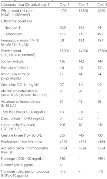
Table 1Initial blood laboratory data of the three patients at Taichung Veterans General Hospital
Laboratory data (On arrival, day 1) Case 1 Case 2 Case 3
White blood cell count
(4,500–11,000/mm3) 4,700 12,300 9,300
Differential count (%)
Neutrophil 76.9 84.7 44
Lymphocyte 15.2 7.6 45.2
Hemoglobin (male: 14–18, female 12–16 g/dL)
5.8 13.1 15.4
Platelet count
(150,000–400,000/mm3) 17,000 14,000 17,000
Sodium (mEq/L) 136 142 144
Potassium (mEq/L) 4.0 4.0 3.7
Blood urea nitrogen (5–25 mg/dL)
12 14 24
Creatinine (0.7–1.4 mg/dL) 0.7 1.4 0.9
Alanine aminotransferase (male: 10–50, female: 10–35 U/L)
28 26 21
Aspartate aminotransferase (8–38 U/L)
30 43 26
Total bilirubin (0.2–1.6 mg/dL) 1.3 0.8 –
Direct bilirubin (0–0.3 mg/dL) 0 0.2 –
Lactate dehydrogenase (120–240 U/L)
206 181 242
Creatine kinase (10–160 U/L) 855 745 155
Prothrombin time (seconds) >169 >164 >164
Activated partial thromboplastin time (s)
>224 >224 >224
Fibrinogen (200–400 mg/dL) 130 – 149.1
D-dimer (<0.55μg/mL) >1 – –
Fibrinogen degradation products (FDPs < 10μg/mL)
The leg wound was complicated with necrotizing fasciitis that required repetitive debridement on days 21 and 31 and supplemental hyperbaric oxygen ther-apy. The wound cultures obtained during surgery revealed S. aureus, Enterococcus spp. and Bacteroides fragilis. After antibiotic treatment, her wound infec-tion improved and split-thickness skin grafting was performed after debridement on day 31. The patient was discharged on day 47 post-bite with good func-tional recovery of the leg.
Case 3
A 69-year-old previously healthy woman was bitten on the left middle finger by a snake while collecting fire-wood. Painful swelling, tissue ecchymosis, and oozing from the wound developed a few minutes later. The pa-tient was sent to VGH-TC 2 h post-bite. The dead snake brought in by the patient was identified asD. acutus. On arrival, her BP was 200/120 mmHg, pulse 95/min, re-spiratory rate 22/min, and body temperature 36.7 °C. Physical examination revealed swelling of left hand. A low platelet count (17,000/mm3), PT > 169 s, aPTT > 224 s, fibrinogen level 149.1 mg/dL and FDPs > 40 μg/ mL were noted in laboratory analyses.
Four vials of monovalent antivenom forD. acutus and 1000 mL of FFP were administered. Seven hours post-bite, both PT and aPTT normalized, and the platelet count in-creased to 230,000/mm3. Because of recurrent oozing from the wound, another four vials of antivenom (two at 12 h intervals) were administered without examination of PT and aPTT levels in the following 24 h. Although the surgeon recommended partial finger amputation due to finger necrosis, the patient declined and insisted on being discharged against medical advice on day 5. No recurrent coagulopathy was noted prior to discharge; however, the patient did not return for follow-up.
Discussion
T. stejnegeri and P. mucrosquamatus account for more than 70% of snakebite cases each year in Taiwan, which share similar clinical manifestations as well as the same treatment with bivalent specific antivenom [6, 8]. Sporadic cases with INR above 1.67 and platelet counts of 36,000/ mm3were reported inP. mucrosquamatusenvenomation, and none manifested systemic bleeding [11, 14]. In con-trast, severe coagulopathy and thrombocytopenia are the main laboratory findings of D. acutus envenomation in addition to serious wound complications and systemic bleeding [11–13]. Valenta et al. [15] reported a victim ofD. acutusbite who developed incoagulable blood between 1.5 and 7 h post-bite. Hung et al. [13] reported the case of a man with persistent thrombocytopenia 44 h after en-venomation; his platelet level was normal 30 min post-bite. In our observation, rapid-onset and severe coagulopathy and thrombocytopenia developed as early as 2 to 3 h post-bite may persist if correct antivenom is not administered.
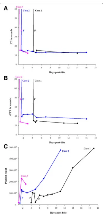
D. acutusvenom is composed of several hemotoxins, in-cluding the thrombin-like enzymes (TLEs), anticoagulant toxins, platelet aggregation inhibitors, hemorrhagins, and enzymes that facilitate venom spreading [16–19]. The anticoagulation effect of TLEs occurs rapidly, and circulat-ing fibrinogen levels start to fall within 30 min, reachcirculat-ing 9% of the normal value within 2 h [16]. Anticoagulant toxins inactivate prothrombin, tissue factor and coagula-tion factors V and IX/X, resulting in a transient but marked Fig. 1aTrend of PT in three patients.bTrend of aPTT in three patients.
prolongation of blood coagulation within 5 min after in-jection [16, 20, 21]. The platelet inhibitors, mainly adeno-sine diphosphatase, inhibit platelet aggregation in the presence of adenosine diphosphate or collagen [17]. The hemorrhagins (e.g., snake venom metalloproteinases) cause extensive vascular damage and increase vascular permeability [22]. These effects may explain the remark-able bleeding tendency and wound complications in D. acutusenvenomation.
The Taiwan government produces four types of anti-venom against the six major anti-venomous snakebites [8]. Con-cerning the monovalent antivenom forD. acutus, each vial roughly neutralizes 52 mg of venom. The Taiwan Poison Control Center thus recommends that 2 to 4 vials of anti-venom be administered in a enanti-venomed patient because the mean amount ofD. acutusvenom injected is 105.1 mg [8]. However, this recommendation was not validated and there is no standard dosing regimen available in Taiwan. According to our findings, the current recommendation is 2 to 4 vials of antivenom to be administered as the first dose and repeated at 6 to 8 h intervals if coagulopathy or thrombocytopenia persists (e.g., INR > 3, aPTT > 50s, and platelet level < 20,000/mm3) [23, 24]. Although cross-neutralization occurs between bivalent antivenom for T. stejnegeri and P. mucrosquamatus and monovalent anti-venom forD. acutus, substitution should be avoided as the bivalent antivenom has only a weak cross-reactivity with theD. acutusvenom [13].
All of our cases had a rapid improvement in coagulop-athy after receiving specific antivenom. In case 2, s slightly prolonged aPTT persisted for 4 days without clinical thrombosis or bleeding tendency. Although a workup for prolonged aPTT was not performed in the case, it was probably unrelated to the venom because no apparent response was observed after repetitive anti-venom administration [25]. In case 1, thrombocytopenia did not show improvement after antivenom therapy, which was probably attributable to uncontrolled infec-tion. Nevertheless, Valenta et al. [15] reported a single case of D. acutusenvenomation who manifested severe coagulopathy in the absence of thrombocytopenia. Therefore, the exact mechanism of thrombocytopenia in
D. acutussnakebite remains unclear.
Evidence suggests that, when compared with anti-venom alone, early FFP replacement therapy shortens coagulopathy in patients suffering from hemotoxic snake envenomation and, theoretically, lower the risk of major bleeding [26]. The bleeding risk may be pronounced in cases of severe thrombocytopenia [10]. However, in cases of venom-induced thrombotic microangiopathy, platelet transfusion may be problematic [27]. In our study, no elevation of blood bilirubin or lactate dehydro-genase was found, and the first two cases had received platelet concentrate transfusions, resulting in good
responses. It appears to be safe to use the blood compo-nent therapy duringD. acutus envenomation. In case 3, the platelet count spontaneously normalized after 24 h with FFP and specific antivenom administration alone. This effect may have occurred because of the relatively modest injury in this case. However, the patient received a higher dosage (eight vials) of antivenom, which was sufficient to counteract the venom effects on platelets.
Conclusion
Severe coagulopathy and thrombocytopenia could occur as early as 2 to 3 h after D. acutusenvenomation. The current recommendation for antivenom therapy is 2 to 4 vials as the first dose and repeat it every 6 to 8 h if coag-ulopathy or thrombocytopenia persists. Our observation may be helpful to first-line medical personnel in the timely diagnosis and management ofD. acutusbites.
Abbreviations
aPTT:activated partial thromboplastin time; BP: Blood pressure; FDPs: Fibrinogen degradation products; FFP: Fresh frozen plasma; INR: International normalized ratio; PT: Prothrombin time; TLEs: Thrombin-like enzymes; VGH-TC: Taichung Veterans General Hospital
Acknowledgments
Not applicable.
Publisher’s Note
Springer Nature remains neutral with regard to jurisdictional claims in published maps and institutional affiliations.
Funding
Not applicable.
Authors’contributions
The first two authors, CLC and YCM interpreted the clinical findings and drafted the manuscript. The third to the fifth authors, PYL, LCC, and SCL provided professional opinions in bacteriology of snakebite and snake venomics and antivenomics, and revised the manuscript. The correspondent author CCY designed this study, interpreted the clinical findings and revised the manuscript. All authors read and approved the final manuscript.
Competing interests
The authors declare that they have no competing interests.
Consent for publication
Written informed consent was obtained from the patients for publication of this case report.
Ethics approval and consent to participate
The study protocol was approved by the Institutional Review Board of Taichung Veterans General Hospital (IRB, CE14202A).
Author details
1Department of Emergency Medicine, Kaohsiung Armed Forces General
Hospital, Taipei, Taiwan.2Department of Emergency Medicine, Division of
Clinical Toxicology, Taichung Veterans General Hospital, Taipei, Taiwan.
3Department of Medicine, Division of Clinical Toxicology and Occupational
Medicine, Taipei Veterans General Hospital, 201 Sec. 2, Shipai Road., Taipei 112, Taiwan.4Institute of Environmental and Occupational Health Sciences,
School of Medicine, National Yang-Ming University, Taipei, Taiwan.
5Department of Medicine, Division of Infection, Taichung Veterans General
Hospital, Taichung, Taiwan.6National Tsing Hua University, College of Life
Sciences, Hsinchu, Taiwan.7National Health Research Institutes, National
8Department of Emergency Medicine, Chang Guang Memorial Hospital,
Taipei, Taiwan.
Received: 30 September 2016 Accepted: 21 March 2017
References
1. World Health Organization (WHO). Snake antivenoms. Fact sheet N° 337. Reviewed February 2015. Retrieved from http://www.who.int/mediacentre/ factsheets/fs337/en/. Accessed 23 Oct 2016.
2. Wei CB, Chen J, Li JH. Acutolysin C, a weak hemorrhagic toxin from the venom ofAgkistrodon acutuswith leucoagglutination activity. J Venom Anim Toxins incl Trop Dis. 2011;17(1):34–41.
3. Wei CB, Chen J. A novel lipocalin homologue from the venom gland of
Deinagkistrodon acutussimilar to mammalian lipocalins. J Venom Anim Toxins incl Trop Dis. 2012;18(1):16–23.
4. Chieh-Fan C, Tzeng-Jih L, Wen-Chi H, Hua-Wei Y. Appropriate antivenom doses for six types of envenomations caused by snakes in Taiwan. J Venom Anim Toxins incl Trop Dis. 2009;15(3):479–90.
5. Ratanabanangkoon K, Tan KY, Eursakun S, Tan CH, Simsiriwong P, Pamornsakda T, et al. A simple and novel strategy for the production of a pan-specific antiserum against elapid snakes of Asia. PLoS Negl Trop Dis. 2016;10(4):e0004565.
6. Mao YC, Hung DZ. Epidemiology of snake envenomation in Taiwan. Clinical Toxinology in Asia Pacific and Africa. 2015. p. 3–22. http://link.springer.com/ referenceworkentry/10.1007/978-94-007-6386-9_45. Accessed 21 Feb 2017. 7. Uetz P, Hošek J. The Reptile Database.Retrieved from
http://www.reptile-database.org/. Accessed 8 Dec 2014.
8. Mao YC, Hung DZ. Management of snake envenomation in Taiwan. Clinical Toxinology in Asia Pacific and Africa. 2015. p. 23–52. http://link.springer. com/referenceworkentry/10.1007/978-94-007-6386-9_43. Accessed 21 Feb 2017.
9. Ansell J, Hirsh J, Hylek E, Jacobson A, Crowther M, Palareti G, et al. Pharmacology and management of the vitamin K antagonists: American College of Chest Physicians evidence-based clinical practice guidelines. Chest. 2008;133(6_Suppl):160S–98.
10. Williamson DR, Albert M, Heels-Ansdell D, Arnold DM, Lauzier F, Zarychanski R, et al. Thrombocytopenia in critically ill patients receiving thromboprophylaxis: frequency, risk factors, and outcomes. Chest. 2013;144(4):1207–15.
11. Shen MC. Afibrinogenemia and thrombocytopenia following crotalid snake bites in Taiwan. J Formos Med Assoc. 1983;82(2):239–44.
12. Rao DS. Clinical observation of 21 cases ofAgkistrodon acutus(Guenther) bite complicated by disseminated intravascular coagulation (author’s transl). Zhonghua Nei Ke Za Zhi. 1981;20(11):670–2.
13. Hung DZ, Wu TC, Deng JF. The painful experience of inappropriate therapy of snake bites: a report of two cases. Zhonghua Yi Xue Za Zhi (Taipei). 1997; 60(6):326–30.
14. Chen YW, Chen MH, Chen YC, Hung DZ, Chen CK, Yen DHT, et al. Differences in clinical profiles of patients withProtobothrops mucrosquamatusandViridovipera stejnegerienvenoming in Taiwan. Am J Trop Med Hyg. 2009;80(1):28–32. 15. Valenta J, Stach Z, Michalek P. Envenoming by Crotalid Snake Chinese
MoccasinAgkistrodon AcutusBite - A Case Report. Prague Med Rep. 2015; 116(2):155–60.
16. Ouyang C, Teng CM.In vivoeffects of the purified thrombin-like and anticoagulant principles ofAgkistrodon acutus(Hundred pace snake) venom. Toxicon. 1978;16(6):583–93.
17. Ouyang C, Huang TF. Platelet aggregation inhibitors fromAgkistrodon acutussnake venom. Toxicon. 1986;24(11–12):1099–106.
18. Xu X, Wang C, Liu J, Lu Z. Purification and characterization of hemorrhagic components fromAgkistrodon acutus(hundred pace snake) venom. Toxicon. 1981;19(5):633–44.
19. Xu X, Wang XS, Xi XT, Liu J, Huang JT, Lu ZX. Purification and partial characterization of hyaluronidase from five pace snake (Agkistrodon acutus) venom. Toxicon. 1982;20(6):973–81.
20. Ouyang C, Teng CM. The effect of the purified anticoagulant principle of
Agkistrodon acutusvenom on blood coagulation. Toxicon. 1973;11(3):287–92. 21. Ouyang C, Teng CM, Huang TF. Characterization of the purified principles of
Formosan snake venoms which affect blood coagulation and platelet aggregation. J Formos Med Assoc. 1982;81(7):781–90.
22. Kamiguti AS. Platelets as targets of snake venom metalloproteinases. Toxicon. 2005;45(8):1041–9.
23. Gold BS, Dart RC, Barish RA. Bites of venomous snakes. N Engl J Med. 2002; 347(5):347–56.
24. Boyer LV, Seifert SA, Cain JS. Recurrence phenomena after immunoglobulin therapy for snake envenomations: Part 2. Guidelines for clinical management with crotaline Fab antivenom. Ann Emerg Med. 2001;37(2):196–201. 25. Chng WJ, Sum C, Kuperan P. Causes of isolated prolonged activated partial
thromboplastin time in an acute care general hospital. Singapore Med J. 2005;46(9):450–6.
26. Isbister GK, Buckley NA, Page CB, Scorgie FE, Lincz LF, Seldon M, et al. A randomized controlled trial of fresh frozen plasma for treating venom-induced consumption coagulopathy in cases of Australian snakebite (ASP-18). J Thromb Haemost. 2013;11(7):1310–8.
27. Isbister GK. Snakebite doesn’t cause disseminated intravascular coagulation: coagulopathy and thrombotic microangiopathy in snake envenoming. Semin Thromb Hemost. 2010;36(4):444–51.
• We accept pre-submission inquiries
• Our selector tool helps you to find the most relevant journal
• We provide round the clock customer support
• Convenient online submission
• Thorough peer review
• Inclusion in PubMed and all major indexing services
• Maximum visibility for your research
Submit your manuscript at www.biomedcentral.com/submit