Effective Apical Infection of Differentiated
Human Bronchial Epithelial Cells and
Induction of Proinflammatory Chemokines
by the Highly Pneumotropic Human
Adenovirus Type 14p1
Elena Lam1,2, Mirja Ramke1¤, Gregor Warnecke3, Sonja Schrepfer4, Verena Kopfnagel5,
Thomas Dobner2, Albert Heim1*
1Institute of Virology, Hannover Medical School, Hannover, Germany,2Heinrich-Pette-Institute,
Department Viral Transformation, Hamburg, Germany,3Department of Cardiothoracic, Transplantation and Vascular Surgery, Hannover Medical School, Hannover, Germany,4University Heart Center Hamburg, Transplant and Stem Cell Immunobiology Laboratory, Universitäts Klinikum Eppendorf, Hamburg, Germany, 5Department of Dermatology, Hannover Medical School, Hannover, Germany
¤ Current address: Harvard Medical School, Massachusetts Eye and Ear Infirmary, Boston, Massachusetts, United States of America
Abstract
Background
Only a few pneumotropic types of the human adenoviruses (e.g. type B14p1) cause severe lower respiratory tract infections like pneumonia and acute respiratory distress syndrome (ARDS) even in immunocompetent patients. By contrast, many other human adenovirus (HAdV) types (e.g. HAdV-C5) are associated mainly with upper respiratory tract infections. This is in accordance with a highly physiological cell culture system consisting of differenti-ated primary human bronchial epithelial cells which are little susceptible for apical HAdV-C5 infections.
Objective and Methods
We hypothesized that a pneumotropic and highly pathogenic HAdV type infects differenti-ated human bronchial epithelial cells efficiently from the apical surface and also induces proinflammatory cytokines in order to establish ARDS and pneumonia. Therefore, the apical infection of differentiated primary human bronchial epithelial cells with the pneumotropic and virulent type HAdV-B14p1 was investigated in comparison to the less pneumotropic HAdV-C5 as a control.
Results
Binding of HAdV-B14p1 to the apical surface of differentiated human bronchial epithelial cells and subsequent internalization of HAdV DNA was 10 fold higher (p<0.01) compared to the a11111
OPEN ACCESS
Citation:Lam E, Ramke M, Warnecke G, Schrepfer S, Kopfnagel V, Dobner T, et al. (2015) Effective Apical Infection of Differentiated Human Bronchial Epithelial Cells and Induction of Proinflammatory Chemokines by the Highly Pneumotropic Human Adenovirus Type 14p1. PLoS ONE 10(7): e0131201. doi:10.1371/journal.pone.0131201
Editor:Michael Nevels, University of Regensburg, GERMANY
Received:February 5, 2015
Accepted:May 29, 2015
Published:July 13, 2015
Copyright:© 2015 Lam et al. This is an open access article distributed under the terms of theCreative Commons Attribution License, which permits unrestricted use, distribution, and reproduction in any medium, provided the original author and source are credited.
Data Availability Statement:All relevant data are within the paper and its Supporting Information files.
Funding:This work was supported in part by Robert Koch Institut (RKI) grants FKZ 380 and 1369-461. Elena Lam received Hannover biomedical research school stipend funding. The funders had no role in study design, data collection and analysis, decision to publish, or preparation of the manuscript.
less-pneumotropic C5 one hour after infection. Overall, the replication cycle of HAdV-B14p1 following apical infection and including apical release of infectious virus progeny was about 1000-fold more effective compared to the non-pneumotropic HAdV-C5 (p<0.001). HAdV-B14p1 infected cells expressed desmoglein 2 (DSG2), which has been described as potential receptor for HAdV-B14p1. Moreover, HAdV-B14p1 induced proinflammatory che-mokines IP-10 and I-Tac as potential virulence factors. Interestingly, IP-10 has already been described as a marker for severe respiratory infections e.g. by influenza virus A H5N1.
Conclusions
The efficient "apical to apical" replication cycle of HAdV-B14p1 can promote endobronchial dissemination of the infection from the upper to the lower respiratory tract. Simultaneous induction of proinflammatory cytokines probably contributes to the high virulence of HAdV-B14p1.
Introduction
Only four types (type 4 of species HAdV-E, types 3, 7 and 14p1 of species HAdV-B) of the 71 human adenovirus (HAdV) types frequently cause lower respiratory tract infections, present-ing as pneumonia and acute respiratory distress syndrome (ARDS).
HAdV-B14 was first described as respiratory pathogen in Dutch military recruits in the late 1950s [1] and found to be associated with pharyngoconjunctival fever in college students but was not associated with severe clinical diseases [2]. Subsequently, the significance of the other pneumotropic types HAdV-E4 and -B7 for severe lower respiratory tract infections (including ARDS) in military recruits was recognized in the 1960s and a vaccine for these types was devel-oped [3].
The re-emerging HAdV-B14p1 was first isolated in the US, related to fatal pneumonia out-breaks [4] and predominated beginning from 2006 [5]. HAdV-B14p1 causes lower respiratory tract infections not only in military recruits (as HAdV-E4 and -B7) but also in the civilian pop-ulation affecting infants, young adults, and elderly individuals with and without preexisting medical conditions [4]. These findings indicated a higher virulence of the re-emergent HAdV-B14p1 even compared to HAdV-E4 and HAdV-B7. Recently HAdV-B14p1 was also isolated in Canada, China, Ireland and Scotland from pneumonia patients [6–9].
So far, the organo-tropism and virulence factors of HAdV-B14p1 are not yet fully eluci-dated. Probably, all HAdV types can be transmitted by droplets and replicate in the upper respiratory tract. Efficient endobronchial (luminal) spread of the HAdV-B14p1 infection to the lower respiratory tract and induction of inflammatory cytokines may be essential for a rapid onset of pneumonia. Animal models to study HAdV pneumonia like the cotton rat [10] have drawbacks due to the species specificity of HAdV. Their replication in rodents is inefficient, expression of their late genes is incomplete [11] and the release of infectious virus progeny is aborted. Therefore, the application of high titer viral inoculums (e.g. 106to 1010plaque forming units/ml) was required to establish a pneumonia phenotype in animal models [10].
the coxsackie and adenovirus receptor (CAR) is mainly expressed on the basolateral side. This may limit the luminal, endobronchial spread of the HAdV-C5 infection from the upper to the lower respiratory tract. In accordance with this finding, the few cases of HAdV-C5 pneumonias have been limited to immunocompromised patients and may be the result of lower respiratory tract infections by viremia [15].
In the present study, the in vitro model system of differentiated human bronchial epithelial cells was used for the first time to study HAdV-B14p1 infection. Apical HAdV-B14p1 infection was found to be more effective compared to HAdV-C5 infection and resulted in efficient apical release of infectious virus progeny. In vivo, this may promote the luminal (endobronchial) spread of HAdV-B14p1 from the upper to the lower respiratory tract. Furthermore, HAdV-B14p1 infection induced proinflammatory responses by inducing chemokines IP-10 and I-Tac. The attracting of T-cells and macrophages to the side of infection may result in a massive immune response leading to severe pneumonia and ARDS.
Materials and Methods
Cell culture model of differentiated human bronchial epithelial cells
Primary human bronchial epithelial cells were isolated from lung explants of patients suffering from pulmonary fibrosis, pulmonary embolism and chronic obstructive pulmonary disease as described previously (Fig 1A) [16,17]. Written informed consent was provided by the tissue donors. The project was approved from the ethical review committees (Ethikkommission der
Fig 1. Schematic diagram of primary human bronchial epithelial cell isolation and cultivation. (A)Isolation procedure for primary human bronchial epithelial cells(B)Primary human bronchial epithelial cells cultured under submerged (left) and air-liquid interface conditions (right).
Ärztekammer Hamburg, WF-011/13; Ethikkommssion der Medizinischen Hochschule Han-nover 122–2007). Cells were seeded onto collagen I coated, semipermeable 0.33 cm2polyester (PET) filter membrane inserts (0.4μm pore size, Corning Costar, New York) for 24-well plates.
Cells from a single donor were used for each experimental set (including HAdV-B14p1, -C5 and mock infection) in order to limit inter-donor differences. Experiments were repeated with bronchial epithelial cells derived of 4 different donors. Cells were seeded at a density of 105 cells per insert. After cells had reached confluence (with about 5 x 10⁵cells per insert), the air-way medium was removed from the upper compartment and cells were cultured under air-liq-uid interface conditions (Fig 1B). Subsequently, the epithelial cells were cultured for 6 weeks for differentiation until differentiation was observed by expression of cilia (S1 Fig) and high transepithelial electrical resistance (TEER)>400Ωcm2.
Virus stocks
HAdV-C5 (ATCC VR-5) and a clinical isolate of HAdV-B14p1 (kindly provided by Michael Carr, National Virus Reference Laboratory, University College, Dublin, Ireland) [6] were prop-agated on A549 (ATCC CCL-185) cells and harvested with three freeze and thaw cycles at 70–
80% cytopathic effect (CPE) for production of crude virus stocks. Ratio of particles/infectious units as measured by the tissue culture infectious dose 50% method (TCID50) was 4.35 for
HAdV-C5 (TCID50/ml: 3.16 x 10¹⁰; particles/ml: 1.33 x 10¹¹) and 19.7 for HAdV-B14p1
(TCID50/ml: 1010; particles/ml: 1.97 x 10¹¹).
Adenovirus infection
Quadruplicates of differentiated airway epithelial cells (5 x 10⁵cells/ insert, size 0.33 cm2) were infected with HAdV on the apical surface at a multiplicity of infection (moi) of 10 TCID50/cell
for 1 h at 37°C (Fig 2). On day 1, 4 and 8 post infection (p.i.) virus was collected by washing the apical surface with 100μl DMEM-. These samples and medium samples from the lower
com-partment were titered for infectious virus progeny by the TCID50technique on A549 cells.
Quantitative HAdV PCR
Quantitative polymerase chain reaction (qPCR) was used to quantify viral genomes in the stock virus preparation and in infected cells and supernatants. Cells were sampled at 1 h and
Fig 2. Schematic diagram of the infection and experimental procedure of differentiated human bronchial epithelial cells.
48 h p.i. after unbound virus was removed by washing. DNA was extracted using the DNA Blood Kit (Qiagen, Hilden, Germany) according to the manufacturer´s protocol. Quantitative PCR was performed using the Platinum Quantitative PCR SuperMix-UDG (Invitrogen, Darm-stadt, Germany) and specific forward (5´-GCCACGGTGGGGTTTCTAAACTT-3´, Adeno-quant-1) and reverse (5´-GCCCCAGTGGTCTTACATGCACATC -3´, Adenoquant-2) primers and probe (FAM 5´-TGCACCAGACCCGGGCTCAGGTACTCCGA -3´ TAMRA) (Eurogentec, Seraign, Belgium) [18].
Immunostaining and confocal microscopy
Differentiated cells were fixated on the PET membranes as described previously [19]. The PET membrane was kept attached to the insert during the whole staining and washing procedure. The primary antibody was applied as a 30μl drop on a parafilm to the PET membrane and
additionally 50μl of the antibody was applied to the apical side of the cell layer. The washing
steps were carried out by placing the insert into a 24 well plate and adding PBS to the upper and the lower compartment. Secondary antibodies were applied as the primary antibodies. Nuclei were counterstained with DAPI. After final washing the PET membrane was cut from the insert and mounted on a glass slide with the cells facing the cover slip using Mowiol (Sigma-Aldrich, St. Louis, MO) as mounting medium. Primary antibodies used were a poly-clonal rabbit occludin (OCLN) antibody (1:40 diluted; Invitrogen, Paisley, UK), a monopoly-clonal mouse CAR antibody (RmcB, 1:100 diluted, kindly provided by M. Bergelson), a monoclonal mouse desmoglein 2 (DSG2) antibody (Clone 6D8, 1:50 diluted, Santa Cruz Biotechnology, Santa Cruz, CA), and a FITC conjugated monoclonal adenovirus antibody (Millipore, Bilerica, MA). Secondary antibodies were anti rabbit conjugated with FITC and anti mouse conjugated with dsRed (1:200 diluted, Jackson, Immunoresearch, West Grove, PA). Images were acquired with a Leica DM IRB Laser Scanning Confocal Microscope.
Microarray-based mRNA expression analysis
Samples for the“Whole Human Genome Oligo Microarray V2”(ID 026652, Agilent, Santa Clara, CA) were prepared and the microarray was processed as described in the“One-Color Microarray-Based Gene Expression Analysis Protocol V5.7”(Agilent). Scanning was con-ducted with the Agilent Micro Array Scanner G2565CA, data extraction was performed with the“Feature Extraction Software V10.7.3.1”(extraction protocol: GE1_107_Sep09.xml).
Processed signals of the green channel (“gPS”) were normalized by linear scaling: All gPS values of one sample were multiplied by a scaling factor calculated as: 1500 / 75th percentile of the respective array. All normalized gPS values that fell below an intensity border of 15 were substituted by the respective surrogate value of 15.
Quantification of CXCL10 and CXCL11 chemokine expression
Proinflammatory chemokines CXCL10 (IP-10) and CXCL11 (I-Tac) protein levels were quan-tified by enzyme immunoassay (Quantikine Immunoassay CXCL10 and CXCL11, R&D sys-tems, Minneapolis, MN).
Results
Apical HAdV infection of differentiated bronchial epithelial cells
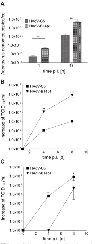
After apical infection of differentiated human bronchial epithelial cells, cell associated
HAdV-B14p1 DNA concentrations were significantly higher (p<0.01) compared to HAdV-C5
of HAdV-B14p1 (Fig 3A). HAdV genome replication resulted in elevated levels of HAdV-B14p1 DNA compared to HAdV-C5 DNA at 48 h p.i. (Fig 3A).
Apical and basal release of infectious virus progeny
The release of infectious HAdV progeny at the apical surface of differentiated human bronchial epithelial cells was monitored with help of the TCID50method. Release of HAdV-B14p1 was
sig-nificantly higher compared to HAdV-C5 on day 4 p.i. (p<0.01) and 8 d p.i. (p<0.001) (Fig 3B).
To compare directed basal to apical release after apical HAdV-C5 and HAdV-B14p1 infec-tion, the infectious virus progeny release at the basal surface of differentiated human bronchial epithelial cells was additionally monitored. HAdV-C5 reached significant higher titers com-pared to HAdV-B14p1 on day 4 p.i. (p<0.001) and day 8 p.i. (p<0.05) (Fig 3C).
CPE was finally observed on day 12 p.i. and transepithelial resistance dropped (S2 Fig) as cells were lysed and tight junctions disrupted. An early and temporary decrease of TEER was only observed in HAdV-C5 infection but cells were morphologically unchanged and viable.
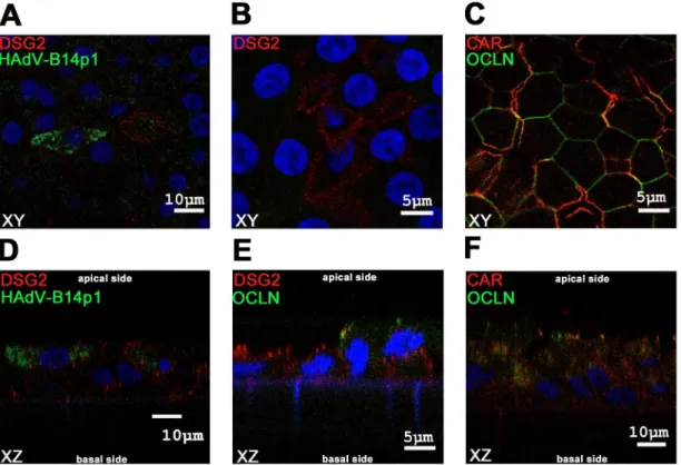
Desmoglein 2 (DSG2) expression
Bronchial epithelial cells were immunostained for HAdV hexon antigens and DSG2, the recently described novel adenoviral apical receptor [20], at day 4 post infection. DSG2 positive cells were found to be HAdV-B14p1 infected (Fig 4A–4D). Staining for HAdV-B14p1 was mainly cytoplasmic whereas DSG2 staining was mainly cell membrane associated. DSG2 was expressed close to the apical side of differentiated bronchial epithelial cells (Fig 4B–4E). For comparison, CAR, the main receptor for HAdV-C5, was expressed on the basolateral side and partially colocalized with the tight junction marker occludin (OCLN) (Fig 4C–4F).
Induction of proinflammatory chemokines
The induction of the chemokine genes IP-10 (CXCL10) and I-Tac (CXCL11) and of the proin-flammatory cytokine gene IL-6 was observed in a genome wide mRNA microarray analysis at 48 h post HAdV-B14p1 infection (S1 Table), but not in HAdV-C5 infected control cultures. Subsequently the release of chemokines IP-10 and I-Tac by HAdV-B14p1 by infected cell cul-tures was confirmed on the protein level by ELISA (Fig 5A and 5B).
Discussion
less-pneumotropic HAdV-C5 (Fig 3A). This correlated well with the expression of DSG2, the recently described receptor for HAdV-B14p1 at the distal end of intercellular junctions [20]. Thus DSG2 should be accessible from the apical side, probably facilitating entry by opening intercellular junctions, whereas most other non pneumotropic HAdV types (including HAdV-C5) bind to CAR [26]. This may limit the infection efficiency of HAdV-C5 from the endobronchial (apical) side because only a low abundance splice variant of CAR has been observed on the apical surface of human bronchial epithelial cells [19].
Although not every cell positive for DSG2 was infected with HAdV-B14p1 in our model sys-tem, all infected cells were indeed located at the apical surface, indicating apical binding and entry of HAdV-B14p1 (Fig 4D). In addition, a few bronchial epithelial cells negative for DSG2 were also found to be HAdV-B14p1 infected (data not shown). This finding may be explained with down regulation of DSG2 expression in HAdV-B14p1 infected cells. On the other hand, initial infection of the differentiated pseudostratified layer may be facilitated by DSG2 but not limited to DSG2 positive cells suggesting the relevance of other cellular receptors for
HAdV-B14p1. DSG2 expression has been described on polarized BT474, T84 and CaCo-2 cells [20]. In the present study DSG2 expression was detected for the first time on differentiated
100because the TCID50values measured on day 1 are probably remaining viral particles originating from the virus inoculum.
doi:10.1371/journal.pone.0131201.g003
Fig 4. Immunofluorescence staining of differentiated human bronchial epithelial cells.Confocal microscopy immunofluorescence analysis of differentiated human bronchial epithelial cells. Figs A, B, C show XY planes, Figs D, E, F show XZ planes.(A, D)Cells were infected from the apical side with HAdV-B14p1 at a moi of 10 (TCID50/cell), fixated 4 days p.i. and stained for HAdV in green (FITC conjugated antibody) and the desmoglein 2 (DSG2 receptor) in red (dsRed antibody), the nucleus was counterstained in blue (DAPI).(B, E)Differentiated human bronchial epithelial cells stained for DSG2 receptor in red (dsRed) and occludin (OCLN) a tight junction marker in green (FITC), nucleus was counterstained in blue (DAPI).(C, F)Differentiated human bronchial epithelial cells were stained for CAR receptor in red (dsRed) and the tight junction marker OCLN in green (FITC) and the nucleus in blue (DAPI).
primary bronchial epithelial cells. However, the direct binding of HAdV-14p1 to DSG2 was not studied, thus the receptor usage of HAdV-14p1 on bronchial epithelial cells needs to be confirmed in a future study.
Other species HAdV-B types, like HAdV-B3 or -B7 were not included in present study, as these frequently isolated respiratory pathogens cause pneumonia only in a small subset of cases. Moreover, reports on the receptor usage of HADV-B3 are partially contradictory report-ing the bindreport-ing to CD46 [27] or CD80/86 [28] as their main receptor. Probably different HAdV-B3 strains could use different receptors which may be related to their different virulence and pneumotropism.
The temporary drop of the TEER (S2 Fig) observed on day 1–3 p.i. in HAdV-C5 infection might be explained by a faint "early CPE" due to free capsid proteins in the virus stocks [29]. An early CPE is caused mainly by the penton base protein [30]. Additionally, HAdV-C fiber pro-teins, produced in excess during virus replication, are able to disrupt tight junctions, allowing HAdV-C progeny to escape to the apical surface of a differentiated bronchial epithelium by a paracellular pathway [31]. This previous study already reported the efficient primary release of HAdV-C2 on the basolateral side, similar to our results with HAdV-C5 (Fig 3C). The overall (apical plus basal) release of infectious virus progeny was not significantly different between HAdV-C5 and HAdV-B14p1 when surveyed on day 4 and 8 p.i. More efficient genome replica-tion and secondary cell to cell spread may have minimized the differences between
HAdV-B14p1 and HAdV-C5 binding and entry. In spite of these effects, the apical to apical rep-lication cycle of the highly virulent HAdV-B14p1 was far more effective and resulted in an about 1000-fold higher virus titer compared to HAdV-C5 infection (day 8 p.i., p<0.001) (Fig 3B).
The induction of proinflammatory and chemotactic cytokines may also be essential for severe inflammation of the lower respiratory tract. Induction of CXCL10 (IP-10) and IL-8 by an NF-kB pathway has been observed as a response of the cell to adenoviral infections [32]. In case of the pneumotropic type HAdV-B7, the induction of IP-10 was observed in type I and type II alveolar epithelial cells whereas induction of IL-8 was only observed in type I alveolar
Fig 5. Induction of chemokines after apical HAdV infection of differentiated human bronchial epithelial cells. (A)IP-10 concentration in cell culture medium on day 4 and 8 p.i. as determined by ELISA(B)I-Tac concentration in cell culture medium on day 4 and 8 p.i. as determined by ELISA (n.s.: not significant,*p<0.05;***p<0.001, two way ANOVA). Values shown are SEM values of quadruplicate infections.
epithelial cells [33]. IL-8 induction seems to be inconsistent between HAdV types and the infected tissues or cells [33–35].
Interestingly, a clinical study found elevated IP-10 levels to be a potential biomarker for severe acute respiratory virus infections [36]. Human airway epithelial cells have already been shown to release IP-10 in response to influenza A H5N1 infection [37]. This is in congruence with the results of this study since HAdV-B14p1 infection of differentiated bronchial epithelial cells resulted in a significant IP-10 and additionally I-Tac induction and release. The induction of these chemokines by HAdV-B14p1 infection, may result in the infiltration of the infected lung with macrophages, activated T cells and NK cells and subsequently result in the expres-sion of multiple proinflammatory cytokines as observed in ARDS [38,39].
In conclusion, the“apical to apical”replication cycle of pneumotropic HAdV-B14p1 could promote the endobronchial (luminal) spread of HAdV-B14p1 to the lower respiratory tract. Subsequent induction of proinflammatory cytokines by HAdV-B14p1 may lead to severe pneumonia and ARDS.
Supporting Information
S1 Fig. Scanning electron microscopy depicting of cilia on the apical surface of differenti-ated bronchial epithelial cells cultured for 6 weeks on the air—liquid interface.
(TIF)
S2 Fig. TEER of differentiated human bronchial epithelial cells during HAdV infection.
TEER values were measured on differentiated human bronchial epithelial cells after HAdV infection from day 1 to day 15 p.i. An initial drop in resistance (day 1–3 p.i.) observed with HAdV-C5 infection might be due to a slight, reversible early CPE caused by the virus inoculum. (TIF)
S1 Table. Microarray data: All genes found to be upregulated (threshold value 2.0) in differ-entiated human bronchial epithelial cells by HAdV-B14p1 or HAdV-C5 infection compared to mock infection.Multiple listing of gene names (for example, see CXCL10 (IP-10), CXCL11 (I-TAC) and IL-6) indicated that the upregulation was detected by multiple, different probes. Genes were listed by relative signal intensity in HAdV-B14p1 infection vs. mock infection. (XLS)
Acknowledgments
Microarray data used or referred to in this publication was generated by the Research Core Unit Transcriptomics of the Hannover Medical School. RmcB antibody was kindly provided by J. M. Bergelson, Boston, MA [26]. A clinical isolate of HAdV-B14p1 was kindly provided by Michael Carr, Dublin, Ireland.
Author Contributions
Conceived and designed the experiments: AH EL. Performed the experiments: EL VK MR. Analyzed the data: AH EL VK. Contributed reagents/materials/analysis tools: GW SS TD. Wrote the paper: AH EL GW TD.
References
2. Kendall EJ, Riddle RW, Tuck HA, Rodan KS, Andrews BE, McDonald JC. Pharyngo-conjunctival fever; school outbreaks in England during the summer of 1955 associated with adenovirus types 3, 7, and 14. Br Med J. 1957; 2: 131–136. PMID:13436877
3. Top FH Jr., Grossman RA, Bartelloni PJ, Segal HE, Dudding BA, Russell PK, et al. Immunization with live types 7 and 4 adenovirus vaccines. I. Safety, infectivity, antigenicity, and potency of adenovirus type 7 vaccine in humans. J Infect Dis. 1971; 124: 148–154. PMID:4330997
4. Louie JK, Kajon AE, Holodniy M, Guardia-LaBar L, Lee B, Petru AM, et al. Severe pneumonia due to adenovirus serotype 14: a new respiratory threat? Clin Infect Dis. 2008; 46: 421–425. doi:10.1086/ 525261PMID:18173356
5. Metzgar D, Osuna M, Kajon AE, Hawksworth AW, Irvine M, Russell KL. Abrupt emergence of diverse species B adenoviruses at US military recruit training centers. J Infect Dis. 2007; 196: 1465–1473. PMID:18008225
6. O'Flanagan D, O'Donnell J, Domegan L, Fitzpatrick F, Connell J, Coughlan S, et al. First reported cases of human adenovirus serotype 14p1 infection, Ireland, October 2009 to July 2010. Euro Surveill. 2011; 16.
7. Huang G, Yu D, Zhu Z, Zhao H, Wang P, Gray GC, et al. Outbreak of febrile respiratory illness associ-ated with human adenovirus type 14p1 in Gansu Province, China. Influenza Other Respir Viruses. 2013; 7: 1048–1054. doi:10.1111/irv.12118PMID:23692915
8. Girouard G, Garceau R, Thibault L, Oussedik Y, Bastien N, Li Y. Adenovirus serotype 14 infection, New Brunswick, Canada, 2011. Emerg Infect Dis. 2013; 19: 119–122. doi:10.3201/eid1901.120423PMID:
23260201
9. Parcell BJ, McIntyre PG, Yirrell DL, Fraser A, Quinn M, Templeton K, et al. Prison and community out-break of severe respiratory infection due to adenovirus type 14p1 in Tayside, UK. J Public Health (Oxf). 2014.
10. Prince GA, Porter DD, Jenson AB, Horswood RL, Chanock RM, Ginsberg HS. Pathogenesis of adeno-virus type 5 pneumonia in cotton rats (Sigmodon hispidus). J Virol. 1993; 67: 101–111. PMID:8380066
11. Young AM, Archibald KM, Tookman LA, Pool A, Dudek K, Jones C, et al. Failure of translation of human adenovirus mRNA in murine cancer cells can be partially overcome by L4-100K expression in vitro and in vivo. Mol Ther. 2012; 20: 1676–1688. doi:10.1038/mt.2012.116PMID:22735379
12. Walters RW, Grunst T, Bergelson JM, Finberg RW, Welsh MJ, Zabner J. Basolateral localization of fiber receptors limits adenovirus infection from the apical surface of airway epithelia. J Biol Chem. 1999; 274: 10219–10226. PMID:10187807
13. Zabner J, Zeiher BG, Friedman E, Welsh MJ. Adenovirus-mediated gene transfer to ciliated airway epi-thelia requires prolonged incubation time. J Virol. 1996; 70: 6994–7003. PMID:8794344
14. Zabner J, Freimuth P, Puga A, Fabrega A, Welsh MJ. Lack of high affinity fiber receptor activity explains the resistance of ciliated airway epithelia to adenovirus infection. J Clin Invest. 1997; 100: 1144–1149. PMID:9276731
15. Ganzenmueller T, Heim A. Adenoviral load diagnostics by quantitative polymerase chain reaction: tech-niques and application. Rev Med Virol. 2012; 22: 194–208. doi:10.1002/rmv.724PMID:22162042 16. Lam E, Ramke M, Groos S, Warnecke G, Heim A. A differentiated porcine bronchial epithelial cell
cul-ture model for studying human adenovirus tropism and virulence. J Virol Methods. 2011; 178: 117– 123. doi:10.1016/j.jviromet.2011.08.025PMID:21907242
17. Karp PH, Moninger TO, Weber SP, Nesselhauf TS, Launspach J, Zabner J, et al. An in vitro model of differentiated human airway epithelia. In: Wise C, editor. Methods in molecular biology. Totowa, NJ; 2002. pp. 115–137.
18. Heim A, Ebnet C, Harste G, Pring-Akerblom P. Rapid and quantitative detection of human adenovirus DNA by real-time PCR. J Med Virol. 2003; 70: 228–239. PMID:12696109
19. Excoffon KJ, Gansemer ND, Mobily ME, Karp PH, Parekh KR, Zabner J. Isoform-specific regulation and localization of the coxsackie and adenovirus receptor in human airway epithelia. PLoS One. 2010; 5: e9909. doi:10.1371/journal.pone.0009909PMID:20361046
20. Wang H, Li ZY, Liu Y, Persson J, Beyer I, Moller T, et al. Desmoglein 2 is a receptor for adenovirus sero-types 3, 7, 11 and 14. Nat Med. 2011; 17: 96–104. doi:10.1038/nm.2270PMID:21151137
21. Ryu JS, Cho JH, Han HS, Jung MH, Yoon YH, Song ES, et al. Acute respiratory distress syndrome induced by adenovirus in an otherwise healthy woman. Yonsei Med J. 2003; 44: 732–735. PMID:
12950134
23. Barraza EM, Ludwig SL, Gaydos JC, Brundage JF. Reemergence of adenovirus type 4 acute respira-tory disease in military trainees: report of an outbreak during a lapse in vaccination. J Infect Dis. 1999; 179: 1531–1533. PMID:10228076
24. Dudding BA, Top FH Jr., Scott RM, Russell PK, Buescher EL. An analysis of hospitalizations for acute respiratory disease in recruits immunized with adenovirus type 4 and type 7 vaccines. Am J Epidemiol. 1972; 95: 140–147. PMID:4334127
25. Top FH Jr., Dudding BA, Russell PK, Buescher EL. Control of respiratory disease in recruits with types 4 and 7 adenovirus vaccines. Am J Epidemiol. 1971; 94: 142–146. PMID:4327997
26. Bergelson JM. Receptors mediating adenovirus attachment and internalization. Biochem Pharmacol. 1999; 57: 975–979. PMID:10796067
27. Sirena D, Lilienfeld B, Eisenhut M, Kalin S, Boucke K, Beerli RR, et al. The human membrane cofactor CD46 is a receptor for species B adenovirus serotype 3. J Virol. 2004; 78: 4454–4462. PMID:
15078926
28. Short JJ, Vasu C, Holterman MJ, Curiel DT, Pereboev A. Members of adenovirus species B utilize CD80 and CD86 as cellular attachment receptors. Virus Res. 2006; 122: 144–153. PMID:16920215 29. Pereira HG. A protein factor responsible for the early cytopathic effect of adenoviruses. Virology. 1958;
6: 601–611. PMID:13616174
30. Schrader E, Wigand R. Neutralization of adenovirus infectivity and cytotoxin in various cell cultures. J Virol Methods. 1981; 2: 321–330. PMID:7021571
31. Walters RW, Freimuth P, Moninger TO, Ganske I, Zabner J, Welsh MJ. Adenovirus fiber disrupts CAR-mediated intercellular adhesion allowing virus escape. Cell. 2002; 110: 789–799. PMID:12297051
32. Borgland SL, Bowen GP, Wong NC, Libermann TA, Muruve DA. Adenovirus vector-induced expression of the C-X-C chemokine IP-10 is mediated through capsid-dependent activation of NF-kappaB. J Virol. 2000; 74: 3941–3947. PMID:10756005
33. Wu W, Booth JL, Duggan ES, Patel KB, Coggeshall KM, Metcalf JP. Human lung innate immune cyto-kine response to adenovirus type 7. J Gen Virol. 2010; 91: 1155–1163. doi:10.1099/vir.0.017905-0 PMID:20071488
34. Yoon JS, Kim HH, Lee Y, Lee JS. Cytokine induction by respiratory syncytial virus and adenovirus in bronchial epithelial cells. Pediatr Pulmonol. 2007; 42: 277–282. PMID:17245736
35. Moro MR, Bonville CA, Suryadevara M, Cummings E, Faddoul D, Kobayaa H, et al. Clinical features, adenovirus types, and local production of inflammatory mediators in adenovirus infections. Pediatr Infect Dis J. 2009; 28: 376–380. PMID:19319023
36. Sumino KC, Walter MJ, Mikols CL, Thompson SA, Gaudreault-Keener M, Arens MQ, et al. Detection of respiratory viruses and the associated chemokine responses in serious acute respiratory illness. Tho-rax. 2010; 65: 639–644. doi:10.1136/thx.2009.132480PMID:20627924
37. Chan MC, Cheung CY, Chui WH, Tsao SW, Nicholls JM, Chan YO, et al. Proinflammatory cytokine responses induced by influenza A (H5N1) viruses in primary human alveolar and bronchial epithelial cells. Respir Res. 2005; 6: 135. PMID:16283933
38. Kiehl MG, Ostermann H, Thomas M, Muller C, Cassens U, Kienast J. Inflammatory mediators in bronchoalveolar lavage fluid and plasma in leukocytopenic patients with septic shock-induced acute respiratory distress syndrome. Crit Care Med. 1998; 26: 1194–1199. PMID:9671368