Proteomic Analysis of Pure Human Airway
Gland Mucus Reveals a Large Component of
Protective Proteins
Nam Soo Joo1*, Idil Apak T. Evans2, Hyung-Ju Cho1¤a, Il-Ho Park1¤b, John F. Engelhardt2,
Jeffrey J. Wine1
1The Cystic Fibrosis Research Laboratory, Stanford University, Stanford, CA, 94305, United States of America,2Department of Anatomy and Cell Biology, Carver College of Medicine, University of Iowa, Iowa City, IA, 52242, United States of America
¤a Current Address: Department of Otorhinolaryngology, Yonsei University College of Medicine, Seoul, Republic of Korea
¤b Current Address: Department of Otorhinolaryngology-Head and Neck Surgery, College of Medicine, Guro Hospital, Korea University, Seoul, Korea
Abstract
Airway submucosal glands contribute to innate immunity and protect the lungs by secreting mucus, which is required for mucociliary clearance and which also contains antimicrobial, anti-inflammatory, anti-proteolytic and anti-oxidant proteins. We stimulated glands in trache-al trimmings from three lung donors and collected droplets of uncontaminated mucus as they formed at the gland orifices under an oil layer. We analyzed the mucus using liquid chromatography-tandem mass spectrometry (LC-MS/MS). Analysis identified 5486 peptides and 441 proteins from across the 3 samples (269–319 proteins per subject). We focused on 269 proteins common to at least 2 0f 3 subjects, of which 102 (38%) had protective or innate immunity functions. While many of these have long been known to play such roles, for many others their cellular protective functions have only recently been appreciated in addition to their well-studied biologic functions (e.g. annexins, apolipoproteins, gelsolin, hemoglobin, his-tones, keratins, and lumican). A minority of the identified proteins are known to be secreted via conventional exocytosis, suggesting that glandular secretion occurs via multiple mecha-nisms. Two of the observed protective proteins, major vault protein and prohibitin, have not been observed in fluid from human epithelial cultures or in fluid from nasal or bronchoalveolar lavage. Further proteomic analysis of pure gland mucus may help clarify how healthy airways maintain a sterile environment.
Introduction
Human airways are constantly challenged by multiple viral, fungal, and bacterial pathogens. Airway innate defenses allow us to maintain near-sterile airways. The mechanisms include mucociliary and cough clearance, antimicrobial properties of airway mucus or‘airway surface
OPEN ACCESS
Citation:Joo NS, Evans IAT, Cho H-J, Park I-H, Engelhardt JF, Wine JJ (2015) Proteomic Analysis of Pure Human Airway Gland Mucus Reveals a Large Component of Protective Proteins. PLoS ONE 10(2): e0116756. doi:10.1371/journal.pone.0116756
Academic Editor:Jon M. Jacobs, Pacific Northwest National Laboratory, UNITED STATES
Received:October 8, 2014
Accepted:December 12, 2014
Published:February 23, 2015
Copyright:© 2015 Joo et al. This is an open access article distributed under the terms of theCreative Commons Attribution License, which permits unrestricted use, distribution, and reproduction in any medium, provided the original author and source are credited.
Data Availability Statement:All relevant data are within the paper and its Supporting Information files.
liquid’, and resident leukocytes [1,2,3]. When innate mucosal defenses are compromised, as in cystic fibrosis (CF) and chronic bronchitis [2,4], chronic respiratory bacterial and fungal infec-tions occur, and when the adaptive immune system attempts to combat these, inflammation and tissue damage result.
Airway submucosal glands provide abundant mucus to the human upper airways [5]. The secretory part of the gland consists of two major cells types, the mucous cells, which secrete mainly mucin 5B (MUC5B), and the serous cells, which secrete electrolytes, water and a host of protective and innate defense proteins or“anti-proteins”because of their microbial, anti-proteolytic, anti-oxidant, and anti-inflammatory properties. Serous cells have been described as“immobilized neutrophils”[1]. Serous cells express the cystic fibrosis transmembrane con-ductance regulator (CFTR) [6], a protein kinase A (PKA) and adenosine triphosphate (ATP)-activated anion channel that conducts chloride and bicarbonate across apical membranes. Loss of CFTR function leads to CF lung infections by compromising the multiple roles it plays in in-nate immunity [7,8,9,10], including glandular secretion [11,12,13,14,15,16,17,18].
Prior proteomic analyses of human bronchial alveolar lavage fluid/BALF [19,20,21,22,23, 24,25], fluid from primary human tracheobronchial epithelial cell cultures [26,27] fluid from the Calu-3 cell line model of human airway gland serous cells [28], human nasal lavage fluid/ NLF [29,30,31], and LC-MS/MS analyses of BALF from pigs [32] and mice [33] have been reported. The consensus from these studies is that airway and nasal fluids contain multiple pro-teins that help airways maintain near-sterility.
Because glandular mucus secretion is compromised in CF, it has been hypothesized that gland-derived innate defense molecules might be reduced in amount or bioavailability [3,10,34]. It is also possible that the profile of expressed proteins might become different as a result of the genetic change or secondarily as infection and inflammation proceed. A first step toward ad-dressing this issue is to look at proteins in gland mucus that has not been mixed with the complex array of compounds secreted by surface epithelial and airway resident leukocytes. Here, we report the results of our attempts to do that. To obtain the purest samples of human submucosal mucus practicable, excised tracheal segments were stimulated with a mixture of forskolin and carbachol and mucus droplets were collected directly from gland orifices under oil. Under these conditions, the mucus doesn’t contact the epithelia surface, which has been cleaned and dried prior to apply-ing the oil. The resultapply-ing mucus should contain only glandular secretions.
Experimental Procedures
Human airway tissues
All written consents from the donors were obtained for the use of tissue samples in the present study. The protocol for handling human airway tissues was approved (protocol#: 11638) by the Institutional Review Board of Stanford University, Stanford, CA 94305. Tracheal tissues uti-lized in the present study were obtained from tracheal trimmings (surgical scrap) from donor lungs at the time of transplant. Human airway tissues were placed in cold PhysioSol solution (Abbott Laboratories, IL, USA) and were used within 12 hours of the tissue acquisition.
Glandular mucus collection
Tracheal mucosae were dissected from the underlying cartilage in cold Krebs Ringer bicarbon-ate buffer oxygenbicarbon-ated with 95% O2and 5% CO2. The buffer composition was 115 mM NaCl,
2.4 mM K2HPO4, 0.4 mM KH2PO4, 25 mM NaHCO3, 1.2 mM MgCl2, 1.2 mM CaCl2, and
10 mM Glucose (pH 7.4 at 37°C) adjusted to*290 mosM with a Wescor vapor pressure
os-mometer (Logan, UT, USA). Indomethacin (1μM) was present in the buffer solution to
mini-mize endogenous prostaglandin production. Methods for agonist-stimulation were described Competing Interests:The authors have declared
previously [28,35]. Briefly a*1.5 cm2tissue sample was mounted mucosal side in a 35 mm
Petri dish lined with pliable silicone so that the glands were bathed in buffer while the surface was dry and could be covered with oil. Glandular secretion was stimulated with 10μM
forsko-lin + 10μM carbachol (to achieve adequate amounts of mucus) and mucus bubbles that formed
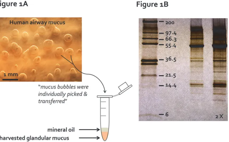
in the oil (Fig. 1A), collected individually using sterilized microforceps and transferred into a sterilized Eppendorf tube containing*50μL water-saturated mineral oil. The collected mucus
bubbles were stored at -20°C until use.
Protein gels and liquid chromatography in-line with tandem mass
spectrometry (LC-MS/MS)
Some of the mucus samples were run on Tris-Tricine 4–20% SDS polyacrylamide gradient gels followed by silver staining to separate protein bands (5*200 kDa). Samples were diluted 1:10
with sample buffer containing 0.5%β-mercaptoethanol and incubated at 95°C at a Perkin Elmer thermocycler (Santa Clara, CA, USA) for 10 min. After gel-electrophoresis, the gel was exposed to Bio-Rad silver staining solution (Hercules, CA, USA) for 15–20 min.
Gland mucus samples from three healthy human donors (HN1-3) were sent to University of Iowa for LC-MS/MS experiments. The 3 donor subjects were male and 36.7 ± 14.8 (mean ± S.D.) years old. Water-saturated mineral oil in the gland mucus samples was removed by
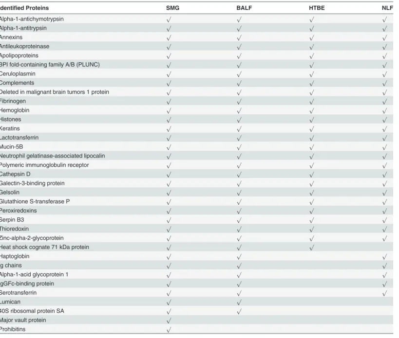
Fig 1. Primary pure human gland mucus collection. (A)Individual gland mucus bubbles, stimulated by a combination of 10μM carbachol and 10μM forskolin, formed under the water-saturated mineral oil on the tracheal mucosa, were picked up with sterilized microforceps and transferred into a sterilized Eppendorf tube for proteomics analysis.(B)A representative image of silver-stained protein bands from a human gland mucus sample collected as (A) on a Tris-Tricine 4–20% SDS polyacrylamide gradient gel. Protein molecular weight standard are shown on the left lane of the gel. Twice the same sample volumes were loaded on the far right lane.
centrifugation. Each mucus sample was transferred to a new sterile Eppendorf tube and pro-teins were denatured in 100μl of 50 mM Tris pH 8.5 buffer containing 8 M urea and 100 mM β-mercaptoethanol. Samples were lysed by passage through a 25 gauge needle using a 1 mL sy-ringe and 50μg total protein was prepared for MS analysis. Samples were reduced with 10 mM
dithiothreitol for 1 hour at 37°C and then alkylated with 55 mM iodoacetamide for 1 hour at room temperature in the dark. After alkylation, samples were diluted with 50 mM Tris buffer pH 8.5 to have 0.5 M final urea concentration (1/16 dilution) and digested with trypsin over-night. Peptides were acidified with o-phosphoric acid and 1% acetonitrile was added. Samples were desalted using a C18 microspin cartridge (The Nest Group, MA, USA). Desalted samples were then applied to a SCX microspin cartridge (The Nest Group, MA, USA) and eluted using 4 different salt concentrations (20, 40, 60, and 120 mM KCl) according to manufacturer’s pro-tocol. Eluted peptides were desalted and applied to an LC column in-line with the mass spec-trometer. After separation of peptides, MS data were recorded on a LTQ-XL linear ion trap mass spectrometer (Thermo Fisher Scientific, MA, USA). A 90 minute gradient was applied for all the peptide separations on the LC column. Proteins were identified via two automated data-bases (a subset of the SwissProt 2012_09 and the SwissProt 2012_09 datadata-bases, 20,235 entries) and two search engines (Mascot Search Engine (version 2.4.0, Matrix Science, MA, USA) and X! Tandem (version cyclone, GPM)). The identified proteins by LC-MS/MS analysis with a minimum 2 peptides and 0% false discovery rate (FDR) were afforded by Mascot Search En-gine. Maximum missed cleavages for trypsin were set to 2. Cysteine carbamodimethylation was used as fixed and methionine oxidation was used as variable modification. Mass tolerance for the parent ions was set to 1.8 Da and mass tolerance for fragment ions was set to 0.4 Da. The search results were compiled using Scaffold software (version 3.6.5, PS Inc., OR, USA). The set-tings of Scaffold were adjusted to 99.0% protein threshold and 90.0% peptide threshold. There-fore, all the proteins that are identified and presented in the manuscript have a False Discovery Rate (FDR) of 0% with 0 decoy proteins. To determine reproducibility, the same protein sam-ple was analyzed 3x using the identical LC-MS/MS method. The initial run identified 68 pro-teins (100%), the second run, 62 propro-teins (91%) and the third run, 67 propro-teins (98%).
Reagents
All reagents and drugs used in the present study were purchased from Sigma-Aldrich unless stated otherwise. Forskolin was dissolved in dimethyl sulfoxide, indomethacin in absolute etha-nol and carbachol in sterile double distilled water. Aliquots of stock solutions were stored at -20°C and diluted to 1:1,000 with Krebs Ringer buffer solution before use.
Results and Discussion
Identification of proteins in pure human airway gland mucus
Mucus bubbles were collected as shown inFig. 1A. To produce as much gland mucus as possi-ble from the small tissue samples, we typically stimulated with forskolin + carbachol for 2–4 hours. This allowed collection of*10μL of uncontaminated gland mucus. Our earlier study
with porcine airways indicated that each agonist used alone produced mucus with comparable protein bands when run on SDS-PAGE with silver staining [28]. A representative example of human airway glandular mucus run on Tris-Tricine 4–20% gradient SDS-PAGE (Fig. 1B) shows more than 50 identifiable silver-stained protein bands.
(S4 Table). Of these common proteins, 102/269 (38%) have been shown to have protective/in-nate immunity (Fig. 2B). Other overlapping biological activities of the 269 common proteins included (number of proteins): biological regulation (152); metabolic process (170); multicellu-lar organismal process (118); developmental process (104); and establishment of localization (83) (S5 Table). InTable 1, the 102 proteins with protective roles were condensed into their re-spective families (e.g. 17 keratins represented by one family) even though not all members of a given family have been shown to play protective roles.Table 1includes proteins with well-established innate immune roles and others for which antimicrobial activity was well-established subsequent to the assignment of other functions. Eleven of these are highlighted below.
Examples of common gland proteins initially known for functions remote
from innate defense
Annexinsare cytosolic or membrane-attached proteins involved in vesicle transport during exocytosis and endocytosis, but annexins A1, A2, and A5 have been detected extracellularly [36]. An antifungal role has been demonstrated for annexin A1 [37] and annexin A1 is among the proteins whose expression is markedly increased in primary human airway epithelial cells exposed toPseudomonas aeruginosa[38]. Annexin A2 helps to clear bacteria and viruses by fa-cilitating their docking to macrophages and thus increasing macrophage phagocytosis [39].
Apolipoproteinsare the major structural component of lipoproteins, which are mainly known for mediating lipid uptake and transport. However, apolipoproteins A1 and H also have anti-inflammatory and anti-oxidant activities [40], and apolipoprotein A1 is a also a po-tent anti-microbial against Gram positiveStaphylococcus epidermidis[41]. Apolipoprotein H and peptides derived from it showed anti-microbial activity against both Gram positive Staphy-lococcus aureusandS. pyogenesand Gram negativeE. coli[42].
BPI (bactericidal/permeability-increasing) proteins, also known as PLUNC (palate, lung and nasal epithelium clone) proteins are mainly expressed in exocrine glands of mouth, nose and the upper airways. Both BPI fold-containing protein family A1 (SPLUNC1, short PLUNC1) and family B1 (LPLUNC1, long PLUNC1) were detected in the HN1-3 gland mucus samples. BPI proteins have selective anti-microbial actions against Gram negative bacteria [43,44], and A1/SPLUNC1 has potent anti-proteolytic activity that endogenously regulates ENaC in airways [45] in a manner that should increase the volume of ASL.
DMBT 1(deleted in malignant brain tumors 1 protein) belongs to the scavenger receptor cysteine-rich, SRCR, superfamily [46] and the SRCR domain is known to bind various patho-gens includingStreptococci, H. pylori[47], andHIV[48]. The broad range of pathogen binding affinity of DMBT 1 allows it to serve as a pattern recognition molecule and to play a role in in-nate immunity. DMBT-1 can be found both in extracellular secretions and bound to the plas-ma membrane [49].
Gelsolinsevers actin-filaments to aid actin assembly and disassembly. It is also found extra-cellularly, where it increases DNase-I activity by interfering with actin inhibition of DNase-1; resulting in a thinning of DNA-rich CF sputum [50]. A gelsolin-derived peptide, PBP-10, has antimicrobial activity againstE. coli, Pseudomonas aeruginosa, andS. pneumoniae[51].
Fig 2. Proteins identified in human tracheal submucosal gland mucus by LC-MS/MS. (A)A modified Venn diagram of showing distribution of proteins among three healthy human (HN) donors. Across the 3 donors, 5486 peptides and 441 proteins were identified. Of these, 61% (269 proteins, the shaded area) and 35% (154 proteins) were found in 2–3 and all 3 HN gland mucus samples, respectively.(B)Among these 269 proteins, 102 (38%) were categorized as proteins with“anti”-activities: antimicrobial, antiinflammatory, antiproteolytic and antioxidant. Other biological activities include: biological regulation, metabolic/multi-organismal/developmental processes, binding activity, and establishment of localization.
Histonesare highly basic nuclear proteins and the principal structural component of chro-matin. Their antimicrobial activity was accidentally discovered more than 5 decades ago during isolation of a bactericidal substance called phagocytin [58].Buforin I, a well characterized toad stomach antimicrobial peptide, andparasin I, another bactericidal peptide, are derived from Histone 2A [59,60,61]. Histone 2A is also secreted into the amniotic fluid where it is antimicro-bial and endotoxin neutralizing [62]. Histone H4 released from human sebaceous glands sup-pressesStaphylococcus aureusandPropionibacterium acnes growth[63].
Keratins, orcytokeratins, are fibrous structural proteins abundant in epithelial tissues and skin. Fourteen type I and II keratins, including keratin II 6A, constituted a large portion of structural proteins common to glands. Recently, Tomet al. discovered broad spectrum antimi-crobial activity of glycine-rich C-terminal peptides from keratin 6A [64], with both Staphylo-coccus aureusandPseudomonas aeruginosabeing susceptible. Consistent with a role in innate immunity, multiple keratin genes in human primary airway epithelial cells show markedly
Table 1. Protein families with protective functions found in gland mucus.
Access # MW Identified Proteins Access # MW Identified Proteins
P05091 56 Aldehyde dehydrogenase P01876 38 Ig chains (5)
Q04828 37 Aldo-keto reductase Q9Y6R7 572 IgGFc-binding protein
P02763 24 α-1-acid glycoprotein 1 P02538 60 Keratins (16)
P01011 48 α-1-antichymotrypsin P02788 78 Lactotransferrin
P01009 47 α-1-antitrypsin P30740 43 LE inhibitor
P07355 39 Annexins (5) P51884 38 Lumican
P03973 14 Antileukoproteinase P61626 17 Lysozyme C
P02647 31 Apolipoproteins (4) Q14764 99 Major vault protein
P61769 14 β-2-microglobulin Q9HC84 596 Mucins (2)
Q8TDL5 52 BPI fold-containing family A/B P05164 84 Myeloperoxidase
P07339 45 Cathepsins (3) P59665 10 Neutrophil defensin 1
P00450 122 Ceruloplasmin P80188 23 NGAL-lipocalin
P01024 187 Complements (4) Q06830 22 Peroxiredoxins (2)
P28838 56 Cytosol aminopeptidase P01833 83 Polymeric Ig receptor
Q9UGM3 261 DMBT-1 Q99623 33 Prohibitins (2)
P07099 53 Epoxide hydrolase 1 P06454 12 Prothymosin alpha
P02671 95 Fibrinogen chains (3) Q8IWL1 26 PS-A2
P21333 281 Filamins (2) P08865 33 40S RP (7)
Q08380 65 Galectins/GBP (3) P06703 10 S100-proteins (7)
P06396 86 Gelsolin P02787 77 Serotransferrin
P09211 23 Glutathione S-transferase P P29508 45 Serpin B3
P00738 45 Haptoglobin P0DJI8 14 SA A-1 protein
P11142 71 HSC 71 P04179 25 Superoxide dismutase 2
P68871 16 Hemoglobin P10599 12 Thioredoxin
P62805 11 Histones (2) P25311 34 Zinc-α-2-glycoprotein
P30508 41 HLA class I antigen
At least one member of these families of proteins with protective functions was found in 2–3 samples of human mucus, with (n) indicating the total number of family members found in human gland mucus. MW is shown as kDa. The full list of 102 protective proteins is found inS3 TableandS4 Table.
Abbreviations: GBP, Galectin binding protein; DMBT-1, Deleted in malignant brain tumors 1 protein; HSC 71, Heat shock cognate 71 kDa protein; LE inhibitor, Leukocyte elastase inhibitor; PS-A2, pulmonary surfactant A2; 40S RP, 40S ribosomal proteins; and SA A-1, Serum amyloid A-1. Accession # is from the UniProt databasehttp://www.uniprot.org/uniprot/.
increased expression after exposure toPseudomonas aeruginosa[38]. It is unknown how many keratins and keratin-derived peptides have antimicrobial activity.
Lumicanis an extracellular matrix proteoglycan, containing N-terminal leucine-rich do-main, also found in skin, intestine and cornea. Lumican-deficient mice have impaired innate immunity when challenged with bacterial lipopolysaccharide [65]; they also exhibit defective clearance ofPseudomonas aeruginosafrom the lung [66] and the cornea [67].
It has long been hypothesized that balanced activities of proteases and anti-proteases plays a key role in airway fluid and tissue homeostasis [16,28]. As shown inS5 Table, multiple anti-proteases, includingα-1 antitrypsin,α-1 antichymotrypsin, antileukoproteinase, serpin, and leukocyte elastase inhibitor, and several proteases, including cathepsin, calpain and fibulin, were detected in at least 2 of 3 human gland mucus samples. The balance might be tipped to-ward proteolysis in CF because of abundant neutrophil-derived proteases coupled with reduced bioavailability of gland-derived anti-proteases.
Origins of proteins in human airway submucosal gland mucus
Of the common 269 proteins, 194 (72%) are considered to be cytosolic/membrane proteins and 75 proteins (28%) are categorized as secreted proteins. Although great care was taken to insure that the collected mucus came directly from the duct orifices, the large number of pro-teins that are typically intracellular or membrane bound was unexpected and raises several possibilities.
First, although the mucus is‘pure’in the sense that it is only of glandular origin, it is not cell-free. Various types of cells have been observed in mucus while still within the gland ducts [68,69], and membrane proteins that are not in the apical membrane, such as Na+, K+, ATPase, were found in all samples. Because cells were expected to be a small component overall, we did not attempt to deplete them from the mucus for this initial proteomic analysis, but their pres-ence needs to be kept in mind when considering the possible origins of the proteins.
A second possibility is that these cellular proteins are in the mucus because they have been secreted by pathways other than exocytosis. As discussed above, some of cellular proteins, e.g. keratins, histones, hemoglobin, peroxiredoxin and prohibitin have been identified previously in extracellular secretions, but because their secretion mechanisms are not understood they have not been classified as secreted proteins. In addition to exocytosis, at least four alternate means exist for exporting proteins: holocrine secretion, apocrine secretion, exosome release and microvesicle release. Holocrine secretion is poorly understood and in human seems to be limited to sebaceous and meibomian glands; recent evidence suggests that apoptosis may be the mechanism [70]. Apocrine secretion involves a portion of the apical membrane blebbing and then budding off, as in milk-protein secretion by the mammary gland [71]. Apocrine se-cretion has been observed in mouse airways [72,73,74] and may well occur in submucosal glands. In support of this hypothesis, CFTR-dependent secretion of mucus from skin glands of Xenopus laevis results in the loss of>75% of the mucous cell mass [75].
to be exclusively intracellular until recently are in fact transported into airway mucus, where they or their peptide fragments participate in innate defense of the airways. We do not have a ready explanation for how the many cellular proteins have evolved additional mucosal defense functions. As more mucosal proteins are identified, it will be interesting to see if testable hy-potheses on this point can be generated.
A comparative analysis of innate defense and protective proteins of
gland mucus with those of other airway fluids
To what extent does our sample of the human gland mucus innate defense proteome overlap that of other airway fluids? Data from LC-MS/MS analyses have been reported for nasal lavage fluid (NLF), apical fluid from primary human tracheobronchial epithelial cell culture (HTBE) [20,21,22,23,24,26,27,29,31] and bronchial alveolar lavage fluid (BALF) [20,21,22,23,24,26,27, 29,31]. These proteomics studies of airway fluids have identified from 42 to>1,500 proteins
(n = 3*18 healthy controls, BALF), 38 to 267 proteins (n = 6*29 controls, NLF) and 132 to
186 proteins in apical fluid of primary human airway epithelial cultures.
For comparison we selected 35 protective proteins that were commonly detected in our 3 HN mucus samples(Table 2). Among these, 18 were detected in all 4 sources of airway secre-tions (Table 2, top section) and the others were not reported for one or more sources. The larg-est overlap was with BALF, which is to be expected because BALF should include SMG mucus. Only two of the 35 proteins were absent from BALF: major vault protein (MVP) and prohibi-tin, both of which protect against bacterial infections in human and mice [84,85,86]. It is possi-ble that these proteins were not detected in BALF because of the dilution factor. Sixteen of the proteins were not reported for apical fluid from airway cell cultures, which lack glands and also presumably lack proteins that might enter gland mucus via transcytosis or transudation, for ex-ample haptoglobin, immunoglobulin chains, and serotransferrin. Five proteins were not re-ported for nasal lavage fluid: heat-shock cognate 71 kDa protein, lumican, 40S-ribosomal protein SA, MVP and prohibitins. As with BALF, most of the NLF innate defense and protec-tive proteins overlap SMG proteins. Nasal submucosal glands and anterior nasal glands are likely the major sources of these proteins.
Limitations of this study
The small number of subjects and the limited amount of uncontaminated gland mucus that could be collected from each one will bias toward an underestimate of the gland mucus prote-ome—especially for low molecular weight proteins. Because we were stimulating the glandsex vivo, we would also underestimate proteins that are transcytosed from the blood. Conversely, we did not attempt to deplete resident cells from the mucus and as discussed, these probably contributed to the proteins we detected. If these are the cells that are normally found in glands, for example leukocytes that enter the airways via the glands, their inclusion may be informa-tive. However, we used a high concentration of secretory agonists to insure adequate samples of mucus, which, together with unintended damage from dissection and an unavoidable level of hypoxia inex vivotissues, could have led to higher than normal levels of epithelia sloughing.
Summary and conclusions
[8,14,87,88]. We hypothesized a role for airway glands in innate host defense, based on the rec-ognized role of mucus clearance in protecting the airways [2] and the glandular source of much mucus [5]. Because glands express CFTR [6], we sought and found evidence that gland secre-tion is defective in CF [3], and suggested this was one reason why CF lungs are vulnerable to in-fections. This study supports those hypotheses by showing that a substantial proportion of the proteins in human airway gland mucus have protective functions.
Table 2. A comparative analysis of anti-proteins of 4 airway secretions.
Identified Proteins SMG BALF HTBE NLF
Alpha-1-antichymotrypsin p p p p
Alpha-1-antitrypsin p p p p
Annexins p p p p
Antileukoproteinase p p p p
Apolipoproteins p p p p
BPI fold-containing family A/B (PLUNC) p p p p
Ceruloplasmin p p p p
Complements p p p p
Deleted in malignant brain tumors 1 protein p p p p
Fibrinogen p p p p
Hemoglobin p p p p
Histones p p p p
Keratins p p p p
Lactotransferrin p p p p
Mucin-5B p p p p
Neutrophil gelatinase-associated lipocalin p p p p
Polymeric immunoglobulin receptor p p p p
Cathepsin D p p p p
Galectin-3-binding protein p p p p
Gelsolin p p p p
Glutathione S-transferase P p p p p
Peroxiredoxins p p p p
Serpin B3 p p p p
Thioredoxin p p p p
Zinc-alpha-2-glycoprotein p p p p
Heat shock cognate 71 kDa protein p p p
Haptoglobin p p p
Ig chains p p p
Alpha-1-acid glycoprotein 1 p p p
IgGFc-binding protein p p p
Serotransferrin p p p
Lumican p p
40S ribosomal protein SA p p
Major vault protein p
Prohibitins p
The data represent a summary of our comparative analysis of 35 common protective HN anti-proteins in the present study to 3 other airwayfluids. Abbreviations: SMG, airway submucosal gland mucus; BALF, bronchial alveolar lavagefluid; HTBE, apicalfluid of primary human tracheobronchial epithelial cell culture; and NLF, nasal lavagefluid. The data from other airwayfluids are referenced in the text.
The present study suggests two ways in which mucus is protective beyond its critical role in mechanical clearance [2]. First, in direct support of physical entrapment and removal of patho-gens, mucus has a host of anti-microbial compounds and proteins that kill pathogens or inhibit their growth. Importantly, these anti-microbial mechanisms are extremely diverse and some-times synergistic, which should make it more challenging for resistance to develop. Second, to deal with toxic compounds that are either inhaled or generated to confront pathogens, mucus also contains a host of anti-proteolytic, anti-oxidant, or anti-inflammatory proteins that help protect airway tissue. Finally, although not directly studied here, mucus contains cells like neu-trophils and macrophages that kill pathogens with yet another set of overlapping mechanisms.
The sophisticated orchestration the innate mucosal defenses contrast in two fundamental ways with the use of synthetic antibiotics in most clinical settings. First, as the term‘innate’ em-phasizes, these defenses are ever present in the airways, ready to act as soon as a single patho-gen makes contact with the mucus, rather than after infections have become established. Second, as emphasized above, our natural defenses are multi-factorial, unlike the mono-antibi-otic therapies that still dominate clinical practice.
To conclude, we established the feasibility of carrying out a proteomics analysis of‘pure’
human airway gland mucus, and showed that a large proportion of the proteins in mucus have protective roles. Many of the proteins encountered have traditionally been considered to play exclusively intracellular roles, but it is increasingly clear that cells can export these proteins to the extracellular space, where they or their peptide fragments take on new roles. It seems likely that additional protective roles will be uncovered for many of the other proteins that are resi-dent in airway mucus.
Supporting Information
S1 Table. Total peptide fragments found in 3 HN mucus samples.Peptides (and proteins in S 2) were identified via two databases, a subset of theSwissProt2012_09 and theSwissProt 2012_09 databases with 20,235 entries, and two search engines,Mascot Search EngineandX! Tandem. The detailed set up for the search engines: Fragment Tolerance: 0.40 Da (monoisoto-pic), Parent Tolerance: 1.8 Da (monoisoto(monoisoto-pic), Fixed Modifications: +57 on C (carbamido-methyl), Variable Modifications: +16 on M (oxidation), and Digestion Enzyme: trypsin. The peptide thresholds ofScaffoldsoftware were adjusted to 90% minimum.
(XLSX)
S2 Table. Unique peptides observed for each HN sample and relative abundance of 441 HN proteins.The peptide thresholds ofScaffoldsoftware were adjusted to 99% minimum and two peptides minimum. The 441 HN proteins were arranged as a rank order of their relative abun-dance (#1, the most abundantly detected), based on Total Ion Current (TIC) values of individu-al proteins cindividu-alculated byScaffoldafter normalizing the dataset for TIC.
(XLSX)
S3 Table. Proteins found in 3 of 3 HN mucus samples, top 65 proteins have protective properties.The protective and the other group proteins (total 154 proteins) are arranged ac-cording to their relative abundance (RA), fromS2 Table, in each group.
(XLSX)
S4 Table. Proteins found in 2 of 3 HN mucus samples, top 37 proteins have protective properties.Additional 114 proteins detected in 2 of 3 HN samples, other than the ones listed inS3 Table, are arranged according to their relative abundance (RA) in each group.
S5 Table. Biological activities of 269 proteins found in at least 2 of 3 HN mucus samples.
The list of 269 proteins is arranged according to their relative abundance. (XLSX)
S6 Table. HN peptides with additional MS/MS information.
(XLSX)
S7 Table. HN Proteins with additional MS/MS information.
(XLSX)
Acknowledgments
We thank the Stanford transplant team, including doctors, nurses, and staffs. We thank Jin Hyeok Jeong and Zachary Sellers for useful discussions and Kim Tran, Sara Modlin, Jessica Char, Marlene Kennedy, and Lesje Atkinson for their technical assistance. We also thank the Carver Trust sponsored Proteomics Facility at the University of Iowa. We are especially grate-ful to organ donors whose contribution makes this study possible.
Author Contributions
Conceived and designed the experiments: NSJ JJW. Performed the experiments: NSJ IATE HJC IHP. Analyzed the data: NSJ IATE JFE JJW. Contributed reagents/materials/analysis tools: NSJ IATE HJC IHP JFE JJW. Wrote the paper: NSJ IATE JFE JJW.
References
1. Basbaum CB, Jany B, Finkbeiner WE (1990) The serous cell. Annu Rev Physiol 52: 97–113. PMID: 2158772
2. Knowles MR, Boucher RC (2002) Mucus clearance as a primary innate defense mechanism for mam-malian airways. J Clin Invest 109: 571–577. PMID:11877463
3. Wine JJ, Joo NS (2004) Submucosal glands and airway defense. Proc Am Thorac Soc 1: 47–53. PMID:16113412
4. Bals R, Hiemstra PS (2004) Innate immunity in the lung: how epithelial cells fight against respiratory pathogens. Eur Respir J 23: 327–333. PMID:14979512
5. Ballard ST, Trout L, Bebok Z, Sorscher EJ, Crews A (1999) CFTR involvement in chloride, bicarbonate, and liquid secretion by airway submucosal glands. Am J Physiol 277: L694–699. PMID:10516209 6. Engelhardt JF, Yankaskas JR, Ernst SA, Yang Y, Marino CR, et al. (1992) Submucosal glands are the
predominant site of CFTR expression in the human bronchus. Nat Genet 2: 240–248. PMID:1285365 7. Gustafsson JK, Ermund A, Ambort D, Johansson ME, Nilsson HE, et al. (2012) Bicarbonate and
func-tional CFTR channel are required for proper mucin secretion and link cystic fibrosis with its mucus phe-notype. J Exp Med 209: 1263–1272. doi:10.1084/jem.20120562PMID:22711878
8. Pezzulo AA, Tang XX, Hoegger MJ, Alaiwa MH, Ramachandran S, et al. (2012) Reduced airway sur-face pH impairs bacterial killing in the porcine cystic fibrosis lung. Nature 487: 109–113. doi:10.1038/ nature11130PMID:22763554
9. Quinton PM (2008) Cystic fibrosis: impaired bicarbonate secretion and mucoviscidosis. Lancet 372: 415–417. doi:10.1016/S0140-6736(08)61162-9PMID:18675692
10. Dajani R, Zhang Y, Taft PJ, Travis SM, Starner TD, et al. (2005) Lysozyme secretion by submucosal glands protects the airway from bacterial infection. Am J Respir Cell Mol Biol 32: 548–552. PMID: 15746432
11. Cho HJ, Joo NS, Wine JJ (2011) Defective fluid secretion from submucosal glands of nasal turbinates from CFTR-/- and CFTR (DeltaF508/DeltaF508) pigs. PLoS One 6: e24424. doi:10.1371/journal. pone.0024424PMID:21935358
13. Ianowski JP, Choi JY, Wine JJ, Hanrahan JW (2007) Mucus secretion by single tracheal submucosal glands from normal and cystic fibrosis transmembrane conductance regulator knockout mice. J Physiol 580: 301–314. PMID:17204498
14. Jayaraman S, Joo NS, Reitz B, Wine JJ, Verkman AS (2001) Submucosal gland secretions in airways from cystic fibrosis patients have normal [Na(+)] and pH but elevated viscosity. Proc Natl Acad Sci U S A 98: 8119–8123. PMID:11427704
15. Joo NS, Cho HJ, Khansaheb M, Wine JJ (2010) Hyposecretion of fluid from tracheal submucosal glands of CFTR-deficient pigs. J Clin Invest 120: 3161–3166. doi:10.1172/JCI43466PMID:20739758 16. Joo NS, Irokawa T, Robbins RC, Wine JJ (2006) Hyposecretion, not hyperabsorption, is the basic
de-fect of cystic fibrosis airway glands. J Biol Chem 281: 7392–7398. PMID:16410244
17. Joo NS, Irokawa T, Wu JV, Robbins RC, Whyte RI, et al. (2002) Absent secretion to vasoactive intesti-nal peptide in cystic fibrosis airway glands. J Biol Chem 277: 50710–50715. PMID:12368280 18. Sun X, Sui H, Fisher JT, Yan Z, Liu X, et al. (2010) Disease phenotype of a ferret CFTR-knockout
model of cystic fibrosis. J Clin Invest 120: 3149–3160. doi:10.1172/JCI43052PMID:20739752 19. Chen J, Ryu S, Gharib SA, Goodlett DR, Schnapp LM (2008) Exploration of the normal human
bronch-oalveolar lavage fluid proteome. Proteomics Clin Appl 2: 585–595. doi:10.1002/prca.200780006 PMID:21136857
20. Gharib SA, Nguyen E, Altemeier WA, Shaffer SA, Doneanu CE, et al. (2010) Of mice and men: compar-ative proteomics of bronchoalveolar fluid. Eur Respir J 35: 1388–1395. doi:10.1183/09031936. 00089409PMID:20032019
21. Kosanam H, Sato M, Batruch I, Smith C, Keshavjee S, et al. (2012) Differential proteomic analysis of bronchoalveolar lavage fluid from lung transplant patients with and without chronic graft dysfunction. Clin Biochem 45: 223–230. doi:10.1016/j.clinbiochem.2011.11.015PMID:22206736
22. Merkel D, Rist W, Seither P, Weith A, Lenter MC (2005) Proteomic study of human bronchoalveolar la-vage fluids from smokers with chronic obstructive pulmonary disease by combining surface-enhanced laser desorption/ionization-mass spectrometry profiling with mass spectrometric protein identification. Proteomics 5: 2972–2980. PMID:16075419
23. Plymoth A, Yang Z, Lofdahl CG, Ekberg-Jansson A, Dahlback M, et al. (2006) Rapid proteome analysis of bronchoalveolar lavage samples of lifelong smokers and never-smokers by micro-scale liquid chro-matography and mass spectrometry. Clin Chem 52: 671–679. PMID:16497942
24. Magi B, Bini L, Perari MG, Fossi A, Sanchez JC, et al. (2002) Bronchoalveolar lavage fluid protein com-position in patients with sarcoidosis and idiopathic pulmonary fibrosis: a two-dimensional electropho-retic study. Electrophoresis 23: 3434–3444. PMID:12373774
25. Wu J, Kobayashi M, Sousa EA, Liu W, Cai J, et al. (2005) Differential proteomic analysis of bronchoal-veolar lavage fluid in asthmatics following segmental antigen challenge. Mol Cell Proteomics 4: 1251–
1264. PMID:15951573
26. Candiano G, Bruschi M, Pedemonte N, Musante L, Ravazzolo R, et al. (2007) Proteomic analysis of the airway surface liquid: modulation by proinflammatory cytokines. Am J Physiol Lung Cell Mol Physiol 292: L185–198. PMID:17215433
27. Kesimer M, Kirkham S, Pickles RJ, Henderson AG, Alexis NE, et al. (2009) Tracheobronchial air-liquid interface cell culture: a model for innate mucosal defense of the upper airways? Am J Physiol Lung Cell Mol Physiol 296: L92–L100. doi:10.1152/ajplung.90388.2008PMID:18931053
28. Joo NS, Lee DJ, Winges KM, Rustagi A, Wine JJ (2004) Regulation of antiprotease and antimicrobial protein secretion by airway submucosal gland serous cells. J Biol Chem 279: 38854–38860. PMID: 15234967
29. Casado B, Pannell LK, Iadarola P, Baraniuk JN (2005) Identification of human nasal mucous proteins using proteomics. Proteomics 5: 2949–2959. PMID:15996010
30. Lindahl M, Stahlbom B, Tagesson C (2001) Identification of a new potential airway irritation marker, pal-ate lung nasal epithelial clone protein, in human nasal lavage fluid with two-dimensional electrophoresis and matrix-assisted laser desorption/ionization-time of flight. Electrophoresis 22: 1795–1800. PMID: 11425234
31. Tomazic PV, Birner-Gruenberger R, Leitner A, Obrist B, Spoerk S, et al. (2014) Nasal mucus proteomic changes reflect altered immune responses and epithelial permeability in patients with allergic rhinitis. J Allergy Clin Immunol 133: 741–750. doi:10.1016/j.jaci.2013.09.040PMID:24290289
32. Bartlett JA, Albertolle ME, Wohlford-Lenane C, Pezzulo AA, Zabner J, et al. (2013) Protein composition of bronchoalveolar lavage fluid and airway surface liquid from newborn pigs. Am J Physiol Lung Cell Mol Physiol 305: L256–266. doi:10.1152/ajplung.00056.2013PMID:23709621
34. Clarke LL, Gawenis LR, Bradford EM, Judd LM, Boyle KT, et al. (2004) Abnormal Paneth cell granule dissolution and compromised resistance to bacterial colonization in the intestine of CF mice. Am J Phy-siol Gastrointest Liver PhyPhy-siol 286: G1050–1058. PMID:14715526
35. Joo NS, Wu JV, Krouse ME, Saenz Y, Wine JJ (2001) Optical method for quantifying rates of mucus se-cretion from single submucosal glands. Am J Physiol Lung Cell Mol Physiol 281: L458–468. PMID: 11435221
36. Gerke V, Moss SE (2002) Annexins: from structure to function. Physiol Rev 82: 331–371. PMID: 11917092
37. Lilly EA, Yano J, Fidel PL Jr (2010) Annexin-A1 identified as the oral epithelial cell anti-Candida effector moiety. Mol Oral Microbiol 25: 293–304. doi:10.1111/j.2041-1014.2010.00579.xPMID:20618702 38. Vos JB, van Sterkenburg MA, Rabe KF, Schalkwijk J, Hiemstra PS, et al. (2005) Transcriptional re-sponse of bronchial epithelial cells to Pseudomonas aeruginosa: identification of early mediators of host defense. Physiol Genomics 21: 324–336. PMID:15701729
39. Swisher JF, Khatri U, Feldman GM (2007) Annexin A2 is a soluble mediator of macrophage activation. J Leukoc Biol 82: 1174–1184. PMID:17715360
40. Barter PJ, Nicholls S, Rye KA, Anantharamaiah GM, Navab M, et al. (2004) Antiinflammatory properties of HDL. Circ Res 95: 764–772. PMID:15486323
41. Tada N, Sakamoto T, Kagami A, Mochizuki K, Kurosaka K (1993) Antimicrobial activity of lipoprotein particles containing apolipoprotein Al. Mol Cell Biochem 119: 171–178. PMID:8455579
42. Nilsson M, Wasylik S, Morgelin M, Olin AI, Meijers JC, et al. (2008) The antibacterial activity of peptides derived from human beta-2 glycoprotein I is inhibited by protein H and M1 protein from Streptococcus pyogenes. Mol Microbiol 67: 482–492. PMID:18093092
43. Canny G, Levy O, Furuta GT, Narravula-Alipati S, Sisson RB, et al. (2002) Lipid mediator-induced ex-pression of bactericidal/ permeability-increasing protein (BPI) in human mucosal epithelia. Proc Natl Acad Sci U S A 99: 3902–3907. PMID:11891303
44. Horwitz AH, Williams RE, Liu PS, Nadell R (1999) Bactericidal/permeability-increasing protein inhibits growth of a strain of Acholeplasma laidlawii and L forms of the gram-positive bacteria Staphylococcus aureus and Streptococcus pyogenes. Antimicrob Agents Chemother 43: 2314–2316. PMID:10471588 45. Garcia-Caballero A, Rasmussen JE, Gaillard E, Watson MJ, Olsen JC, et al. (2009) SPLUNC1
regu-lates airway surface liquid volume by protecting ENaC from proteolytic cleavage. Proc Natl Acad Sci U S A 106: 11412–11417. doi:10.1073/pnas.0903609106PMID:19541605
46. Mollenhauer J, Wiemann S, Scheurlen W, Korn B, Hayashi Y, et al. (1997) DMBT1, a new member of the SRCR superfamily, on chromosome 10q25.3-26.1 is deleted in malignant brain tumours. Nat Genet 17: 32–39. PMID:9288095
47. Madsen J, Tornoe I, Nielsen O, Lausen M, Krebs I, et al. (2003) CRP-ductin, the mouse homologue of gp-340/deleted in malignant brain tumors 1 (DMBT1), binds gram-positive and gram-negative bacteria and interacts with lung surfactant protein D. Eur J Immunol 33: 2327–2336. PMID:12884308 48. Chu Y, Li J, Wu X, Hua Z, Wu Z (2013) Identification of human immunodeficiency virus type 1 (HIV-1)
gp120-binding sites on scavenger receptor cysteine rich 1 (SRCR1) domain of gp340. J Biomed Sci 20: 44. doi:10.1186/1423-0127-20-44PMID:23815775
49. Ligtenberg AJ, Karlsson NG, Veerman EC (2010) Deleted in malignant brain tumors-1 protein (DMBT1): a pattern recognition receptor with multiple binding sites. Int J Mol Sci 11: 5212–5233. doi: 10.3390/ijms1112521PMID:21614203
50. Davoodian K, Ritchings BW, Ramphal R, Bubb MR (1997) Gelsolin activates DNase I in vitro and cystic fibrosis sputum. Biochemistry 36: 9637–9641. PMID:9289015
51. Bucki R, Pastore JJ, Randhawa P, Vegners R, Weiner DJ, et al. (2004) Antibacterial activities of rhoda-mine B-conjugated gelsolin-derived peptides compared to those of the antimicrobial peptides cathelici-din LL37, magainin II, and melittin. Antimicrob Agents Chemother 48: 1526–1533. PMID:15105101 52. Yang F, Friedrichs WE, Navarijo-Ashbaugh AL, deGraffenried LA, Bowman BH, et al. (1995) Cell
type-specific and inflammatory-induced expression of haptoglobin gene in lung. Lab Invest 73: 433–440. PMID:7564277
53. Newton DA, Rao KM, Dluhy RA, Baatz JE (2006) Hemoglobin is expressed by alveolar epithelial cells. J Biol Chem 281: 5668–5676. PMID:16407281
54. Eaton JW, Brandt P, Mahoney JR, Lee JT Jr (1982) Haptoglobin: a natural bacteriostat. Science 215: 691–693. PMID:7036344
56. Hodson D, Hirsch JG (1958) The antibacterial activity of hemoglobin. J Exp Med 107: 167–183. PMID: 13491755
57. Parish CA, Jiang H, Tokiwa Y, Berova N, Nakanishi K, et al. (2001) Broad-spectrum antimicrobial activi-ty of hemoglobin. Bioorg Med Chem 9: 377–382. PMID:11249130
58. Hirsch JG (1958) Bactericidal action of histone. J Exp Med 108: 925–944. PMID:13598820
59. Kim HS, Park CB, Kim MS, Kim SC (1996) cDNA cloning and characterization of buforin I, an antimicro-bial peptide: a cleavage product of histone H2A. Biochem Biophys Res Commun 229: 381–387. PMID: 8954908
60. Park IY, Park CB, Kim MS, Kim SC (1998) Parasin I, an antimicrobial peptide derived from histone H2A in the catfish, Parasilurus asotus. FEBS Lett 437: 258–262. PMID:9824303
61. Kim HS, Yoon H, Minn I, Park CB, Lee WT, et al. (2000) Pepsin-mediated processing of the cytoplasmic histone H2A to strong antimicrobial peptide buforin I. J Immunol 165: 3268–3274. PMID:10975843 62. Kim HS, Cho JH, Park HW, Yoon H, Kim MS, et al. (2002) Endotoxin-neutralizing antimicrobial proteins
of the human placenta. J Immunol 168: 2356–2364. PMID:11859126
63. Lee DY, Huang CM, Nakatsuji T, Thiboutot D, Kang SA, et al. (2009) Histone H4 is a major component of the antimicrobial action of human sebocytes. J Invest Dermatol 129: 2489–2496. doi:10.1038/jid. 2009.106PMID:19536143
64. Tam C, Mun JJ, Evans DJ, Fleiszig SM (2012) Cytokeratins mediate epithelial innate defense through their antimicrobial properties. J Clin Invest 122: 3665–3677. doi:10.1172/JCI64416PMID:23006328 65. Wu F, Vij N, Roberts L, Lopez-Briones S, Joyce S, et al. (2007) A novel role of the lumican core protein
in bacterial lipopolysaccharide-induced innate immune response. J Biol Chem 282: 26409–26417. PMID:17616530
66. Shao H, Lee S, Gae-Scott S, Nakata C, Chen S, et al. (2012) Extracellular matrix lumican promotes bacterial phagocytosis, and Lum-/- mice show increased Pseudomonas aeruginosa lung infection se-verity. J Biol Chem 287: 35860–35872. doi:10.1074/jbc.M112.380550PMID:22865855
67. Shao H, Scott SG, Nakata C, Hamad AR, Chakravarti S (2013) Extracellular matrix protein lumican pro-motes clearance and resolution of Pseudomonas aeruginosa keratitis in a mouse model. PLoS One 8: e54765. doi:10.1371/journal.pone.0054765PMID:23358433
68. Rock JR, Randell SH, Hogan BL (2010) Airway basal stem cells: a perspective on their roles in epitheli-al homeostasis and remodeling. Dis Model Mech 3: 545–556. doi:10.1242/dmm.006031PMID: 20699479
69. Tamada T, Sasaki T (2001) [The role of airway submucosal glands in the airway mucosal defense sys-tem]. Nihon Kokyuki Gakkai Zasshi 39: 157–165. PMID:11431907
70. Liman N, Alan E (2013) The process of apoptosis in a holocrine gland as shown by the avian uropygial gland. Anat Rec (Hoboken) 296: 504–520. doi:10.1002/ar.22645PMID:23362229
71. Helminen HJ, Ericsson JL (1968) Studies on mammary gland involution. I. On the ultrastructure of the lactating mammary gland. J Ultrastruct Res 25: 193–213. PMID:5715762
72. Etherton JE, Purchase IF, Corrin B (1979) Apocrine secretion in the terminal bronchiole of mouse lung. J Anat 129: 305–322. PMID:115821
73. Pack RJ, Al-Ugaily LH, Morris G, Widdicombe JG (1980) The distribution and structure of cells in the tracheal epithelium of the mouse. Cell Tissue Res 208: 65–84. PMID:6248229
74. Aryal G, Kimula Y, Koike M (2003) Ultrastructure of Clara cells stimulated by isoproterenol. J Med Dent Sci 50: 195–202. PMID:15074357
75. Engelhardt JF, Smith SS, Allen E, Yankaskas JR, Dawson DC, et al. (1994) Coupled secretion of chlo-ride and mucus in skin of Xenopus laevis: possible role for CFTR. Am J Physiol 267: C491–500. PMID: 7521128
76. Valadi H, Ekstrom K, Bossios A, Sjostrand M, Lee JJ, et al. (2007) Exosome-mediated transfer of mRNAs and microRNAs is a novel mechanism of genetic exchange between cells. Nat Cell Biol 9: 654–659. PMID:17486113
77. Raposo G, Stoorvogel W (2013) Extracellular vesicles: exosomes, microvesicles, and friends. J Cell Biol 200: 373–383. doi:10.1083/jcb.201211138PMID:23420871
78. Kesimer M, Scull M, Brighton B, DeMaria G, Burns K, et al. (2009) Characterization of exosome-like vesicles released from human tracheobronchial ciliated epithelium: a possible role in innate defense. FASEB J 23: 1858–1868. doi:10.1096/fj.08-119131PMID:19190083
79. van Niel G, Raposo G, Candalh C, Boussac M, Hershberg R, et al. (2001) Intestinal epithelial cells se-crete exosome-like vesicles. Gastroenterology 121: 337–349. PMID:11487543
81. Ogawa Y, Miura Y, Harazono A, Kanai-Azuma M, Akimoto Y, et al. (2011) Proteomic analysis of two types of exosomes in human whole saliva. Biol Pharm Bull 34: 13–23. PMID:21212511
82. Choi JY, Khansaheb M, Joo NS, Krouse ME, Robbins RC, et al. (2009) Substance P stimulates human airway submucosal gland secretion mainly via a CFTR-dependent process. J Clin Invest 119: 1189–
1200. doi:10.1172/JCI37284PMID:19381016
83. Inglis SK, Corboz MR, Taylor AE, Ballard ST (1997) In situ visualization of bronchial submucosal glands and their secretory response to acetylcholine. Am J Physiol 272: L203–210. PMID:9124370
84. Kowalski MP, Dubouix-Bourandy A, Bajmoczi M, Golan DE, Zaidi T, et al. (2007) Host resistance to lung infection mediated by major vault protein in epithelial cells. Science 317: 130–132. PMID: 17615361
85. Sharma A, Qadri A (2004) Vi polysaccharide of Salmonella typhi targets the prohibitin family of mole-cules in intestinal epithelial cells and suppresses early inflammatory responses. Proc Natl Acad Sci U S A 101: 17492–17497. PMID:15576509
86. Theiss AL, Vijay-Kumar M, Obertone TS, Jones DP, Hansen JM, et al. (2009) Prohibitin is a novel regu-lator of antioxidant response that attenuates colonic inflammation in mice. Gastroenterology 137: 199–
208, 208 e191–196. doi:10.1053/j.gastro.2009.03.033PMID:19327358
87. Matsui H, Grubb BR, Tarran R, Randell SH, Gatzy JT, et al. (1998) Evidence for periciliary liquid layer depletion, not abnormal ion composition, in the pathogenesis of cystic fibrosis airways disease. Cell 95: 1005–1015. PMID:9875854