RESUMO RESUMORESUMO
RESUMORESUMO.-.-.-.-.- [Comparação de t rês mét odos de diagnóst ico paraComparação de t rês mét odos de diagnóst ico paraComparação de t rês mét odos de diagnóst ico paraComparação de t rês mét odos de diagnóst ico paraComparação de t rês mét odos de diagnóst ico para det ecção de lept ospiras em rins de camundongos selvagens det ecção de lept ospiras em rins de camundongos selvagensdet ecção de lept ospiras em rins de camundongos selvagens det ecção de lept ospiras em rins de camundongos selvagensdet ecção de lept ospiras em rins de camundongos selvagens (((((M us m uscul usM us m uscul usM us m uscul usM us m uscul usM us m uscul us).).).] Foram capt urados 41 camundongos ().). M us musculus) na região urbana, próximo à ferrovia da cidade de
Santa Fé, Argentina. Os rins de cada animal capturado foram removidos para estudos bacteriológicos e histológicos. Um dos rins foi imerso em meio semi-sólido de Fletcher para iso-lament o de lept ospiras, as quais foram serologicament e tipificadas. O outro rim foi microscopicamente examinado por coloração de cortes histológicos pela hematoxilina-eosina, impregnação pela prat a e imunohist oquímica. Lept ospiras pertencentes ao serogrupo Ballum foram isoladas em 16 (39%) das 41 amostras availadas. A presença do agente foi observa-da em 18 (44%) e 19 (46%) observa-das 41 amostras avaliaobserva-das por im-pregnação pela prata e imunohistoquímica, respectivamen-te. Leptospiras foram detectadas em grande numero na su-perfície apical das células epiteliais e no lumen dos túbulos medulares e foram menos frequentemente encontradas na superficie apical de células epiteliais ou no lúmen dos túbulos corticais, o que é considerado achado raro em animais porta-dores. Lesões microscópicas consistindo de nefrite
mononu-Comparison of t hree diagnost ic t echniques for t he det ect ion of
Comparison of t hree diagnost ic t echniques for t he det ect ion of
Comparison of t hree diagnost ic t echniques for t he det ect ion of
Comparison of t hree diagnost ic t echniques for t he det ect ion of
Comparison of t hree diagnost ic t echniques for t he det ect ion of
lept ospires in t he kidneys of wild house mice (
lept ospires in t he kidneys of wild house mice (
lept ospires in t he kidneys of wild house mice (
lept ospires in t he kidneys of wild house mice (
lept ospires in t he kidneys of wild house mice (
M us musculus
M us musculus
M us musculus
M us musculus
M us musculus
)))))
11111Carlos A. Rossetti2,5*, Bibiana N. Vanasco3, Noemí Pini4 and Julio C. Carfagnini5
ABSTRACT ABSTRACTABSTRACT ABSTRACT
ABSTRACT.-.-.-.- Rossetti C.A., Vansco B.N., Pini, N & Carfagnini J.C. 2004. Comparison of t hree.- Comparison of t hreeComparison of t hreeComparison of t hreeComparison of t hree diagnost ic t echniques for t he det ect ion of lept ospires in t he kidneys of wild house mice diagnost ic t echniques for t he det ect ion of lept ospires in t he kidneys of wild house micediagnost ic t echniques for t he det ect ion of lept ospires in t he kidneys of wild house mice diagnost ic t echniques for t he det ect ion of lept ospires in t he kidneys of wild house mice diagnost ic t echniques for t he det ect ion of lept ospires in t he kidneys of wild house mice. Pesqui sa Vet er i nár i a Br asi lei r a 24(1):6-10. Inst it ut o de Pat obiología, Cent ro Nacional de Investigaciones Agropecuarias (CNIA) del Instituto Nacional de Tecnología Agropecuaria (INTA), CC 25 (1712) Castelar, Buenos Aires, Argentina. E-mail: [email protected]
Forty-one wild house mice (Mus musculus) were trapped in an urban area, near railways, in Santa Fe city, Argentina. Both kidneys from each mouse were removed for bacteriological and histological examination. One kidney was inoculated into Fletcher semi-solid medium and isolates were serologically typed. The other kidney was microscopically examined after hematoxylin-eosin, silver impregnation and immunohistochemical stains. Leptospires, all of them belonging to the Ballum serogroup, were isolated from 16 (39%) out of 41 samples. The presence of the agent was recorded in 18 (44%) and in 19 (46%) out of 41 silver impregnated and immunohistochemically stained samples respectively. Additionally, leptospires were detected in high number on the apical surface of epithelial cells and in the lumen of medullary tubules and they were less frequently seen on the apical surface of epithelial cells or in the lumen of the cortical tubules, which represents an unusual finding in carrier animals. Microscopic lesions consisting of focal mononuclear interstitial nephritis, glomerular shrinkage and desquamation of tubular epithelial cells were observed in 13 of 19 infected and in 10 of 22 non-infected mice; differences in presence of lesions between infected and non-infected animals were not statistically significant (P= 0,14). The three techniques, culture, silver impregnation and immunohistochemistry, had a high agreement (k³0.85) and no significant differences between them were detected (P> 0.05). In addition, an unusual location of leptospires in kidneys of carrier animals was reported, but a relationship between lesions and presence of leptospires could not be established.
INDEX TERMS: Leptospires, diagnostic techniques, M us musculus, lesions.
1Received on April 28, 2003.
Accepted for publication on December 8, 2003.
2Inst it ut o de Pat obiología, Cent ro Nacional de Invest igaciones Agro -pecuarias (CNIA) del Instituto Nacional de Tecnología Agropecuaria (INTA), CC 25 (1712) Castelar, Buenos Aires, Argentina. *Author for correspondence. E-mail: crossetti@ cicv.inta.gov.ar
3Instituto Nacional de Enfermedades Respiratorias “ E. Coni” , Blas Parera 8260 (3000), Santa Fe, Argentina.
4Instituto Nacional de Enfermedades Virales Humanas “ Dr. J. Maiztegui” , Monteagudo 2510 (2700), Pergamino, Buenos Aires, Argentina.
musculus, lesões.
INTRODUCTION INTRODUCTIONINTRODUCTION INTRODUCTIONINTRODUCTION
Rodents have been reported as chronic carriers of pathogenic serovars of lept ospires (Babudieri 1958). The presence of lept ospires can be det ect ed by cult ure or st ain of renal sect ions by silver impregnat ion or immunohist ochemical techniques (Thiermann 1977, Ellis et al. 1983) and histological al t er at i ons can be obser ved w i t h l i ght m i cr oscopy by hematoxylin-eosin stain (Scanziani et al. 1989). The isolation of lept ospires from kidneys of wild house mice has been reported (Brown et al. 1960, Brockie 1977, Songer et al. 1983, Vanasco et al. 2000). However, there is scarce information about detection of leptospires in kidneys of wild house mice by silver impregnation staining (Songer et al. 1983, Zamora et al. 1995) and detection by immunohistochemical staining apparently has not been reported. In addition, only a brief description of microscopic findings in kidneys of wild house mice infected by leptospires has been published (Songer et al. 1983). The purpose of t his st udy was t o evaluat e t he agreement among three different techniques for detection of leptospires in kidneys of wild house mice and to determi-n e t he asso ci at i o determi-n o f l ep t o sp i r e d et ect i o determi-n w i t h histopathological findings.
MA MA MA MA
MATERIALS AND METHODSTERIALS AND METHODSTERIALS AND METHODSTERIALS AND METHODSTERIALS AND METHODS
Animals. Animals. Animals. Animals.
Animals. Forty-one wild house mice (M us musculus) were trapped
alive in an urban area, near railways, in Santa Fe city, Argentina, using Sherman and Tomahawk traps. They were anaesthetised with ethyl ether, euthanized by cer vical dislocation, and both kidneys were collected.
Bact eriological procedures. Bact eriological procedures. Bact eriological procedures. Bact eriological procedures.
Bact eriological procedures. One kidney was aseptically removed, macerated and inoculated into Fletcher semi-solid medium supple-mented with 5 fluorouracil (200ug/ml) and neomycin (300 ug/ml), to inhibit the growth of contaminants. Cultures were incubated at 28ºC and examined weekly by dark-field microscopy during 2 months. The isolat es were serot yped t o serogroup level by microscopic agglut inat ion t est (MAT) per formed wit h rabbit immune sera, following the technique described by Faine (1982). Rabbit immune sera, prepared according to the standard procedures (Int. Comm. Syst. Bacteriol. 1984) and representing the following 23 leptospires serogroups, were used: Aust ralis, Aut umnalis, Ballum, Bat aviae, Can i co l a, Cel l ed o n i , Cyn o p t er i , Dj asi m an , Gr i p p o t yp ho sa, Hebdomadis, Ict erohaemorrhagiae, Javanica, Louisiana, Mini,
of the strain isolated from a wild house mouse and another one from a hamst er serologically negat ive and free from lept ospire infection were used respectively as positive and negative controls for silver staining and IHC. The presence of the agent detected by WS and IHC stained, was recorded as (+ + + : > 60%), (+ + : 40 to 60%) or (+ : < 40%) according to the percentage of tubules filled with the agent in 10 different (5 cortical and 5 medullary) microscopic fields. Histopathological findings were also recorded as (+ + + ), (+ + ) or (+ ) according t o t he following paramet ers: (+ + + ): > of 6 inflammat or y focus, > of 20% of t ubules wit h epit helial cells desquamated and > of 10% of glomerular shrinkage; (+ + ): 4 to 6 inflammat or y focus, 10 t o 20% of t ubules wit h epit helial cells desquamated and 5 to 10% of glomerular shrinkage; and (+ ): < of 4 inflammat or y focus, < of 10% of t ubules wit h epit helial cells desquamated and < of 5% of glomerular shrinkage obser ved in each tissue section. Percentages of tubular epithelial cells desquamated and glomerular shrinkage were det ermined aft er count ing 10 different (5 cortical and 5 medullary) microscopic fields. Mice were considered infected if leptospires were detected in kidneys at least by one of the three diagnostic techniques used.
St at ist ical analysis. St at ist ical analysis. St at ist ical analysis. St at ist ical analysis.
St at ist ical analysis. The significance of association between infection and microscopic lesions was calculated using Chi square test (X2), performed by Statistics for Windows version 2.0 (Analytical Software, Tallahassee, Florida, USA, 1998); P value of < 0,05 was considered statistical significant. The association between isolation, silver impregnation and IHC was determined by Kappa test (k) (Landis & Koch 1977).
RESUL RESULRESUL RESULRESULTSTSTSTSTS
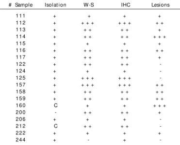
Leptospires were isolated from 16 (39%) samples and their presence was recorded in 18 (44%) and in 19 (46%) out of 41W-S and IHC stained samples, respectively (Table 1). The three techniques had a high agreement (k³0.85) and no significant differences between them were detected (P> 0.05). According to the agglutination titers, all of the isolates were assigned t o t he Ballum serogroup. The presence of t he agent was recorded as black filiform organisms in silver impregnated (Fig. 1) and labeled of a brown color in IHC stained samples (Fig. 2).
Onl y one of t he k i dneys col l ect ed (# 160) show ed macroscopic alterations consisting of numerous gray-white focal depressions in t he cort ex no great er t han 1 mm in
diameter. Six H-E stained kidney sections were recorded as (+ + + ), 8 as (+ + ) and 9 samples as (+ ) (Table 2). Lesions were observed in 13 out of 19 infected and in 10 out of 22 non-infected rodents; differences observed in the proportion of mice having kidney lesions among infect ed and non-infected groups were not statistically significant (P= 0.14). Lesions were confined to the cortex and they were similar in infected and non-infected animals. Focal interstitial infiltration of macrophages with minor proportion of lymphocytes and plasma cells, glomerular shrinkage and desquamat ion of tubular epithelial cells were the principal alterations observed. Tubules filled with leptospires had a normal epithelial cell layer and lesions were not associated with them. It was not possible to establish any relationship between degree of lesion and the presence of leptospires (Table 2).
1
3
2
Fig. 1. Silver impregnation of a kidney of wild house mouse carrier of leptospires. The agent is black stained on the apical surface of epithelial cells and in the lumen of the renal tubules. Warthin-Starry technique.
Fig. 2. Immunohistochemical detection of leptospires on wild house mouse kidney. Leptospires are brown stained on the apical surface of epithelial cells and in the lumen of renal tubules. Note the intact epithelial cell layer in tubules infected and the absence of inflam-mat or y react ion. Immunohist ochemical st ain, heinflam-mat oxylin counterstain.
Fig. 3. Localization of leptospires in a kidney of wild house mice naturally infected. Leptospires are present in high number (+ + + , brown color) in medullar tubules and less frequently in cortical ones. Immunohistochemical stain, hematoxylin counterstain.
T TT T
Tabl e 1. Com par at i ve r esul t s am ong t he t hr ee di agnost i cabl e 1. Com par at i ve r esul t s am ong t he t hr ee di agnost i cabl e 1. Com par at i ve r esul t s am ong t he t hr ee di agnost i cabl e 1. Com par at i ve r esul t s am ong t he t hr ee di agnost i cabl e 1. Com par at i ve r esul t s am ong t he t hr ee di agnost i c t echni ques for t he det ect i on of l ept ospi r es and t he t echni ques for t he det ect i on of l ept ospi r es and t he t echni ques for t he det ect i on of l ept ospi r es and t he t echni ques for t he det ect i on of l ept ospi r es and t he t echni ques for t he det ect i on of l ept ospi r es and t he pr esence of m i cr oscopi c l esi ons i n k i dneys of i nfect ed pr esence of m i cr oscopi c l esi ons i n k i dneys of i nfect ed pr esence of m i cr oscopi c l esi ons i n k i dneys of i nfect ed pr esence of m i cr oscopi c l esi ons i n k i dneys of i nfect ed pr esence of m i cr oscopi c l esi ons i n k i dneys of i nfect ed m i ce (C: cont am i nat ed; - : negat i ve; + , + + , + + + : m i ce (C: cont am i nat ed; - : negat i ve; + , + + , + + + :m i ce (C: cont am i nat ed; - : negat i ve; + , + + , + + + : m i ce (C: cont am i nat ed; - : negat i ve; + , + + , + + + : m i ce (C: cont am i nat ed; - : negat i ve; + , + + , + + + : di f f er ent degr ee of posi t i vi sm
di f f er ent degr ee of posi t i vi sm di f f er ent degr ee of posi t i vi sm di f f er ent degr ee of posi t i vi sm
di f f er ent degr ee of posi t i vi sm [see t ext for det ai l s])[see t ext f or det ai l s])[see t ext f or det ai l s])[see t ext f or det ai l s])[see t ext f or det ai l s])
# Sample Isolat ion W-S IHC Lesions
111 + + + +
112 + + + + + + + + +
113 + + + + + +
114 + + + + + + + +
115 + + + +
116 + + + + + + +
117 + + + + + +
122 + + + + +
-124 + + +
-125 + + + + + + +
-157 + + + + + + + + +
158 + + + + + + +
159 + + + + + + +
160 C + + + + +
200 - + + + + +
206 + + +
-212 C + + + +
-222 + + + +
244 + - +
-T TT T
Tabl e 2. Rabl e 2. Rabl e 2. Rabl e 2. Rabl e 2. Relat i onshi p bet w een t he degr ee of m i cr oscopi cel at i onshi p bet w een t he degr ee of m i cr oscopi cel at i onshi p bet w een t he degr ee of m i cr oscopi cel at i onshi p bet w een t he degr ee of m i cr oscopi cel at i onshi p bet w een t he degr ee of m i cr oscopi c lesi ons (H/E st ai n) and t he presence of t he agent (IHC lesi ons (H/E st ai n) and t he presence of t he agent (IHC lesi ons (H/E st ai n) and t he presence of t he agent (IHC lesi ons (H/E st ai n) and t he presence of t he agent (IHC lesi ons (H/E st ai n) and t he presence of t he agent (IHC st ai n) obser ved i n ki dneys’ sect i on of 41 w i l d house m i ce st ai n) obser ved i n ki dneys’ sect i on of 41 w i l d house m i cest ai n) obser ved i n ki dneys’ sect i on of 41 w i l d house m i ce st ai n) obser ved i n ki dneys’ sect i on of 41 w i l d house m i cest ai n) obser ved i n ki dneys’ sect i on of 41 w i l d house m i ce
(- (- (- (-
(- negat ive; + , + + , + + + : different degree of posit ivismnegat ive; + , + + , + + + : different degree of posit ivismnegat ive; + , + + , + + + : different degree of posit ivismnegat ive; + , + + , + + + : different degree of posit ivismnegat ive; + , + + , + + + : different degree of posit ivism [see t ext for det ai ls])
[see t ext for det ai ls]) [see t ext for det ai ls]) [see t ext for det ai ls]) [see t ext for det ai ls])
Degree of lesions Presence of the agent
- + + + + + +
- 1 2 3 2 1
+ 3 3 3
-+ -+ 3 - 3 2
-of posit ive cases t han using a monoclonal serum, but t he identification of the agent is up to the genus level (Lept ospi ra spp).
IHC yielded more posit ive result s t han t he ot her met hods used, but t he difference wit h ot her t echniques was not significant . Agreement bet ween W-S and IHC st aining in t his study to detect leptospires on the epithelial cells or in the lumen of renal tubules was similar to that obtained by Scanziani et al. (1989) in pig kidneys and greater than that described by Yener and Keles (2001) in bovine kidneys. In the present study, there was only one case in which t he presence of t he agent was weakly detected by IHC from an infected mouse and were not observed by W-S st aining. This could have been due t o low number of lept ospires present in t he sample t hat were not det ect ed by W-S st ain which is considered a less sensit ive diagnost ic met hod t han IHC. Hamst ers were used inst ead of mice as posit ive and negat ive cont rols of WS and IHC st ains because t hey showed higher suscept ibilit y t o infect ion wit h the strains isolated (data not shown). The lowest rate of infection was obtained by leptospiral isolation. This result was expected since lept ospires are fast idious microorganisms, difficult t o grow in vit ro. However, the percentage of isolates reached was high, probably because samples were t aken and processed immediat ely post -mort em. Delays in sample collect ion could have decreased leptospire isolation because tissue biochemical post mort em changes rapidly reduce t he number of viable leptospires (Faine 1982). The association observed in this study between IHC and isolation was similar to that obtained by Ellis et al. (1983) st udying nat urally infect ed porcine kidneys. Conversely, the association between kidney silver staining and culture was greater in the present study than in those conducted by Thiermann (1977) in rat s and Songer et al. (1983) in house mice.
There is general agreement t hat lept ospires are localized on t he apical surface of epit helial cells and in t he lumen of proximal convolut ed t ubules of kidneys in carrier animals (Babudieri 1958, Miller & Wilson 1967, Marshall 1974, Sterling & Th i er m an n 1981). St er l i n g an d Th i er m an n (1981) hypot hesized t hat in t his locat ion lept ospires could not only obtain nutrients from fluids to fulfill requirements for growth and replicat ion, but also be prot ect ed from t he host immune syst em. In t he present st udy, lept ospires were primarily seen on t he apical surface of epit helial cells and in t he lumen of
(1983) (68 vs. 57%).
Lesions observed were consistent with those produced by leptospires in carrier animals (Sterling & Thiermann 1981, Scanziani et al. 1989, Kener & Yeles 2001), but according to the nature of this study, it could not be ascertained that lesions in infect ed kidneys were t he consequence of t he present infection of leptospires. Lesions in non-infected kidneys could have different origins. For instance, Songer et al. (1983) found organisms morphologically ident ical t o Klosi ella mur i s in kidneys of non-infected wild mice with histological lesions compatible with leptospirosis, and Babudieri (1958) theorized that lesions observed could be the consequence of previous infection by leptospires in animals recovered from the disease. Conversely, infected kidneys without histopathological lesions could be explained by the small area of each sample examined or by t he absence of damage in kidneys of some carrier animals of leptospires (Babudieri 1958).
In conclusion, the three techniques evaluated showed a strong agreement for detecting wild M us musculus carrier of leptospires. In addition, an unusual location of leptospires in k i dneys of car r i er ani m al s w as r epor t ed; how ever, a relat ionship bet ween lesions and presence of lept ospires could not be established.
Acknowledgement s.- Acknowledgement s.- Acknowledgement s.- Acknowledgement s.-
Acknowledgement s.- The authors want to thank Dr. Gabriel Sequeira, National Institute of Respiratory Diseases (INER), Santa Fe, Argentina, for helping in rodent s capt ure; Dr. Sherif Zaki, of CDC, At lant a, USA, for his t echnical assistance and Dr. Luis Calvinho, National Institute of Livestock and Agriculture Technology (INTA), Rafaela, Argentina, for reviewing of the manuscript.
REFERENCES
Babudieri B. 1958. Animal reservoirs of leptospires. Annals N.Y. Acad. Sci. 70:393-413.
Brockie R.E. 1977. Leptospiral infections of rodents in the North Island. N. Z. Vet. J. 25:89-96.
Brown R.Z. & Gorman G.W. 1960. The occurrence of leptospiral infections in feral rodents in southwestern Georgia. Am. J. Public Health 50:682-688. Ellis T.M., Robertson G.M., Hustas L. & Kirby M. 1983. Detection of leptospires
in t issue using an immunoperoxidase st aining procedure. Aust . Vet . J. 60:364-367.
Faine S. 1965. Silver staining of spirochaetes in single tissue sections. J. Clin. Pathol. 18:381-382.
Int ernat ional Commit t ee on Syst emat ic Bact eriology, Subcommit t ee on the Taxonomy of Lept ospi ra 1984. Minutes of the Meeting, August 6-10,
1982, Bost on, Massachuset t s, USA. Int . J. Syst . Bact eriol. 34:258-259. Landis J.R. & Koch, G.G. 1977. The measurement of observer agreement
for cat egorical dat a. Biomet rics 33:159-174.
Marshall R.B. 1974. Ult rast ruct ural changes in renal t ubules of sheep following experiment al infect ion wit h Lept ospi r a i nt er r ogans serot ype pomona. J. Med. Microbiol. 7:505-508.
M i l l er N.G. & Wi l son R.B. 1967. El ect r on m i cr oscopi c st udy of t he relat ionship of Lept ospi r a pomona t o t he renal t ubules of t he hamst er
during acut e and chronic lept ospiosis. Am. J. Vet . Res. 28:225-235. Scanziani E., Siponi G. & Mandelli G. 1989. Immunoperoxidase studies on
leptospiral nephritis of swine. Vet. Pathol. 26:442-444.
Seibold H.R., Keech H. & Bokelman D.L. 1961. Histopathologic and serologic st udy of subclinical lept ospirosis among cat t le. J. Am.Vet . Med. Assoc. 138:424-430.
Songer J.G., Chilelli C.J., Reed R.E. & Trautman R.J. 1983. Leptospirosis in rodents from an arid environment. Am. J. Vet. Res. 44:1973-1976. St erling C.R. & Thiermann A.B. 1981. Urban rat s as chronic carriers of
leptospirosis: An ultrastructural investigation. Vet. Pathol. 18:628-637. Thiermann A.B. 1977. Incidence of leptospirosis in the Detroit rat population.
Am. J. Trop. Med. Hyg. 26:970-974.
Vanasco B.N., Rossetti C.A., Sequeira G., Sequeira M.D., Calderón G. & Tarabla H. 2000. First isolations of leptospires serogroup Ballum serovar arborea in Argentina. Vet. Rec. 147:245-246.
Yener Z. & Keles H. 2001. Immunoperoxi dase and hi st opat hologi cal examinat ions of lept ospiral nephrit is in cat t le. J. Vet . Med. A 48:441-447.