28
Turkish Journal of Cerebrovascular Diseases 2014; 20 (1): 28-31 Türk Beyin Damar Hastalıkları Dergisi 2014; 20 (1): 28-31 doi: 10.5505/tbdhd.2014.70288
CASE REPORT OLGU SUNUMU
ANTERIOR COMMUNICATING ARTERY ANEURYSMS AND AN UNUSUAL VARIATIONS OF ANTERIOR
COMMINICATING ARTERY DETECTED BY 3-D CT ANGIOGRAPHY
Hasan Emre AYDIN*, Emre ÖZKARA**, Zühtü ÖZBEK**, Ali ARSLANTAŞ**
* Eskişehir State Hospital Neurosurgery Clinic, Eskişehir, TURKEY
** Eskişehir Osmangazi University Faculty of Medicine Department of Neurosurgery, Eskişehir, TURKEY
ABSTRACT
Aneurysmal subarachnoid haemorrhage which has a serious mortality and morbitity ratio, occurs approximately 10/100.000 population per year and it is usally caused by rupture of a cerebral artery aneurysm. Aneurysms are classified saccular, fusiform or dissecan by morphological description. Vascular congenital anomalies of the cerebral vessels contribute to saccular aneurysm formation by increasing hemodynamic stress on the vessel wall. Although anterior communicating artery (ACoA) is the most seen site of vascular anomalies associated with aneurysmal formation, the agenesis of the anterior commmunicating artery is very rare. Here, we present unusual anatomical variations of ACoA detected by three-dimensional CT angiography after subarachnoid hemorrhage.
Key Words: Anterior communicating artery, endovascular intervention, intracranial aneursym, subarachnoid hemorrhages, three dimensional computed tomography-angiography, variation.
3-B BT ANJİOGRAFİ İLE TESPİT EDİLEN ANTERİOR KOMMUNİKAN ARTER ANEVRİZMALARI VE
ANTERİOR KOMMUNİKAN ARTERİNNADİR GÖRÜLEN ANATOMİK VARYASYONLARI
ÖZET
Ciddi mortalite ve morbitide oranına sahip anevrizmal subaraknoid kanama insidansı yaklaşık olarak / 000 popülasyon yıldır ve genelde bir serebral arter anevrizmasının rüptürüne bağlı olarak gelişir. Anevrizmalar morfolojik olarak sakküler, füziform ya da dissekan olarak sınıflandırılır. Sakkuler anevrizmalar genelde hemodinamik stresin maksimum olduğu Willis poligonu damar bifürkasyonlarında yer almaktadır. Beyin damarlarının konjenital anomalileri, anomalili damar duvarına olan basınç artışına bağlı olarak sakküler anevrizma oluşumuyla bağlantılıdırlar. Her ne kadar anterior kommunikan arter (ACoA), anevrizma ve konjenital anomali birlikteliğinin en sık görüldüğü damar olsa da anterior kommunikan arter agenezisi çok nadir görülmektedir. Burada, biz subaraknoid kanama sonrası üç boyutlu bilgisayarlı tomografik anjiografi ile tespit ettiğimiz anterior kommunikan arterin anevrizma ile ilişkili nadir görülen anatomik varyasyonlarını sunduk.
Anahtar Sözcükler: Anterior komminikan arter, endovasküler girişim, intrakranial anevrizmalar, subaraknoid kanama, üçboyutlu bilgisayarlı tomografi anjiografi, varyasyon.
INTRODUCTION
Hemorrhages occurring generally by arterial reasons in subarachnoid space surrounding brain and spinal cord are called as subarachnoid hemorrhages (SAH). 75-80% of SAHs which
develop spontaneously are caused by saccular aneurysms (1). Development in physiopathologic information on SAH in recent years, improvement in neuromonitorization techniques in intensive _____________________________________________________________________________________________________________________________ Corresponding author: Dr. Hasan Emre Aydın Eskişehir State Hospital Neurosurgery Clinic Eskişehir, Turkey.
E-mail: dremreaydin@gmail.com
Received: 07.12.2012 Accepted: 29.03.2013
This article should be cited as following: Aydın H.E, Özkara E, Özbek Z, Arslantaş A. Anterior communicating artery aneurysms and an unusual variations of anterior comminicating artery detected by 3-D CT angiography. Turkish Journal of Cerebrovascular Diseases 2014; 20 (1): 28-31. doi:10.5505/tbdhd.2014.70288.
29 Aydın et al
care units and developments especially in microanatomy, microsurgery and studies in interventional neuroradiology have triggered significant developments in the diagnosis and therapy of disease. Mortality and morbidity in SAH are declared as approximately 10–67% and 10-20% respectively (2).
Main point in the treatment and therapy of subarachnoid hemorrhage up to aneurysm is to determine the number, place, size, arterial feeding, adjacency to surrounding vascular and neurological tissues, accompanying anatomic variation (if any) or anomalies of aneurysm. Under all information herein and considering general and neurologic table of patient it is required to decide whether aneurysm is clipped by microsurgical techniques or to cover it with endovascular intervention. For this purpose golden method in diagnosis is conventional angiography (3). Technological developments in recent years have caused computerized tomography 3D angiography to be used practically since it is non-invasive, fast, economic and especially has high sensivity and it has been shown to be effective in showing vascular anomalies (3,4,5).
In our letter, rare anatomic localization and variation determined together with anterior communicating arterial aneurysm detected by us in the light of three-dimensional computed tomoghraphic angiography (3D BT-Angiography) in the 2 cases that were taken to our clinic due to SAH are discussed.
CASES
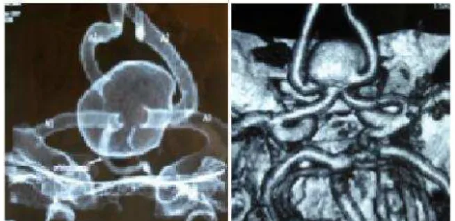
Two female patients were admitted to our clinic due to the severe headache was followed with medical treatments organized upon the detection of an appearance in compliant of SAH with CT scans of brain. In the 3-D CT angiography for the first case, anterior communicating artery was seen to have an apparent dysplastic character and to have turned to an irregular spherical aneurismal excess filling reaching approximately 17-18 mm in diameter. A1 segments of both anterior cerebral arteries were watched to be discharged to aneurysm and A2 segments are watched to originate from posterolateral aneurysm (Figure 1 and 2). In second case, approximately 7x5 mm left-oriented saccular aneurysm has been determined in A1-A2 junction of right anterior cerebral artery in 3-D CT
Turkish Journal of Cerebrovascular Diseases 2014; 20 (1): 28-31
Figure1. 3D-CT scan reveals that A1 segments of both anterior
cerebral arteries are watched to be discharged to aneurysm.
Figure 2. 3D-CT scan reveals that A2 segments are watched to
originate from posterolateral aneurysm.
angiography. Fusiform enlargement has been observed in A2 segment right at the distal of aneurysm. Anterior communicating artery or formation which may represent it was not detected. Left A1-A2 junction was adjacent to aneurysm and left anterior cerebral artery was determined to pass right under aneurysm neck, to turn to A2 segment and to have no connection with right anterior cerebral artery. (Figure 3 and 4).
Figure 3. Fusiform enlargement has been observed in A2
segment right at the distal of aneurysm.
Figure 4. Approximately 7x5 mm left-oriented saccular
aneurysm has been determined in A1-A2 junction of right anterior cerebral artery in 3-D CT angiography.
DISCUSSION
30 basilar artery, anterior and middle cerebral artery bifurcation of internal carotid artery.
Classically anterior communicating artery combines right and left anterior cerebral arteries and has average 4mm length, 1.7 mm diameter and forms the front edge of Willis polygon (7). AcoA complex is an anatomic area where intracranial aneurysms are seen most common. Aneurysms in such area are of different importance in terms of flow dynamics of anterior circulation, usual anatomic variations, deep interhemispheric location and perforator branches causing serious neurologic deficiency in case of damage (8). Vascular anomalies in Wills polygon are commonly related to aneurismal formation (9,10,11). AcoA and its anomalies have been discussed and described by various research makers. Contradiction frequency of anterior communicating artery has been declared as 8% and 20% and highest frequency is stated to be seen in anterior communicating artery aneurysms in the recent studies. Anomaly frequency has been determined to be higher in the cadaverous and microsurgical researches. Although anatomic variations of anterior communicating artery were declared as 60% and 21,4% in the studies of Serizowa et al. and Ogawa et al. respectively, such rates are determined to be less frequent in the radiological studies (7,13).
Anterior Communicating Artery complex starts to be formed as plexiform and primitive in 35th day of embryologic life. Incompletion of plexiform anastomoses are thought to cause fusion anomalies (9,12). Most common anterior communicating artery complex anomalies or variations are; unilateral anterior cerebral artery hypoplasia, multiple vascular channels, dimples, fenestrations, duplications, fusion, median artery of the corpus callosum (MACC) and azygos anterior cerebral artery (10,11). Although a wide range of variations related to intracerebral aneurysms are notified in general terms, in the literature %26.6 percent of cases identified variant related to aneurysm location is A1 hyplosia accompanying anterior communication artery aneurysm (14). Examining literature anterior communicating artery agenesis is a rarely seen anatomic anomaly. Absence of anterior communicating artery is declared to be approximately 0,1% in various series (15). First case presented herein is a different variation since all anterior cerebral artery branches are related to
ACoA aneurysms and its unusual variations
aneurysm, second case is a rarely seen anatomic anomaly together with anterior communicating artery aplasia. Determination of anatomic differences before treatment in detail is important for treatment selection and creation of the protocol for agreed treatment. Endovascular treatment is planned to be applied to both cases presented herein.
Conclusion
In the patients suffering from aneurismal subarachnoid hemorrhage with high mortality and morbidity, place and location of lesion and anomalies accompanying it are very important in terms of treatment protocol and follow-up. It is necessary to know rarely seen variations in terms of surgical decision and strategy. 3-D CT Angiography is considered useful in terms of determination and detailed anatomy of aneurysm.
REFERENCES
1. Greenberg MS, SAH and Aneurysms, Handbook of Neurosurgery. 5th edition. NewYork, Thieme, 2001; 754. 2. Matsubara S, Hadeishi H, Suzuki A, et al. Incidence and risk factors for the growth of unruptured cerebral aneurysms: observation using serial computerized tomography angiography. J. Neurosurgery 2004; 101: 908-914.
3. Alberico RA, Patel MP, Jacobs B, et al. Evalution of the circle of willis with three-dimensional CT angiography in patients with suspected intracranial aneurysms. AJNR 1995; 16:1571-1578.
4. Jayaraman MV, Mayo-Smith WW, Tung GA, et al. Detection of intracranial aneurysms: multi-detector row CT angiography compared with DSA. Radiology 2004; 230(2): 510-8.
5. Dehdashti AR, Rufenacht DA, Delavelle J,et al. Therapeutic decision and management of aneurysmal subarachnoid haemorrhage based on computed tomographic angiography. Br J Neurosurg 2003; 17(1): 46-53.
6. Ingerbrigtsen Ton, Assessment of risk for aneurysm rupture, EANS training course, vasculer neurosurgery, Lisbon 17-21 June 2007.
7. Serizawa T, Saeki N, Yamaura A. Microsurgical anatomy and clinical significance of the anterior communicating artery and its perforating branches Neurosurgery. 1997; 40(6): 1211-1216.
8. Juha Hernesniemi, Reza Dashti, Martin Lehecka, et al. Microneurosurgical management of anterior communicating artery aneurysms. Surgical Neurology 2008; 70; 8–29. 9. Oliveira JG, du Mesnil de Rochemont R, Beck J, et al. A rare anomaly of the anterior communicating artery complex hidden by a large broad-neck aneurysm and disclosed by three-dimensional rotational angiography. Acta Neurochir (Wien) 2008; 150(3): 279-284.
10.Kwak R, Niizuma H, Hatanaka M,et al. Anterior communicating artery aneurysms with associated anomalies. J Neurosurg 1980; 52(2): 162-164.
11.Namiki J, Doumoto Y. Microsurgically critical anomaly of the anterior communicating artery complex during the pterional approach to a ruptured aneurysm: double fenestration of the proximal A2 segments.
31 Aydın et al
Neurol Med Chir (Tokyo) 2003; 43(6): 304-307.
12.San-Galli F, Leman C, Kien P, et al. Cerebral arterial fenestrations associated with intracranial saccular aneurysms. Neurosurgery 1992; 30(2): 279-83.
13.Ogawa A, Suzuki M, Sakurai Y, et al. Vascular anomalies associated with aneurysms of the anterior communicating artery: microsurgical observations. J Neurosurg 1990; 72: 706-709.
Turkish Journal of Cerebrovascular Diseases 2014; 20 (1): 28-31
14.Aydın IH, Kadioglu HH, Tuzun Y, et al. Vascular variations associated with anterior communicating artery aneurysms-an intraoperative study. Minim Invasive Neurosurg. 1997; 40(1): 17-21.