C A SE REPORT
254 J Vasc Bras. 2014 July-Sept.; 13(3):254-256 http://dx.doi.org/http://dx.doi.org/10.1590/jvb.2014.037
Post-kidney transplantation external iliac artery stenosis
due to vascular clamp: report of a case
Estenose de artéria ilíaca externa após transplante renal devido a
clampe vascular: relato de um caso
Murat Ozban1, Cagatay Aydin1, Belda Dursun2, Baki Yagci3, Onur Birsen1, Koray Tekin1
Abstract
We report a case of right external iliac artery stenosis after kidney transplantation surgery caused by vascular clamp application injury. he patient presented with claudication of the ipsilateral lower limb and the lesion was diagnosed angiographically. he patient was treated with endovascular stent placement.
Keywords: clamp stenosis; kidney transplantation; iliac artery.
Resumo
Relatamos um caso de estenose da artéria ilíaca externa direita após cirurgia de transplante renal causada por lesão durante a colocação de um clampe vascular. O paciente apresentou claudicação do membro inferior ipsilateral e a lesão foi diagnosticada angiograicamente. O paciente foi tratado com colocação de stent endovascular.
Palavras-chave: estenose de clampe; transplante de rim; artéria ilíaca.
1Pamukkale University School of Medicine, Department of General Surgery, Kinikli, Denizli, Turkey. 2Pamukkale Univeristy School of Medicine, Department of Nephrology, Kinikli, Denizli, Turkey. 3Pamukkale University School of Medicine, Department of Radiology, Kinikli, Denizli, Turkey.
Financial support: None.
Conlicts of interest: No conlicts of interest declared concerning the publication of this article. Submitted: 02.28.14. Accepted: 05.19.14.
Murat Ozban, Cagatay Aydin et al.
255 J Vasc Bras. 2014 July-Sept.; 13(3):254-256
INTRODUCTION
Kidney transplantation is the preferred method for treatment of end stage renal failure. Iliac artery stenosis is one of the rarer complications of this surgery. Most of these lesions are attributed to atherosclerotic vascular damage caused by peripheral arterial disease in the transplant recipient.1 Less
frequently, the cause is intraoperative trauma to the vessel as a result of vascular clamp application.2
Although these lesions are usually suspected because of clinical manifestations such as murmurs, lower extremity claudication, hypertension, and renal allograft dysfunction, they may also be discovered incidentally.3
CASE REPORT
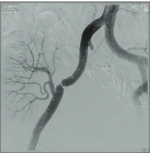
A 52-year-old male patient who had a kidney transplant from a living donor at our center in May 2007. He was admitted to our clinic with right lower extremity pain 6 months after transplantation. On physical examination, there was attenuation of the right lower extremity peripheral pulses and murmur detectable from the renal allograft with auscultation. Pre-transplant patient history, physical examination, laboratory and imaging methods for the assessment of peripheral artery disease were unremarkable. Laboratory results showed BUN at 31 mg / dl and creatinine at 1.54. Angiography showed right external iliac artery stenosis (80%) 1 cm proximal to the renal artery anastomosis and 1 cm in length on a segment (Figure 1). An endovascular stent was placed at the stenosis site. Control angiography showed full vascular patency, transplant renal artery and intrarenal arteries open, and the renal artery anastomosis distal to the stent (Figure 2). The patient was discharged the next day. Two months after the operation, the claudication had resolved. Laboratory control results showed BUN: 22 mg/dl and creatinine: 1.47 mg/dl. On Doppler examination, the transplanted kidney and iliac arteries were normal.
DISCUSSION
The most common late vascular complication is stenosis of the graft artery at the site of anastomosis with the recipient iliac artery, leading to graft dysfunction and renal vascular hypertension.4
However, a number of case reports in the literature described graft dysfunction with low extremity claudication secondary to stenosis of the iliac artery proximal to the graft implantation site.5,6
Most of these stenotic lesions are seen in transplant recipients who have suffered from peripheral arterial
disease with atherosclerotic vascular damage. Less frequently, the cause is vascular clamp injury to the vessel with subsequent intimal hyperplasia.2 In our
case, the location of the stenosis and the normal angiographic appearance of the vessel away from the lesion suggested that this occlusion was most likely due to endothelial trauma by vascular clamp injury during the operation.
Investigation of kidney transplant graft dysfunction includes assessment of possible vascular complications which occur in up to 15% of renal transplant patients and cause graft dysfunction with high morbidity and mortality.4 These complications
Figure 1. Clamp stenosis of the common iliac artery.
Post-kidney transplantation external iliac artery stenosis
256 J Vasc Bras. 2014 July-Sept.; 13(3):254-256
include renal artery thrombosis, renal artery stenosis, renal vein thrombosis and, rarely, iliac artery stenosis. The most common vascular complication is stenosis of the graft artery at the site of anastomosis with the recipient iliac artery, leading to graft dysfunction.4
The prevalence of iliac artery stenosis in kidney transplant recipients varies from 0.37% to 1.5%.7
Posttransplant renal artery stenosis occurs more frequently, affecting 3–4% of all kidney transplants.4
These stenotic lesions may result in reduced blood
low to the ipsilateral lower extremity as well as the
kidney graft, leading to renal dysfunction and leg ischemia, and are usually detected because of clinical manifestations such as elevated serum creatinine levels, bruits, low extremity claudication, and renal vascular hypertension.5 If the condition is suspected,
patients should undergo biochemical work-up consisting of graft dysfunction and radiological investigation of iliac and transplanted kidney vessels using methods such as Doppler US and angiography.
Percutaneous transluminal angioplasty (PTA) of both renal allograft and native iliac artery stenoses has become accepted as the initial treatment of choice.1
Intravascular stents have greater long-term patency rates in randomized studies. The most commonly used stents are the balloon-expandable and self-expandable stents. Vascular clamp-associated lesions like those seen in our case (focal stenosis and short occlusion) are suitable for such interventions.1 In our
case, an endovascular stent was placed at the stenosis site. Control angiography showed that it resulted in full vascular patency of the external iliac artery (Figure 2). The patient was discharged the next day. Two months after the operation, the claudication had resolved and Doppler examination showed that the transplanted kidney and iliac arteries were normal.
In summary, iliac artery stenosis is a rare complication after kidney transplantation and can lead to lower extremity claudication, hypertension and kidney dysfunction of the transplant. In transplant surgery, the possibility that this complication may have occurred due to vascular clamp damage should be considered in suspected patients, even in the absence of peripheral artery disease.
REFERENCES
1. Mullasari AS, Umesan CV, Radhakrishnan N, Lakshmi V. Iliac artery stenosis causing post-renal transplant hypertension: successful management by percutaneous angioplasty and stent implantation. J Invasive Cardiol. 2002;14(4):212-3. PMid:11923579.
2. Khankan AA, Maeda M, Osuga K, Murakami T, Nakamura H. Post-kidney transplantation iliac artery stenosis due to iatrogenic injury: case report. Cardiovasc Intervent Radiol. 2003;26(2):186-8. http://dx.doi.org/10.1007/s00270-002-0471-x. PMid:12616412
3. Ardelean A, Mandry D, Claudon M. Complications vasculaires de la transplantation rénale : conduite diagnostique. J Radiol. 2011;92(4):343-57. http://dx.doi.org/10.1016/j.jradio.2011.02.018. PMid:21549890
4. Aktas S, Boyvat F, Sevmis S, Moray G, Karakayali H, Haberal M. Analysis of vascular complications after renal transplantation. Transplant Proc. 2011;43(2):557-61. http://dx.doi.org/10.1016/j. transproceed.2011.01.007. PMid:21440760
5. Aikimbaev K, Akgul E, Aksungur E, Demir E, Erken U. Iliac artery stenosis as a cause of posttransplant renal failure and claudication. Int Urol Nephrol. 2007;39(4):1273-6. http://dx.doi. org/10.1007/s11255-007-9284-9. PMid:17914659
6. Król R, Ziaja J, Chudek J, et al. Iliac artery stenosis as a cause of posttransplant renovascular hypertension: report of two cases. Ann Transplant. 2005;10(2):66-9. PMid:16218036.
7. Voiculescu A , Hollenbeck M, Plum J, et al. Iliac artery stenosis proximal to a kidney transplant: clinical findings, duplex-sonographic criteria, treatment, and outcome. Clin transplant. 2003;76(2):332-9. http://dx.doi.org/10.1097/01. TP.0000072805.40996.B1. PMid:12883188
Correspondence
Murat Ozban Pamukkale University, General Surgery Department Kinikli, Denizli, Turkey Phone:+905324484403 Email: [email protected]
Author information MO is a vascular and transplant surgeon at Pamukkale University Kidney Transplant Center CA is a vascular and transplant surgeon at Pamukkale University Kidney Transplant Center BD is a nephrologist at Pamukkale University Kidney Transplant Center BY is a interventional radiologist at Pamukkale University OB is a vascular and transplant surgeon at Pamukkale University Kidney Transplant Center KT is a vascular and transplant surgeon at Pamukkale University Kidney Transplant Center
Author contributions