Intraamniotic Inflammation in Women with
Preterm Prelabor Rupture of Membranes
Ivana Musilova1, Radka Kutová2, Lenka Pliskova2, Martin Stepan1, Ramkumar Menon3,
Bo Jacobsson4,5, Marian Kacerovsky1,6*
1Department of Obstetrics and Gynecology, Charles University in Prague, Faculty of Medicine Hradec Kralove, University Hospital Hradec Kralove, Hradec Kralove, Czech Republic,2Institute of Clinical Biochemistry, University Hospital Hradec Kralove, Hradec Kralove, Czech Republic,3Department of Obstetrics and Gynecology, Sahlgrenska Academy, Gothenburg, Sweden,4Department of Genes and Environment, Division of Epidemiology, Norwegian Institute of Public Health, Oslo, Norway,5Department of Obstetrics and Gynecology, Division of Maternal-Fetal Medicine & Perinatal Research, The University of Texas Medical Branch at Galveston, Galveston, Texas, United States of America,6Biomedical Research Center, University Hospital Hradec Kralove, Hradec Kralove, Czech Republic
*kacermar@fnhk.cz
Abstract
Objective
To characterize subgroups of preterm prelabor rupture of membranes (PPROM) and short-term neonatal outcomes based on the presence and absence of intraamniotic inflammation (IAI) and/or microbial invasion of the amniotic cavity (MIAC).
Methods
One hundred and sixty-six Caucasian women with singleton pregnancies were included in this study. Amniotic fluid samples were obtained by transabdominal amniocentesis (n=166) and were assayed for interleukin-6 levels by a lateral flow immunoassay. The presence of Ureaplasmaspecies,Mycoplasma hominis,Chlamydia trachomatis, and 16S rRNA was evaluated in the amniotic fluid. IAI was defined as amniotic fluid IL-6 values, measured by a point of care test, higher than 745 pg/mL.
Results
Microbial-associated IAI (IAI with MIAC) and sterile intraamniotic inflammation (IAI alone) were found in 21% and 4%, respectively, of women with PPROM. Women with microbial-associated IAI had higher microbial loads ofUreaplasmaspecies in the amniotic fluid than women with MIAC alone. No differences in the short-term neonatal morbidity with respect to the presence of microbial-associated IAI, sterile IAI and MIAC alone were found after adjust-ing for the gestational age at delivery in women with PPROM.
OPEN ACCESS
Citation:Musilova I, Kutová R, Pliskova L, Stepan M, Menon R, Jacobsson B, et al. (2015) Intraamniotic Inflammation in Women with Preterm Prelabor Rupture of Membranes. PLoS ONE 10(7): e0133929. doi:10.1371/journal.pone.0133929
Editor:Colette Kanellopoulos-Langevin, Xavier Bichat Medical School, INSERM-CNRS - Université Paris Diderot, FRANCE
Received:March 30, 2015
Accepted:July 2, 2015
Published:July 24, 2015
Copyright:© 2015 Musilova et al. This is an open access article distributed under the terms of the
Creative Commons Attribution License, which permits unrestricted use, distribution, and reproduction in any medium, provided the original author and source are credited.
Data Availability Statement:All relevant data are within the paper.
Funding:This work was supported by 1) a grant from the Ministry of Health of the Czech Republic (NS 13461-4/2012), and 2) Charles University in Prague, the Faculty of Medicine in Hradec Kralove, Czech Republic, project“PRVOUK”P37/10.
Conclusions
Microbial-associated but not sterile intraamniotic inflammation is common in Caucasian women with PPROM. The gestational age at delivery but not the presence of inflammation affects the short-term neonatal morbidity of newborns from PPROM pregnancies.
Introduction
Preterm prelabor rupture of membranes (PPROM) is defined as a rupture of fetal membranes with leakage of amniotic fluid prior to 37 weeks of gestation. PPROM represents a serious preg-nancy complication that is responsible for approximately one-third of all preterm deliveries. Recent studies suggest its mainly non-infectious etiology with inflammation as the major underlying pathophysiology [1–5]. However, it is unclear whether inflammation is a cause or consequence of PPROM and how it is linked to PPROM risk factors [6,7].
PPROM is often complicated by microbial invasion of the amniotic cavity (MIAC) and intraamniotic inflammation (IAI), resulting in the development of histological chorioamnioni-tis. MIAC, which is predominated byUreaplasmaspecies, complicates approximately 25–40% of PPROM, depending on the gestational age at sampling, ethnicity, and detection technique [8,9]. The presence of bacteria in the amniotic fluid is postulated to activate an intraamniotic innate immune response through the system of pattern recognition receptors, resulting in microbial-associated IAI [8–11]. Moreover, the intensity of IAI depends on the type of bacteria and the microbial load [12–14]. Therefore, the presence of a small amount of bacteria with a low virulent potential, suchUreaplasmaspecies, in the amniotic is unlikely to elicit IAI and be associated with worse pregnancy and neonatal outcomes. This scenario is considered coloniza-tion of the amniotic fluid [15]. On the other hand, some endogenous mediators called alarmins (e.g., high mobility group box-1 protein) are released into the amniotic fluid and can trigger intraamniotic inflammation through the same system of pattern recognition receptors as in the infectious scenario [5,16]. This leads to the development of sterile IAI (the presence of IAI without any proven microorganism in the amniotic fluid).
However, the diagnosis of microbial-associated or sterile IAI is very often time consuming and costly due to the need for non-cultivation techniques to prove or rule out the presence of MIAC. Currently, the use of non-cultivation techniques to diagnose MIAC should be considered a gold standard because many amniotic fluid microorganisms are difficult to cultivate [10,17].
A pioneering study by Romero et al. has shown that the incidences of microbial-associated and sterile IAI in pregnancies complicated by PPROM are equal (approximately 30%) [3]. Because racial disparity can affect the levels of intraamniotic mediators, resulting in a different rate of sterile IAI, it would be of interest to evaluate whether this distribution of IAI subtypes is observed in a European population consisting of a majority of Caucasian women [18,19]. Moreover, there is a lack of evidence for whether the presence of both subtypes of IAI and MIAC alone affect short-term neonatal morbidity.
Materials and Methods
The Institutional Review Board at University Hospital Hradec Kralove (March 19, 2008; no. 200804 SO1P) approved the study and written informed consent was obtained from all the par-ticipants. Between January 2012 and July 2014, a prospective cohort study was conducted on pregnant women at gestational ages 24+0 or 36+6 weeks, who were admitted to the Depart-ment of Obstetrics and Gynecology, University Hospital Hradec Kralove, Czech Republic. Pregnant women with singleton pregnancies complicated by PPROM and with a maternal age18 years were invited to participate in the study. Women with signs of fetal growth restriction, the presence of either congenital or chromosomal fetal abnormalities, gestational or pre-gestational diabetes, gestational hypertension, preeclampsia, signs of fetal hypoxia, or sig-nificant vaginal bleeding were excluded from the study.
Gestational ages were established by first-trimester fetal biometry. In the Czech Republic, women with PPROM at less than 34 weeks of gestation are treated with corticosteroids to accelerate lung maturation (two doses of 14 mg of betamethasone, administered intramuscu-larly 24 hours apart), tocolytics for 48 hours, and antibiotics (prophylactic parenteral azithro-mycin were given at admission for maximum 7 days), whereas no treatment except antibiotics is initiated to delay delivery after 34 weeks. The management of PPROM in the Czech Republic is active (except at<28 gestational weeks); the induction of labor or an elective cesarean
sec-tion is initiated no later than 72 hours after the rupture of the membranes, depending on the gestational age of the pregnancy, fetal status, and maternal serum levels of C-reactive protein.
PPROM was diagnosed by examination with a sterile speculum to verify the pooling of amniotic fluid in the vagina, and when necessary, it was confirmed by the presence of insulin-like growth factor binding proteins (ACTIM PROM test; MedixBiochemica, Kauniainen, Fin-land) in the vaginal fluid.
Ultrasound-guided transabdominal amniocentesis was performed at admission, but before the administration of antibiotics, corticosteroids, and tocolytics, approximately 2–3 mL of amni-otic fluid was aspirated. A total of 100μL of non-centrifuged amniotic fluid was used for the bed-side assessment of IL-6 levels. The remaining amniotic fluid was immediately transported to the microbiology laboratory for polymerase chain reaction (PCR) testing forUreaplasmaspecies,
Mycoplasma hominis, andChlamydia trachomatisas well as for evaluating 16S rRNA.
This study was approved by the Institutional Review Board committee (March 19, 2008; No 200804 SO1P), and informed consent was received from all participants.
Amniotic fluid IL-6 levels
The IL-6 levels were assessed with a lateral flow immunoassay Milenia QuickLine IL-6 using the Milenia POCScan Reader (Millenia Biotec, GmbH, Giessen, Germany). The measurement range was 50–10000 pg/mL. The intraassay and interassay variations were 12.1% and 15.5%, respectively.
Definition of IAI
Based on recent studies, IAI in PPROM pregnancies was defined as amniotic fluid IL-6 values, measured by a point of care test, higher than 745 pg/mL [24–26].
Detection of
Ureaplasma
species,
Mycoplasma hominis
, and
Chlamydia
trachomatis
bacterial DNA from biological fluids). Real-time PCR was performed on a Rotor-Gene 6000 instrument (QIAGEN, Hilden, Germany) with the commercial kit AmpliSens C. trachomatis/ Ureaplasma/M. hominis-FRT (Federal State Institution of Science, Central Research Institute of Epidemiology, Moscow, Russia) to detect the DNA fromUreaplasmaspecies,Mycoplasma
hominis, andChlamydia trachomatisin a common PCR tube. As a control, we included a PCR
run for beta-actin, a housekeeping gene, to examine the presence of inhibitors of the polymer-ase chain reaction. The amount ofUreaplasmaspecies DNA in copies/mL was determined by an absolute quantification technique employing an external calibration curve. Plasmid DNA (pCR4, Invitrogen) was used for the preparation of the calibration curve [12,13].
Detection of other bacteria in the amniotic fluid
Bacterial DNA was identified by PCR targeting the 16S rRNA gene with the following primer pars:5`-CCTACGGGNGGCWGCAG-3`(V3 region),5`-GACTA CHVGGGTATCTAATCC-3`
(V4 region), and5`-AGGAGGTGATCCAACCGCA-3`(V7 region),5`-GGTTAAGTCCCGC AACGAGCGC-3`(V9 region) [27,28]. Each individual reaction contained 5μL of target DNA,
500 nM of forward and reverse primers and Mastermix 16S Basic (Molzyme, Bremen, Germany) in a total volume of 25μL. The amplification was performed in a 2720 Thermal Cycler (Applied Biosystems, Foster City, CA, USA). The products were visualized on an agarose gel together with the beta-actin control (309 bp). Positive reactions yielded products of 452 bp, which were subsequently analyzed by sequencing. The 16S PCR products were cleaned and used in sequenc-ing PCR reactions utilizsequenc-ing the above primers and the BigDye Terminator kit, version 3.1. Bacte-ria were then typed using the sequences obtained in BLAST and SepsiTest BLAST.
Diagnosis of MIAC
MIAC was determined based on a positive PCR analysis forUreaplasmaspecies,Mycoplasma
hominisand/or forChlamydia trachomatisand/or by positivity for the 16S rRNA gene.
Diagnosis of histological chorioamnionitis (HCA)
The degree of neutrophil infiltration was separately assessed in the free fetal membranes (amnion and chorion-decidua), placenta (amnion and chorionic plate), and umbilical cord according to the criteria of Salafia et al. [29]. A diagnosis of HCA was determined based on the presence of neutrophil infiltration in the chorion-decidua (grades 3–4), chorionic plate (grades 3–4), umbili-cal cord (grades 1–4), and/or amnion (grades 1–4) [29]. The diagnosis of funisitis was performed based on the presence of neutrophil infiltration in the umbilical cord (grades 1–4).
Diagnosis of severe neonatal morbidity
clinical suspicion of sepsis [the presence of symptoms and elevated C-reactive protein and/or an affected white blood cell count] during the first 72 hours of life and between 4 and 120 days of life, respectively); bronchopulmonary dysplasia (defined by the infant’s oxygen dependence at 36 weeks of age); pneumonia (diagnosed by abnormal findings on chest X-rays); and/or neo-natal death prior to hospital discharge [31].
Statistical analysis
The demographic characteristics were compared using the non-parametric Kruskal-Wallis test for continuous variables, and the data are presented as the medians (range). Categorical vari-ables were compared using the Chi-square test or the Fisher’s exact test and are presented as percentages (%). For the analysis of the amniotic fluid IL-6 levels among the subgroups of PPROM, a non-parametric Kruskal-Wallis test was used. For the analysis of short-term neona-tal morbidity among subgroups, the Fisher’s exact test was used. Spearman partial correlation was used to adjust the data for the gestational age at delivery. For the analyses among four sub-groups, exact 2-tailedp-values were used due to the small sample size in the group with sterile IAI. Allp-values were from two-sided tests, and all statistical analyses were performed using SPSS 19.0 (SPSS Inc., Chicago, IL, USA) and GraphPad Prism 5.03 for Mac OS X (GraphPad Software, La Jolla, CA, USA).
Results
In total, 166 women with PPROM at gestational ages 24+0 to 36+6 weeks were recruited. The demographic and clinical characteristics of the women are shown inTable 1. The overall rates of IAI and MIAC were 25% (41/166) and 34% (57/166), respectively. The presence of micro-bial-associated IAI (both IAI and MIAC) was observed in 21% (34/166) of women, 4% (7/166) had sterile IAI (IAI alone), 14% (23/166) had MIAC alone, and the remaining 61% (102/166) of the women were negative (without both MIAC and IAI). If the diagnosis of IAI was
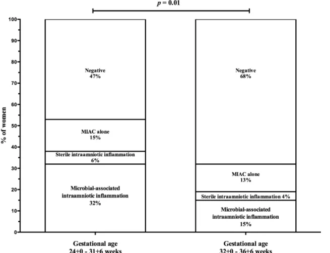
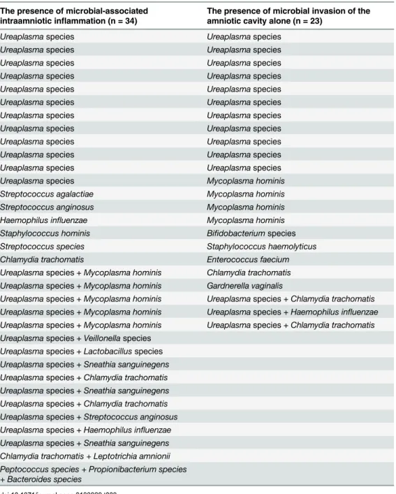
expanded to include both a positive IL-6 and neutrophil infiltration of the amnion (histological amnionitis), then the proportion of women in these four groups was 23%, 10%, 11% and 56%, respectively. Women with PPROM between gestational ages 24+0 and 31+6 weeks had a higher frequency of microbial-associated IAI [32% (17/53) vs. 15% (17/113)], almost the same fre-quencies of sterile IAI [6% (3/53) vs. 4% (4/113%)], almost the same frequency of MIAC alone [15% (8/53) vs. 13% (15/113)], and a lower frequency of negative women [49% (25/53) vs. 71% (77/113);p= 0.01;Fig 1] than women with PPROM between gestational ages 32+0 and 36+6 weeks (Table 2). Women with microbial-associated IAI were more likely to smoke and had higher C-reactive protein levels, white cell blood counts, and rates of HCA and funisitis com-pared to the remaining women (Table 1). Women with microbial-associated IAI also had new-borns with lower birth weights. All of the microorganisms detected in the amniotic fluid are shown inTable 3. The most common bacteria wereUreaplasmaspp., which were identified in 24% (40/166) of the women. Polymicrobial findings were observed in 11% (19/166) of women.
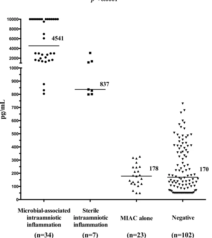
Women with microbial-associated IAI had higher median amniotic fluid IL-6 levels than the remaining women (with microbial-associated IAI: median 4541 pg/mL [range: 804–10000] vs. with sterile IAI: median 837 pg/mL [range 799–3073] vs. with MIAC alone: median 178 pg/ mL [range 50–327] vs. negative: median 170 [range 50–729] pg/mL;p<0.0001;Fig 2).
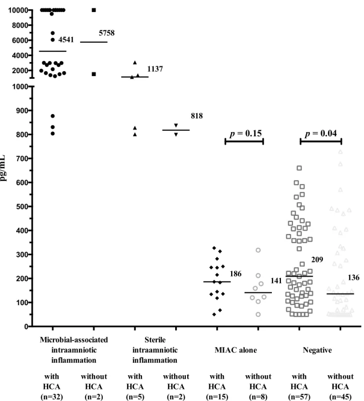
amniotic fluid IL-6 levels when HCA present (with HCA: median 186 pg/mL vs. without HCA: median 141 pg/mL;p= 0.15;Fig 3). Negative women had higher amniotic fluid IL-6 levels when HCA was present (with HCA: median 209 pg/mL vs. without HCA: median 136 pg/mL;
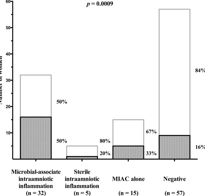
p= 0.04;Fig 3). Women with microbial-associated IAI had a higher rate of HCA with neutro-phil infiltration of the amnion than the remaining women [with microbial-associated IAI: 50% (16/32) vs. with sterile IAI: 20% (1/5) vs. with MIAC alone: 33% (5/15) vs. negative 16% (9/57);
p= 0.0009;Fig 4). The grades of neutrophil infiltration in the chorion-decidua, the chorionic plate, the umbilical cord and the amnion in all subgroup of women are shown inS1 Table.
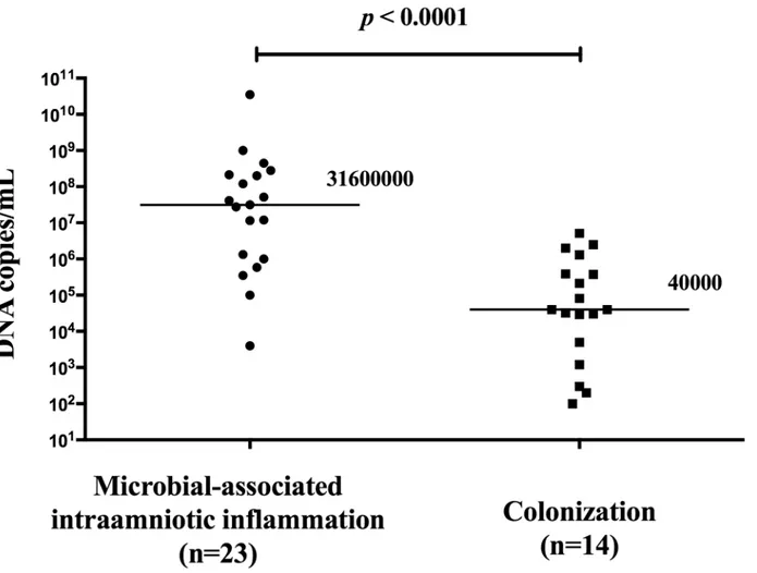
Women with the presence of microbial-associated IAI had a higher load ofUreaplasma spe-cies in the amniotic fluid than women with MIAC alone (with microbial-associated IAI: median 3.2x10⁷copies DNA/mL [range 4.0x10³-.3.5x1010] vs. MIAC alone: median 4.0x10⁴ copies DNA/mL [range 1.0x10²-5.1x10⁶];p<0.0001;Fig 5). There was a strong correlation Table 1. Maternal and neonatal characteristics of preterm prelabor rupture of membrane pregnancies according to the presence or absence of microbial-associated intraamniotic inflammation, sterile intraamniotic inflammation and MIAC alone.
Characteristic Microbial-associated IAI
(n = 34)
Sterile IAI (n = 7)
MIAC alone (n = 23)
Negative (n = 102)
p -value1
p -value2
Maternal age 32 (17–42) 29 (21–36) 32 (18–39) 31 (21–42) 0.89 0.75
Prepregnancy body mass index [kg/m2, median
(range)]
22.5 (16.5–38.0) 24.2 (19.3–
25.7)
22.3 (17.6–
33.5)
22.3 (15.8–
39.0)
0.72 0.62
Smoking [number (%)] 16 (47%) 0 (0%) 5 (22%) 10 (10%) <0.0001 <0.0001
Gestational age at admission [weeks, median (range)]
32+0 (24+2–36+6) 34+6 (25+1–
36+6)
33+0 (25+3–36 +6)
33+6 (24+5–36 +5)
0.16 0.08
Gestational age at delivery [weeks, median (range)]
32+1 (24+5–36+6) 35+0 (25+1–
36+6)
34+6 (26+6–36 +6)
34+2 (25+2–36 +5)
0.04 0.02
Latency from PPROM to amniocentesis [hours, median (range)]
8 (1–97) 7 (2–22) 6 (1–20) 5 (1–72) 0.11 0.06
Latency from amniocentesis to delivery [hours, median (range)]
30 (3–101) 40 (17–90) 37 (7–211) 28 (4–178) 0.66 0.77
Amnioticfluid IL-6 [pg/mL, median (range)] 4541 (804–10000) 837 (799–
3073)
178 (50–327) 170 (50–729) <0.0001 <0.0001
CRP levels at admission [mg/L, median (range)]
12.1 (0.6–106.0) 7.6 (1.0–16.6) 4.7 (0.1–23.0) 5.6 (0.5–44.4) 0.002 0.001
WBC count ad admission [x109L, median (range)]
13.8 (9.2–22.7) 12.1 (8.9–
15.3)
11.9 (8.4–18.2) 11.5 (7.4–20.6) 0.003 0.001
Administration of antibiotics [number (%)] 34 (100%) 7 (100%) 23 (100%) 100 (98%) 1.00 0.57 Administration of corticosteroids [number (%)] 28 (82%) 6 (86%) 16 (70%) 74 (73%) 0.59 0.45
Vaginal delivery [number (%)] 23 (68%) 5 (71%) 17 (74%) 66 (65%) 0.88 0.69
Cesarean section [number (%)] 11 (32%) 2 (29%) 6 (26%) 36 (35%) 0.88 0.69
Birth weight [grams, median (range)] 1800 (550–3390) 2130 (990–
3320)
2150 (780–
3250)
2255 (700–
3350)
0.02 0.008
Histological chorioamnionitis [number (%)] 32 (94%) 5 (71%) 15 (65%) 57 (56%) <0.0001 <0.0001
Funisitis [number (%)] 22 (64%) 2 (29%) 9 (39%) 28 (27%) 0.001 <0.0001
Apgar score<7; 5 minutes [number (%)] 2 (6%) 1 (14%) 0 (0%) 3 (3%) 0.18 0.45
Apgar score<7; 10 minutes [number (%)] 1 (3%) 0 (0%) 0 (0%) 2 (2%) 1.00 0.72
Abbreviations: IAI: Intraamniotic inflammation, MIAC: microbial invasion of the amniotic cavity, IL-6: Interleukin 6, CRP: C-reactive protein, WBC: White blood cells. Statistically significant results are marked in bold.p-value: the comparison among four groups. Continuous variables were compared using the Kruskal-Wallis test (exact 2-tailedp-value). Categorical variables were compared using the Fisher’s exact test (exact 2-tailedp-value).p-value*: the comparison among women with microbial-associated intraamniotic inflammation, microbial invasion of the amniotic cavity alone, and negative women. Continuous variables were compared using the nonparametric Kruskal-Wallis test. Categorical variables were compared using a chi-square test.
(rho 0.71;p<0.0001) between microbial load ofUreaplasmaspecies and IL-6 in the amniotic
fluid (Fig 6).
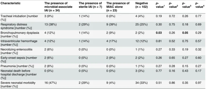
When selected indicators of short-term neonatal morbidity were examined, differences in the rates of the need for tracheal intubation and bronchopulmonary dysplasia were found in the crude analysis. However, there were no differences in the rates of the selected aspects of short-term neonatal morbidity after adjusting for gestational age at delivery (Table 4).
Fig 1. Prevalence of microbial-associated intraamniotic inflammation, sterile inflammation and microbial invasion of the amniotic cavity alone according to the gestational age at sampling.
doi:10.1371/journal.pone.0133929.g001
Table 2. Prevalence of microbial-associated intraamniotic inflammation, sterile inflammation and microbial invasion of the amniotic cavity alone according to the gestational age at sampling.
Gestational age at sampling (weeks+days) Microbial-associated IAI Sterile IAI MIAC alone Negative
24+0–27+6 (n = 11) 5 (46%) 1 (9%) 2 (18%) 3 (27%)
28+0–31+6 (n = 36) 12 (29%) 2 (5%) 6 (14%) 22 (52%)
32+0–33+6 (n = 42) 5 (14%) 0 (0%) 4 (11%) 27 (75%)
34+0–36+6 (n = 77) 12 (16%) 4 (5%) 11 (14%) 50 (65%)
Abbreviations: IAI: Intraamniotic inflammation, MIAC: microbial invasion of the amniotic cavity.
Discussion
Pregnancies with PPROM are often complicated by IAI and MIAC. The management of PPROM mainly depends on the gestational age, severity of the clinical presentation and/or knowledge about the complications. Therefore, a prompt diagnosis of these complications seems to be important for the optimal management of PPROM, appropriate parental counsel-ing and providcounsel-ing adequate neonatal care. Ideally, this information should be available within a short time from the admission of women with PPROM. However, the clinical dilemma for initiating appropriate management is attributed to the lack of proper biochemical or clinical indicators. In this study, we examined subgroups of women with PPROM based on the
Table 3. Microorganism identified in the amniotic fluid of women with preterm prelabor rupture of membranes.
The presence of microbial-associated intraamniotic inflammation (n = 34)
The presence of microbial invasion of the amniotic cavity alone (n = 23)
Ureaplasmaspecies Ureaplasmaspecies
Ureaplasmaspecies Ureaplasmaspecies
Ureaplasmaspecies Ureaplasmaspecies
Ureaplasmaspecies Ureaplasmaspecies
Ureaplasmaspecies Ureaplasmaspecies
Ureaplasmaspecies Ureaplasmaspecies
Ureaplasmaspecies Ureaplasmaspecies
Ureaplasmaspecies Ureaplasmaspecies
Ureaplasmaspecies Ureaplasmaspecies
Ureaplasmaspecies Ureaplasmaspecies
Ureaplasmaspecies Ureaplasmaspecies
Ureaplasmaspecies Mycoplasma hominis
Streptococcus agalactiae Mycoplasma hominis
Streptococcus anginosus Mycoplasma hominis
Haemophilus influenzae Mycoplasma hominis
Staphylococcus hominis Bifidobacteriumspecies
Streptococcus species Staphylococcus haemolyticus
Chlamydia trachomatis Enterococcus faecium
Ureaplasmaspecies +Mycoplasma hominis Chlamydia trachomatis
Ureaplasmaspecies +Mycoplasma hominis Gardnerella vaginalis
Ureaplasmaspecies +Mycoplasma hominis Ureaplasmaspecies +Chlamydia trachomatis
Ureaplasmaspecies +Mycoplasma hominis Ureaplasmaspecies +Haemophilus influenzae
Ureaplasmaspecies +Mycoplasma hominis Ureaplasmaspecies +Chlamydia trachomatis
Ureaplasmaspecies +Veillonellaspecies
Ureaplasmaspecies +Lactobacillusspecies
Ureaplasmaspecies +Sneathia sanguinegens
Ureaplasmaspecies +Chlamydia trachomatis
Ureaplasmaspecies +Sneathia sanguinegens
Ureaplasmaspecies +Chlamydia trachomatis
Ureaplasmaspecies +Streptococcus anginosus
Ureaplasmaspecies +Haemophilus influenzae
Ureaplasmaspecies +Sneathia sanguinegens
Chlamydia trachomatis + Leptotrichia amnionii Peptococcus species + Propionibacterium species + Bacteroides species
Fig 2. Amniotic fluid interleukin-6 concentrations in preterm prelabor rupture of membrane pregnancies that are complicated with the presence or absence of microbial-associated intraamniotic inflammation, sterile intraamniotic inflammation and microbial invasion of the amniotic cavity alone.
Fig 3. Amniotic fluid interleukin-6 concentrations in preterm prelabor rupture of membrane pregnancies that are complicated with the presence or absence of microbial-associated intraamniotic inflammation, sterile intraamniotic inflammation and microbial invasion of the amniotic cavity alone with respect to the presence and absence of histological chorioamnionitis.
Fig 4. Prevalence of histological chorioamnionitis with and without the presence of neutrophil infiltration in the amnion in women with microbial-associated intraamniotic inflammation, sterile intraamniotic inflammation and microbial invasion of the amniotic cavity alone.
presence or absence of IAI and MIAC. We used an AF IL-6 value of>750 pg/ml as the
defini-tion of IAI based on its prognostic utility in women presenting with PPROM [24]. The five principal findings of this study are as follows: i) microbial-associated IAI was a complication in approximately one-fifth of women with PPROM; ii) sterile IAI is rare in PPROM; iii) microbial-associated IAI was associated with a higher rate of neutrophil infiltra-tion of the amnion iv) microbial-associated IAI was related to a higher microbial load of
Urea-plasmaspecies in the amniotic fluid compared to MIAC alone; and v) there were no
differences in the short-term neonatal morbidity with respect to the presence of microbial-associated IAI, sterile IAI, and MIAC alone after adjusting for gestational age at delivery in women with PPROM.
In this study, we found that approximately 21% of PPROM was complicated by IAI. The majority of these cases (83%; 34/41) were complicated by microbial-associated IAI. Sterile IAI was responsible for a small proportion of the pregnancies complicated by IAI (17%; 7/41). This
Fig 5. Amniotic fluid microbial loads ofUreaplasmaspecies in preterm prelabor rupture membrane pregnancies that are complicated by the microbial-associated intraamniotic inflammation and microbial invasion of the amniotic cavity alone.
finding is different from the data reported by Romero et al. in which approximately one-third of women with PPROM had sterile inflammation. In this Romero et al. study where amniotic fluid IL-6 was evaluated by ELISA cut-off level for IAI was 2.6 ng/mL. However, recent studies by Chaemsaithong et al. have shown that 0.745 pg/mL (cut-off used in this study) is equivalent of this cut-off when amniotic fluid IL-6 is measured by a point of care test (used in this study) [24]. It means that the definition for IAI was the same in ours and Romero et al. studies. Differ-ent factors may explain the discrepancy between the studies. First, the difference in the gesta-tional ages of PPROM could contribute to the discrepancy. In our study, only women with PPROM and gestational ages between 24+0 and 36+6 weeks were included, but in Romero’s et al. study, women with gestational ages between 20 and 35 weeks were included. Forty-two percent of women in our study and 90% of women in Romero’s study were at gestational age<33 weeks upon sampling of PPROM. Secondly, the participants’ethnicities and Fig 6. A correlation between microbial load ofUreaplasmaspecies and interleukin-6 in the amniotic fluid from pregnancies complicated by preterm prelabor rupture of membranes.
geographic backgrounds differed between the studies, and amniotic fluid IL-6 has been docu-mented to demonstrate ethnic variations. We do not want to overestimate the rate of sterile IAI in Caucasian woman with pregnancies complicated by PPROM between gestational ages 24 and 37 weeks. Nevertheless, this finding seems to be clinically relevant because only the evalua-tion of amniotic fluid IL-6 levels, without an evaluaevalua-tion of the MIAC, can identify women at high risk of the presence of microbial-associated IAI. This is clinically relevant because the diagnosis of IAI is relatively easy to perform with evaluation of the amniotic fluid IL-6 using broadly available point of care tests. Moreover, the diagnosis of MIAC requires more time and the availability of a laboratory that can perform non-culture-based bacteria detection tests or at least has specific cultivation techniques for genital mycoplasmas. While, these findings are clin-ically relevant and have important clinical implications, we are aware that these findings must be validated on a new independent cohort of women.
The neutrophil infiltration of the amnion is considered as the most advanced stage of HCA related to the highest intraamniotic and fetal inflammatory response [32]. In this study we found that women with microbial-associated IAI had a higher rate of neutrophil infiltration of the amnion than the remaining women with PPROM. It means that this subgroup of PPROM is more often complicated by the most advanced stage of HCA. In addition, it has been shown
Table 4. Neonatal morbidity among preterm prelabor rupture of membrane pregnancies according to the presence or absence of microbial-asso-ciated intraamniotic inflammation, sterile intraamniotic inflammation and microbial invasion of the amniotic cavity alone.
Characteristic The presence of microbial-associate IAI (n = 34)
The presence of sterile IAI (n = 7)
The presence of MIAC alone (n = 23)
Negative (n = 102)
p -value1 p -value2 p -value3 p -value4
Tracheal intubation [number (%)]
3 (9%) 1 (14%) 0 (0%) 4 (4%) 0.19 0.72 0.26 0.77
Respiratory distress syndrome [number (%)]
13 (38%) 2 (29%) 9 (39%) 25 (25%) 0.30 0.75 0.18 0.69
Bronchopulmonary dysplasia [number (%)]
4 (12%) 1 (14%) 2 (9%) 2 (2%) 0.03 0.26 0.05 0.29
Intraventricular hemorrhage [number (%)]
4 (12%) 1 (14%) 4 (17%) 12 (12%) 0.81 0.52 0.75 0.57
Necrotizing enterocolitis [number (%)]
2 (6%) 0 (0%) 0 (0%) 1 (1%) 0.27 0.33 0.19 0.32
Early-onset sepsis [number (%)]
2 (6%) 0 (0%) 2 (9%) 2 (2%) 0.26 0.65 0.27 0.60
Pneumonia [number (%)] 2 (6%) 0 (0%) 0 (0%) 1 (1%) 0.27 0.28 0.15 0.27
Neonatal death before hospital discharge [number (%)]
0 (0%) 0 (0%) 0 (0%) 3 (3%) 0.77 0.16 0.43 0.17
Severe neonatal morbidity [number (%)]
16 (47%) 2 (29%) 9 (4%) 34 (33%) 0.51 0.86 0.35 0.97
Abbreviations: IAI: Intraamniotic inflammation, MIAC: microbial invasion of the amniotic cavity. Severe neonatal morbidity was defined as a need for intubation and/or respiratory distress syndrome and/or pneumonia and/or bronchopulmonary dysplasia and/or intraventricular hemorrhage and/or necrotizing enterocolitis and/or early-onset sepsis and/or late-onset sepsis and/or neonatal death before hospital discharge. Retinopathy of prematurity (n = 1) and late onset sepsis (n = 2) was not considered in the analysis because of low occurrence in the cohort. Categorical variables were compared using the Fisher’s exact test or the chi-square test. Spearman partial correlation was used to adjust the data for the gestational age at delivery. Statistically significant results are marked in bold.p-value1: the comparison among four groups with the Fisher
’s exact tests (exact 2-tailedp-value).p-value2:the
comparison among four groups with the adjustment for gestational age at delivery.p-value3: the comparison among women with microbial-associated intraamniotic inflammation, microbial invasion of the amniotic cavity alone, and negative women with the chi-square test.p-value4: the comparison among women with microbial-associated intraamniotic inflammation, microbial invasion of the amniotic cavity alone, and negative women with the adjustment for gestational age at delivery.
that the presence the neutrophil infiltration of the amnion is a better indicator of the develop-ment of early-onset sepsis than the presence of funisitis [32]. This finding is in line with our observations because all pregnancies complicated by the development of early onset sepsis had the neutrophil infiltration of the amnion.
Genital mycoplasmas (Ureaplasmaspecies andMycoplasma hominis)are the most common microorganisms isolated from amniotic fluid obtained from pregnancies complicated by PPROM. In the subgroup of women with microbial-associated IAI,Ureaplasmaspecies either alone or with other microorganisms was found in 10 women and in 16 women, respectively. In remaining 5 women,Streptococcus anginosus,Streptococcusspecies,Haemophilus influenzae.
Staphylococcus hominis, andChlamydia trachomatiswere found. In the subgroup of women
with MIAC alone, onlyUreaplasmaspecies and onlyMycoplasma hominiswas found in 13 and 4 women, respectively. Three women had the presence ofUreaplasmaspecies with other bacte-ria, and the remaining 6 women hadGardnerella vaginalis,Bifidobacteriumspecies,
Streptococ-cus agalactiae,Staphylococcus haemolyticus,Enterococcus faeciumandChlamydia trachomatis
in the amniotic fluid. Our previous studies have shown that the intensity of intraamniotic inflammation correlates with the microbial load of genital mycoplasmas [12,13]. In this study, there was a higher microbial load ofUreaplasmaspecies in the amniotic fluid of women with microbial-associated IAI than in women with MIAC alone This finding confirms the hypothe-sis that the intraamniotic inflammatory response of the amniotic cavity determined in the pres-ence of MIAC is likely related to the microbial load and type of bacteria. In this study we confirmed our previous observation that intraamniotic inflammatory response toUreaplasma
species is a dose-dependent [12,33].
From a clinical point of view, it is also important to understand the association between the microbial-associated IAI, sterile IAI, MIAC alone and short-term neonatal morbidity. We did not find any evidence suggesting that the presence of these conditions is detrimental to the short-term neonatal outcomes of newborns from PPROM after adjusting for the gestational age at delivery. Therefore, the gestational age at delivery but not the presence of microbial-asso-ciated IAI, sterile IAI or MIAC alone is an important factor for neonatal morbidity. This is in line with previous reports showing that the gestational age both at PPROM and delivery is associated with adverse neonatal outcomes [34,35]. Moreover, a recent paper by Combs et al. reported similar findings in a cohort of women with spontaneous preterm delivery with intact membranes [15]. Our findings confirm that expectant management is justified in pregnancies complicated by PPROM. However, the association between the presence of microbial-associ-ated IAI, sterile IAI, MIAC alone and long-term neonatal outcomes (e.g., cerebral palsy and neurodevelopmental outcomes) should be determined.
An important strength of this study is that MIAC was identified based on non-cultivation techniques, including non-specific PCR (16S rRNA) and specific PCR (Ureaplasmaspecies,
Mycoplasma hominis, andChlamydia trachomatis). The implementation of specific PCR gave
In conclusion, microbial-associated IAI is more common in PPROM, and sterile IAI is rare in Caucasian women with PPROM between gestational ages 24–37 weeks. Evaluation of the amniotic fluid IL-6 without identifying the bacteria in the amniotic fluid can be used to identify PPROM complicated by microbial-associated IAI. The presence of microbial-related IAI or sterile IAI does not affect short-term neonatal outcomes in PPROM.
Supporting Information
S1 Table. Amniotic fluid IL-6 levels, the presence of histological chorioamnionitis, funisi-tis, and the presence of neutrophil infiltration (grades 1–4) in the chorion-decidua, chori-onic plate, umbilical cord, and amnion in preterm prelabor rupture of membrane pregnancies that are complicated with the presence or absence of microbial-associated intraamniotic inflammation, sterile intraamniotic inflammation and microbial invasion of the amniotic cavity alone.
(XLSX)
Author Contributions
Conceived and designed the experiments: IM RK LP RM BJ MS MK. Performed the experi-ments: IM RK LP MS MK. Analyzed the data: IM RK LP RM BJ MS MK. Contributed reagents/materials/analysis tools: IM RK LP MS MK. Wrote the paper: IM RK LP RM BJ MS MK.
References
1. Menon R, Boldogh I, Hawkins HK, Woodson M, Polettini J, Syed TA, et al. (2014) Histological evidence of oxidative stress and premature senescence in preterm premature rupture of the human fetal mem-branes recapitulated in vitro. Am J Pathol 184: 1740–1751. doi:10.1016/j.ajpath.2014.02.011PMID:
24832021
2. Menon R, Polettini J, Syed TA, Saade GR, Boldogh I (2014) Expression of 8-oxoguanine glycosylase in human fetal membranes. Am J Reprod Immunol 72: 75–84. doi:10.1111/aji.12220PMID:24589083 3. Romero R, Miranda J, Chaemsaithong P, Chaiworapongsa T, Kusanovic JP, Dong Z, et al. (2014)
Ster-ile and microbial-associated intra-amniotic inflammation in preterm prelabor rupture of membranes. J Matern Fetal Neonatal Med: 1–16.
4. Romero R, Espinoza J, Goncalves LF, Kusanovic JP, Friel L, Hassan S (2007) The role of inflammation and infection in preterm birth. Semin Reprod Med 25: 21–39. PMID:17205421
5. Bredeson S, Papaconstantinou J, Difford JH, Kechichian T, Syed TA, Saade GR, et al. (2014) HMGB1 Promotes a p38MAPK Associated Non-Infectious Inflammatory Response Pathway in Human Fetal Membranes. PLoS One 9: e113799. doi:10.1371/journal.pone.0113799PMID:25469638
6. Menon R, Taylor RN, Fortunato SJ (2010) Chorioamnionitis—a complex pathophysiologic syndrome. Placenta 31: 113–120. doi:10.1016/j.placenta.2009.11.012PMID:20031205
7. Menon R (2014) Oxidative stress damage as a detrimental factor in preterm birth pathology. Front Immunol 5:567. doi:10.3389/fimmu.2014.00567PMID:25429290
8. Kacerovsky M, Musilova I, Khatibi A, Skogstrand K, Hougaard DM, Tambor V, et al. (2012) Intraamnio-tic inflammatory response to bacteria: analysis of multiple amnioIntraamnio-tic fluid proteins in women with preterm prelabor rupture of membranes. J Matern Fetal Neonatal Med 25: 2014–2019. doi:10.3109/14767058. 2012.671873PMID:22519389
9. Jacobsson B, Mattsby-Baltzer I, Andersch B, Bokstrom H, Holst RM, Nikolaitchouk N, et al. (2003) Microbial invasion and cytokine response in amniotic fluid in a Swedish population of women with pre-term prelabor rupture of membranes. Acta Obstet Gynecol Scand 82: 423–431. PMID:12752072 10. DiGiulio DB, Romero R, Kusanovic JP, Gomez R, Kim CJ, Seok KS, et al. (2010) Prevalence and
diver-sity of microbes in the amniotic fluid, the fetal inflammatory response, and pregnancy outcome in women with preterm pre-labor rupture of membranes. Am J Reprod Immunol 64: 38–57. doi:10.1111/j. 1600-0897.2010.00830.xPMID:20331587
12. Kacerovsky M, Pliskova L, Bolehovska R, Skogstrand K, Hougaard DM, Tsiartas P, et al. (2012) The impact of the microbial load of genital mycoplasmas and gestational age on the intensity of intraamnio-tic inflammation. Am J Obstet Gynecol 206: 342 e341–348.
13. Kacerovsky M, Pliskova L, Bolehovska R, Musilova I, Hornychova H, Tambor V, et al. (2011) The microbial load with genital mycoplasmas correlates with the degree of histologic chorioamnionitis in preterm PROM. Am J Obstet Gynecol 205: 213 e211–217.
14. Jacobsson B, Aaltonen R, Rantakokko-Jalava K, Morken NH, Alanen A (2009) Quantification of Urea-plasma urealyticum DNA in the amniotic fluid from patients in PTL and pPROM and its relation to inflam-matory cytokine levels. Acta Obstet Gynecol Scand 88: 63–70. doi:10.1080/00016340802572646
PMID:19031281
15. Combs CA, Gravett M, Garite TJ, Hickok DE, Lapidus J, Porreco R, et al. (2014) Amniotic fluid infection, inflammation, and colonization in preterm labor with intact membranes. Am J Obstet Gynecol 210: 125 e121–125 e115.
16. Romero R, Chaiworapongsa T, Alpay Savasan Z, Xu Y, Hussein Y, Dong Z, et al. (2011) Damage-associated molecular patterns (DAMPs) in preterm labor with intact membranes and preterm PROM: a study of the alarmin HMGB1. J Matern Fetal Neonatal Med 24: 1444–1455. doi:10.3109/14767058. 2011.591460PMID:21958433
17. Kacerovsky M, Celec P, Vlkova B, Skogstrand K, Hougaard DM, Cobo T, et al. (2013) Amniotic fluid protein profiles of intraamniotic inflammatory response to Ureaplasma spp. and other bacteria. PLoS One 8: e60399. doi:10.1371/journal.pone.0060399PMID:23555967
18. Menon R, Williams SM, Fortunato SJ (2007) Amniotic fluid interleukin-1beta and interleukin-8 concen-trations: racial disparity in preterm birth. Reprod Sci 14: 253–259. PMID:17636239
19. Fortunato SJ, Lombardi SJ, Menon R (2004) Racial disparity in membrane response to infectious sti-muli: a possible explanation for observed differences in the incidence of prematurity. Am J Obstet Gynecol 190: 1557–1562. PMID:15284734
20. Romero R, Yoon BH, Kenney JS, Gomez R, Allison AC, Sehgal PB (1993) Amniotic fluid interleukin-6 determinations are of diagnostic and prognostic value in preterm labor. Am J Reprod Immunol 30: 167–183. PMID:8311926
21. Romero R, Yoon BH, Mazor M, Gomez R, Gonzalez R, Diamond PB, et al. (1993) A comparative study of the diagnostic performance of amniotic fluid glucose, white blood cell count, interleukin-6, and gram stain in the detection of microbial invasion in patients with preterm premature rupture of membranes. Am J Obstet Gynecol 169: 839–851. PMID:7694463
22. Romero R, Kadar N, Miranda J, Korzeniewski SJ, Schwartz AG, Chaemsaithong P, et al. (2014) The diagnostic performance of the Mass Restricted (MR) score in the identification of microbial invasion of the amniotic cavity or intra-amniotic inflammation is not superior to amniotic fluid interleukin-6. J Matern Fetal Neonatal Med 27: 757–769. doi:10.3109/14767058.2013.844123PMID:24028673
23. Chaemsaithong P, Romero R, Korzeniewski SJ, Dong Z, Yeo L, Hassan SS, et al. (2014) A point of care test for the determination of amniotic fluid interleukin-6 and the chemokine CXCL-10/IP-10. J Matern Fetal Neonatal Med: 1–10.
24. Chaemsaithong P, Romero R, Korzeniewski SJ, Martinez-Varea A, Dong Z, Yoon BH, et al. (2015) A point of care test for interleukin-6 in amniotic fluid in preterm prelabor rupture of membranes: a step toward the early treatment of acute intra-amniotic inflammation/infection. J Matern Fetal Neonatal Med: 1–8.
25. Chaemsaithong P, Romero R, Korzeniewski SJ, Martinez-Varea A, Dong Z, Yoon BH, et al. (2015) A rapid interleukin-6 bedside test for the identification of intra-amniotic inflammation in preterm labor with intact membranes. J Matern Fetal Neonatal Med: 1–11.
26. Kacerovsky M, Musilova I, Stepan M, Andrys C, Drahosova M, Jacobsson B (2015) Detection of intraamniotic inflammation in fresh and processed amniotic fluid samples with the interleukin-6 point of care test. Am J Obstet Gynecol doi:10.1016/j.ajog.2015.05.039
27. Greisen K, Loeffelholz M, Purohit A, Leong D (1994) PCR primers and probes for the 16S rRNA gene of most species of pathogenic bacteria, including bacteria found in cerebrospinal fluid. J Clin Microbiol 32: 335–351. PMID:7512093
28. Fouhy F, Deane J, Rea MC, O'Sullivan O, Ross RP, O'Callaghan G, et al. (2015) The effects of freezing on faecal microbiota as determined using MiSeq sequencing and culture-based investigations. PLoS One 10: e0119355. doi:10.1371/journal.pone.0119355PMID:25748176
29. Salafia CM, Weigl C, Silberman L (1989) The prevalence and distribution of acute placental inflamma-tion in uncomplicated term pregnancies. Obstet Gynecol 73: 383–389. PMID:2915862
30. Papile LA, Burstein J, Burstein R, Koffler H (1978) Incidence and evolution of subependymal and intra-ventricular hemorrhage: a study of infants with birth weights less than 1,500 gm. J Pediatr 92: 529–
31. Gomez R, Romero R, Ghezzi F, Yoon BH, Mazor M, Berry SM (1998) The fetal inflammatory response syndrome. Am J Obstet Gynecol 179: 194–202. PMID:9704787
32. Park CW, Moon KC, Park JS, Jun JK, Romero R, Yoon BH (2009) The involvement of human amnion in histologic chorioamnionitis is an indicator that a fetal and an intra-amniotic inflammatory response is more likely and severe: clinical implications. Placenta 30: 56–61. doi:10.1016/j.placenta.2008.09.017
PMID:19046766
33. Kacerovsky M, Musilova I, Hornychova H, Kutova R, Pliskova L, Kostal M, et al. (2014) Bedside assessment of amniotic fluid interleukin-6 in preterm prelabor rupture of membranes. Am J Obstet Gynecol 211: 385 e381–389.
34. Frenette P, Dodds L, Armson BA, Jangaard K (2013) Preterm prelabour rupture of membranes: effect of latency on neonatal and maternal outcomes. J Obstet Gynaecol Can 35: 710–717. PMID:24007706 35. Nayot D, Penava D, Da Silva O, Richardson BS, de Vrijer B (2012) Neonatal outcomes are associated