tion Studies of a New Virus from the Squirrel Monkey.” Lab Anim Care 19: 372-377, 1969. (29) Melendez, L. V., R. D. Hunt, M. D. Daniel, F. G. Garcia, and C. E. 0. Fraser. ‘Herpesvirus saimiri. II. Experimentally Induced Malignant Lymphoma in Primates.” Lab Anim Care 19: 378-386,1969.
(30) Hunt, R.D., L. V. Melendez, N. W. King, C. E. Gilmore, M. D. Daniel, M. E. Williamson, and T. C. Jones. “Morphology of a Disease with Features of Malignant Lymphoma in Marmo- sets and Owl Monkeys Inoculated with Her- pesvints saimiri. ” J Nat Cancer Inst 44: 447-405,1970.
(31) Maurer, B. A., T. Imamura, and J. Minowada.
“Evidence that Cells Derived from Burkitt Lymphoma are Potential EB Virus-Producing cells.” Bacf Proc 154 1969 3 .
(32) Pope, J. H., M. K. Horne, and E. J. Wetters. “Significance of a Complement-Fixing Anti- gen Associated with Herpes-Like Virus and Detected in the Raji Cell Line.” Nature 222: 186-187, 1969.
(33) Zur Hausen, H., and H. Schulte-Holthausen. “Presence of EB Virus Nucleic Acid Homology in a Virus-Free Line of Burkitt Tumour Cells.” Nature 227: 245-248, 1970. (34) Probert, M., and M. A. Epstein. “Morphological
Transformation in vitro of Human Fibroblasts by Epstein-Barr Virus: Preliminary Observa- tions.” Science 175: 202-203, 1972.
TWO NEW HERPESVIRUSES FROM SPIDER MONKEYS
(ATELES GEOFFROYI) 5, b,
L. V. MeKndez, D.V.M.,7 H. H. Barahona, D.V.M., Ph.D.,’ M. D. Daniel, D.V.M., Ph.D.,’ R. D. Hunt, D.V.M.,7 C. E. O.Fraser, D.V.M., Ph.D.,7 F. G. Garcia, D.V.M.,s N. W. King, D.V.M.,’ and H. Castellanos, D.V.M. 8
Experiments with two new viruses isolated from spider monkeys show them to be distinct from the previously known spider monkey herpesvirus (SMHV]. One of the two, Herpesvirus ateles, was found to cause a disease similar to malignant lymphoma with terminal leukemia in inoculated marmosets.
Introduction
Two facts led us to find the new herpes- viruses described in this presentation. One was 5Paper presented at the II International Sympo- sium on Health Aspects of the International Move- ment of Animals (Special Symposium on Viruses of South American Monkeys) held in Mexico City on 11-13 August 1971. Previously published in Scientific Publication PAHO 235 (1972), pp. 145151.
bResearch supported by NIH, USPHS Grant No. FR 00168-09, and by the Pan American Health Organization, Department of Human and Animal Health.
7New England Regional Primate Research Center, Harvard Medical School, Southboro, Massachusetts.
*University of San Carlos, Guatemala City, Guate- mala.
the discovery by Melendez and his associates in 1968 of the first lymphoma virus of monkeys, Herpesvirus saimiri. This virus, derived from squirrel monkeys, proved capable of inducing leukemic or aleukemic malignant lymphoma in several nonhuman primate species as well as in rabbits (1, 2, 3, 4, 5). The other fact was an association between human patients with lymphosarcoma and spider monkeys (A teles geofjoyi) in Guatemala. A detailed description of this association has been presented elsewhere
(0
64 ENGLISH EDITION-BOLETIN DE LA OSP . Vol. VII, No. 1, I973
animal species; instead, two new herpesvirus isolates were found.
It is known that the spider monkey is a host for a distinct herpesvirus. The virus was isolated by Lennette and has been studied and charac- terized by Hull (7). This agent is known as the spider monkey herpesvirus (SMHV), and its detailed description has been presented by Dr. Hull (8).
This paper deals with the isolation and characterization of two new herpesviruses from the spider monkey-Guatemala isolate AT-46 and Herpesvirus ateles-and comparison of them with SMHV.
Guatemala Isolate AT-46
On 22 July 1970 a kidney was obtained by nephrectomy from spider monkey AT-46. This monkey was housed at the School of Medicine in Guatemala City, Guatemala. It had been in close contact with a patient (R.C.) who de- veloped malignant lymphosarcoma. Kidney
cultures were initiated with the renal cortex tissues of this animal. The cells were obtained by trypsinization of the kidney tissues follow- ing standard procedures. The cells obtained were then grown in plastic flasks (Falcon) containing Eagle’s minimum essential medium with 10 per cent heat-inactivated fetal calf serum (MEM-I 0).
Two-day-old cultures presented cytoplasmic vacuolization and bizarre nuclei; very ill-defined nuclear inclusions were observed at this time in hematoxylin eosin (H.E.) stained cultures. The cytoplasmic vacuolization had increased by the eighth day (Photo 1). At this time secondary cultures were prepared, and nine days later these presented a large number of polykaryo- cytes. Some of these cell cultures were scraped, pooled, and collected as isolate El 15F. This isolate was then stored at 4OC, -86OC, and -176OC (liquid nitrogen).
The type of lesion observed in stained cultures was characterized by disruption of the cell layer and the presence of a large number of polykaryocytes. Most of these polykaryocytes had more than 10 nuclei. Very small and poorly
defined intranuclear inclusions were seen in microscopic observation under immersion oil (Photo 2).
Cytopathic Effect (CPE) of Guatemala Isolate AT-46
Isolate AT-44 produced a cytopathic effect (CPE) in continuous cell cultures of rabbit kidney (RKL), in owl monkey kidney 210 (OMK), and in squirrel monkey fetus lung 241 (SMFL). Isolate AT-46 reached the following titers (TCIDsc/ml): 103.5 in RKL and 105.0 in OMK, 21 and 23 days, respectively, after inoculation.9 The alteration in SMFL began with the appearance of large, dense, spindle- shaped cells. These cells were observed to be polykaryocytes with small intranuclear inclu- sions in H.E.-stained preparations. These cells increased in number until the whole monolayer was altered. The final result was cell lysis (Photo 3). A similar type of cell layer disrup- tion was observed in human embryonic lung (HEL) continuous culture.
Spider Monkey Kidney Isolate 810:
Herpesvirus Ateles
A primary kidney culture was prepared on 17 June 1970 from the renal cortex of black spider monkey 810-69, which had been housed in our laboratories for at least one year. The procedures used to culture this kidney tissue were similar to those employed for Guatemala isolate AT-46. A well-grown cell layer de- veloped in six to eight days. This cell layer presented a large number of bizarre cells and polykaryocytes on the eleventh day. Most of these polykaryocytes had become detached from the surface of the plastic flasks. On the sixteenth day the cell layer was scraped, and- together with the culture fluids-was collected as isolate 810. Like isolate AT-46, this was stored at 4OC, -86oC, and -176’-‘C.
Isolate 810, from now on referred to as H.
PHOTO I-Eightdayold AT-46 spi- der monkey kidney primary culture. Observe the polykaryocytes and the cytoplasmic vacuohzation. Final magni- fication 125 X.
PHOTO 2-Ninedayold AT-46 spider monkey kidney secondary culture show- ing a polykaryocyte with several nuclei containing very illdefined nuclear inclu- sions. Final magnification 1000 X.
66 ENGLISH EDITION-BOLETIN DE LA OSP . Vol. VII, No. 1, 1973
ateles, was inoculated undiluted in SMFL cul- tures and caused the development of large spindle-shaped cells in greater numbers than isolate AT-46. However, in the OMK cell line the CPE was characterized by scattered foci of swollen and rounded cells. In both cultures the development of intranuclear inclusion was observed in H.E.-stained preparations, but these inclusions were better outlined in OMK cell cultures.
Spider Monkey Herpesvirus (SMHV): Lennette-Hull Isolate
This known herpesvirus from the spider monkey was originally isolated by Lennette and studied and characterized by Hull (7). SMHV produced almost complete destruction of HEL cultures one day after inoculation with un- diluted virus material and very slight alteration in SMFL five days after inoculation.
Cytopathogenicity of Herpesvirus Ateles
H. ateles strain 810 reached the following titers (TCIDs,-,/ml): 104.5 in OMK, lo4 in SMFL, 103.s in HEL, and 105ao in RKL at 17, 14, 23, and 30 days, respectively, after inocula- tion. The type of CPE that H. ateles produces in SMFL varies, depending upon the number of passages of the virus in OMK or RKL cell cultures. Virus passed at least three times in OMK cultures developed larger nuclear inclu- sions and more polykaryocytes, and was more virulent for SMFL than the virus passed in RKL continuous cultures.
Comparative Cytopathogenicity of the Spider Monkey Herpesviruses
The following in vitro cell cultures were prepared: whole human embryo, human embry- onic lung, hamster heart, goat synovial bursa, goat bursa capsule, OMK 210, and SMFL and RKL continuous cultures. These cultures were inoculated with Guatemala isolate AT-46, H.
ateles, and SMHV (the whole human embryo
TABLE 1 -Cytopathogenicity of spider monkey herpesviruses.
Cell cultures Viruses
H. ateles AT-46 SMHV
Whole human embryo Hamster heart Goat synovial bursa Goat bursa capsule Owl monkey kidney 210 Squirrel monkey fetus hmg Rabbit kidney
Human embryonic lung
+
+ ;
+ *
+ *
+ +
+ +
+ -4
+ +
*Not tested.
TABLE 2-Comparative titrations of spider monkey herpesviruses in homologous kidney cultures.
SMHV H. ateles AT-46
6.0* 4.0* 1.5*
*Titer loglO/l.Oml.
and goat cultures were not tested with SMHV).’ All these herpesviruses produced CPE in each case, differing only in the titers reached and the time needed to develop CPE. This behavior decreased from SMHV to AT-46 isolate. These results are summarized in Table 1. The same range of virulence was observed when these three herpesviruses were inoculated in spider monkey kidney cell culture (Table 2).
Action of Physicochemical Treatments on Spider Monkey Herpesviruses
inoculated with 100 rnp filtrates of any of these viruses. The results are summarized in Table 3.
TABLE 34pider monkey herpesviruses: cyto-
pathogenicity in RKL’k after physiochemical treat- ments.
Treatments
SMHV
VirUSeS
H. ateles AT46
Heat Ether
220 mp* 100 mp*
- - -
7 ? 7
- - -
t Rabbit kidney continuous culture. *220 m and 100 m virus filtrates.
Electron Microscopy Studies
The spider monkey herpesviruses displayed all the ultrastructural features needed for them to be considered members of the herpesvirus group. Herpesvirus was inoculated in RKL cultures, and when the CPE involved almost 70-75 per cent of the cell layer it was collected. The collected fluids were then treated with a sonic vibrator (Bronwill sonifier BP-l 1 at set- ting 50 with standard l/2 inch titanium tip for four cycles of five seconds each). After this sonic disruption, the culture fluids were centri- fuged at 2,500 rpm for 15 minutes in a PR2 refrigerated centrifuge.
The supernatant obtained was then filtered by 220 rnp, 100 rnF.1, and 50 rnF.1 filters (Millipore). Each of these filtrates was then inoculated in RKL cultures. Ten to 14 days later these cultures were examined under the electron microscope. Typical herpesvirus parti- cles were observed only in the cultures inocu- lated with the 220 mp filtrate. A detailed description of the electron microscope proce- dures, as well as the ultrastructural features of this herpesvirus, have been given elsewhere (9).
Plaque-Forming Capacity of Spider Monkey Herpesviruses
The plaque-forming abilities of Guatemala isolate AT-46 and of H. ateles were tested in
the following cultures: RKL, OMK, hamster heart (HH), and SMFL. Both of these agents were able to develop plaques in SMFL cultures approximately 30 days after inoculation and in HH within 33 to 50 days. The details of the plaque-forming capacity of these agents, as well as the plaquing procedures, have been described in a separate report (10).
Preparation of Antisera against Spider Monkey Herpesviruses
Goats and rabbits were used to prepare antisera against the spider monkey herpes- viruses.
Rabbit Inoculation Procedure
H. ateles and AT46 were the viruses em- ployed. Two New Zealand white rabitts were bled before inoculation; then undiluted fresh virus grown in RKL cultures was inoculated, 2.5 ml i!tradermally (ID) and 2.5 ml intra- venously (IV) in each of the animals. The rabbits were bled 14 days after inoculation, and at this time the inoculation was repeated. The animals were exsanguinated 36 or 40 days after inoculation, as long as the 14-day bleeding had a neutralization index (NI) of about 2.0. Whenever the NI was < 1.5, another similar booster inoculation was given.
Goat Inoculation Procedure
All three spider monkey viruses were used to immunize goats. Previously bled animals were inoculated with fresh undiluted viruses grown in RKL cultures. One ml was inoculated ID in each goat. The animals were then bled 7, 14 and 21 days after inoculation. On day 21 a similar 1 ml booster inoculation was given, and the animals were exsanguinated 28 days after inoculation. With SMHV, 2.0 ml were inocu- lated in each animal instead of 1 ml; the booster inoculation was given on day 28, and the animals were bled 44 days after inoculation.
Results
68 ENGLISH EDITION-BOLETIN DE LA OSP . Vol. VII, No. I, 1973
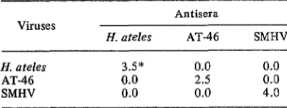
with the highest NI was employed to carry out the cross-neutralization indices test. Each virus was neutralized only by its homologous anti- serum, and no cross-neutralization was de- tected, thus indicating no serologic relationship among the viruses themselves. These results are summarized in Table 4.
TABLE 4-Reciprocal neutralization indices of spider monkey herpesviruses.
Viruses
H. ateles
Antisera
AT-46 SMHV
H. a teles 3.5* 0.0 0.0
AT-46 0.0 2.5 0.0
SMHV 0.0 0.0 4.0
*Neutralization indices.
Nonhuman Primate Inoculation with H. ateks
and AT-46 Isolate
Inoculation with Guatemala Isolate AT-46
Two owl monkeys and three cottontop marmosets were each inoculated intramuscu- larly with approximately 100 TCIDs e of AT-46 isolate. These animals were bled every other week for antibody studies, clinical blood chemistry, and blood counts. The two owl monkeys died approximately two and a half months after inoculation without presenting any previous symptoms or any histopathologic changes. The cottontop marmosets did not present any clinical symptoms or any alteration in their blood pictures for a period of five and a half months, at the end of which they were sacrificed.
Animals Inoculated with Herpesvirus ateles,
Isolate 810
Two owl monkeys and three cottontop marmosets were inoculated in the same way as indicated for AT-46, except that the viral inoculum was approximately 1,580 TCIDse of
H. ateles. Blood was also collected as indicated for the animals inoculated with AT-46. Two of
the marmosets died 28 days after inoculation, and one was sacrificed in a moribund condition 40 days after inoculation. Each animal had malignant lymphoma showing marked simi- larity to H. saimiri lymphoma. The proliferating and invading cells were predominantly large lymphoblasts or recticulum cells. Although no
deviation of peripheral white blood cell count occurred, the animals developed terminal leu- kemia, with up to 31 per cent lymphoblasts in the circulation.
The two owl monkeys were sacrificed in moribund condition 42 days after inoculation. No significant lesions were seen in one; how- ever, in the second animal a focal interstitial collection of reticulum cells and a lesser number of large lymphocytes and eosinophils were present in kidney and lung tissue. A detailed description of these in viuo studies is being published elsewhere (II, 12).
Conclusions
These studies demonstrate that the black spider monkey (Ateles geoffroyi) is a natural reservoir host for two new herpesviruses, the Guatemala isolate AT-46 and Herpesvirus ateles. They also demonstrate that these new agents are very distinct from the spider monkey herpesvirus (SMHV) described previously by Hull (7). This new knowledge undoubtedly is important, particularly since it is not known what public health hazards are involved when this species is employed in biomedical research.
Another very important finding derived from these studies is the discovery of the second lymphoma virus of monkeys, Herpes- virus ateles. This new herpesvirus proved to have an oncogenic capacity in owl and mar- moset monkeys. A disease similar to malignant lymphoma with terminal leukemia developed in one of these species after virus inoculation.
these primate models is of great value to understanding similar disease conditions in man.
ACKNOWLEDGMENTS
We are indebted to Jill Cadwallader, Douglas Jackman, Alice Preville, Eduardo Urizar, and Marjorie Vallee for their valuable assistance in the course of these studies.
SUMMARY
This paper deals with isolation of two new herpesviruses from spider monkeys and de- scribes experiments comparing their behavior with that of spider monkey herpesvirus (SMHV). The two new viruses (Guatemala isolate AT-46 and Herpesvirus ateles) were each found to have a cytopathic effect on various types of animal cell cultures. However, each virus produced a noticeably different effect, which was also distinct from that produced by SMHV.
Isolates of all three viruses appeared inacti- vated by exposure for 30 minutes to a tempera- ture of 56OC, by overnight treatment with ether at 4OC, and by filtration through 100 rnp filters, but not by filtration through 220 rnp filters. Both AT-46 and H. ateles displayed ultrastructural characteristics sufficient to con- sider them members of the herpesvirus group. A number of H. ateles, AT-46, and SMHV antisera were prepared from inoculated rabbits and goats. Cross-neutralization tests showed that each virus was neutralized only by its homologous antiserum. The lack of cross- neutralization indicates that there is no sero- logic relationship between the three viruses.
Additional experiments showed H. ateles
capable of producing lymphomas in marmosets. A disease similar to malignant lymphoma with terminal leukemia developed in three marmo- sets inoculated with the virus.
These studies show that herpesviruses are etiological agents of malignant Iymphoma in primates-and that South American monkeys are valuable models for study of leukemic processes of primates, as well as for under- standing similar disease conditions in man.
REFERENCES
(I) Melendez, L.V., R. D. Hunt, M. D. Daniel, F. G.
Garcia, and C.E.O. Fraser. “Herpes saimiri. II. An Experimentally Induced Malignant Lvmnhoma in Primates.” Lab Anim Care 19: 378-386, 1969.
(2) Hunt, R. D., L. V. Melendez, and M. D. Daniel. “A Disease in Primates Resembling Reticulum Cell Sarcoma Produced by a Herpes Virus.” Presented at the International Academy of Pathology, San Francisco, March 1969. (3) Melindez, L. V., R. D. Hunt, M. D. Daniel, and
C.E.O. Fraser. “DNA Viruses from South American Monkeys: Their Significance in the Establishment of Primate Colonies for Bio- medical Research. IV Symposium ILAR-
ICLA, Washington, DC.” In Defining the Laboratory Animal. National Academy of Sciences, - Washington, D.C., 1971, - pp. 550-563.
(4) Hunt, R. D., L. V. Melendez, N. W. King, C. E. Gilmore, M. D. Daniel, M. E. Williamson, and T. C. Jones. “Morphology of a Disease with Features of Malignant Lymphoma in Mar- ’ mosets and Owl Monkeys Inoculated with
Herpesvirus saimiri.” J Nat Cancer Inst 44: 447-465,1970.
(5) Melendez, L. V., M. D. Daniel, and R. D. Hunt. “Herpesvirus saimiri Induced Malignant Lymphoma: Recovery of the Viral Agent from the Fatally Affected Animals.” In Com- parative Leukemia Research, ed. by Ray M. Dutcher. Karger, 1970, pp. 751-753.
(6) Melendez, L. V., H. Castellanos, R. D. Hunt, H. H. Barahona, M. D. Daniel, C.E.O. Fraser, and F. G. Garcia. “Spider Monkey Herpesviruses and Malignant Lymphomas.” Presented at the 10th Meeting of the Advisory Committee on Medical Research, Pan American Health Organization, 14-18 June 1971.
(7) Hull, R. N. “The Simian Viruses.” In Virology Monographs, Vol.2. Springer-Verlag, New York, 1968, pp. l-66.
(8) Hull, R. N., A. C. Dwyer, A. W. Holmes, E. Nowakowski, F. Deinhardt, E. H. Lennette, and R. W. Emmons. “Recovery and Charac- terization of a New Simian Herpesvirus from a Fatally Infected Spider Monkey.” In Znter- national Movement of Animals, Pan American Health Organization, Scientific Publication !$;;4423.5, Washington, D.C., 1972, pp.
(9) King, N. W., M. D. Daniel, H. H. Barahona, and L. V. Melendez. “Viruses from South American Monkeys: Ultrastructural Studies.” In International Movement of Animals, Pan American Health Organization, Scientific Publication PAHO 235, Washington, D.C.,
1972, pp. 189-209.
(10) Daniel, M. D., L. V. Melendez, and H. H. Barahona. “Plaque Characterization of Viruses from South American Nonhuman Primates.” In International Movement of Animals, Pm American Health Organization, Scientific
70 ENGLISH EDITION-BOLETIN DE LA OSP . Vol. VII, No. I, I973
(II) MelBndez, L. V., R. D. Hunt, M. D. Daniel, C. E. (12) MelBndez, L. V., R. D. Hunt, F. G. Garcia, M. D. 0. Fraser, H. H. Garcia, and N. W. King. Daniel, C. E. 0. Fraser, H. H. Barahona, and “Lymphoma Viruses of Monkeys: Herpesvirus N. W. King. “Herpesvirus ateles, a New saimiri and Herpesvirus ateles. the First Onco- Lymphoma Virus of Monkeys.” Presented at genie Herpesviruses from Primates.” Presented the V International Symposium on Compara- at the Symposium on Oncogenesis and tive Leukemia Research, Padua-Venice, Italy,
Herpes-type Viruses, Cambridge, England, 13-17 September 1971. 20-25 June 1971.
JUNGLE YELLOW FEVER IN BRAZIL
There have been reports of an outbreak of jungle yellow fever in the
Brazilian State of Goias since December 1972. A total of 18 fatal cases,
occurring up to 17 January 1973, have been confirmed.
The first three cases, with onset from 6 to 21 December, occurred in the municipio of Silvinia, near the southern border of the Federal District. An additional three fatal cases occurred during December in the municipios of Luzlnia and Planaltina (south of the Federal District) and that of Go&, on the western border of the state. Twelve fatal cases diagnosed during the first half of January pertained to Silvgnia, Luzilinia, Go&, and areas to the northwest and southwest of the Federal District, including Jatai and Rio Verde near the southwestern border of the State of Coilis.
Since appearance of the first cases, vaccination of the people in the affected areas and surrounding regions has been intensified.
The cases do not constitute a cause of alarm. This disease occurs periodically in enzootic and epizootic areas of Brazil, which include forest areas of low population density situated for the most part in Amazonia, south central Go&, and Mato Grosso.
Even in the affected areas, there is no danger of infecting the populations of urban centers. This type of yellow fever occurs exclusively in rural areas where the mosquito vectors of jungle yellow fever are present, and there is little likelihood of the disease spreading to cities which are free of Aedes negypti.
In areas where people are settling, such as along the Transamazonic Highway, yellow fever vaccination is being required of all settlers and persons desiring to enter the region.
The Manguinhos Institute of the Ministry of Health produces five million doses of yellow fever vaccine annually and has the capacity to manufacture
ten million. Its production has exceeded the needs of the country to the extent that the Institute frequently responds to requests of other countries.
The Ministry of Health maintains a permanent campaign for yellow fever vaccination in all of the enzootic areas of the country. Through this continuing program, the Ministry has already, in the current year, vaccinated about 200,000 persons in the State of Go&.
In Bolivia, an outbreak of jungle yellow fever has occurred in the localities La Victoria and La Esperanza in Chap& Province, Department of Cocha- bamba, since 15 January 1973. As of 16 March, 12 cases had been reported. Laboratory confirmation of the diag’nosis has been obtained for two of the 10 fatal cases.
Recent captures of vectors in the two localities have demonstrated the presence only of mosquitoes of the genus Haemagogus. Yellow fever
vaccination has been intensified in the area. [Weekly Epidemiological Report of the Pan American Health Organization, Vol. 45, Nos. 5, 10, and 14;
January, March, and April 1973; pp. 24, 54, and 73.1