F
REQUENCY
OF
CAMPYLOBACTER JEJUNI AND
OTHER AGENTS OF ACUTE DIARRHEA
IN VENEZUELAN CHILDREN1
Mar&a Isa&Z Urrestarazu,2 Rafael T. Darricarrere T.,2y3 Mireya Phex, 4 Georgette Daoud,5 Nork Serrano,2
MarLa Eugenia Cauazza, 2 and Irene Phez-SchaeZ2
I
NTRODUCTION
Diarrhea1 diseases have great public health significance, being a very common cause of illness and malnutri- tion in young children (l-3). In Vene- zuela, approximately seven children die every day of these diseases (4).
Studies in various parts of the world have shown rotavirus and entero- toxigenic Escherichia coli (henceforth referred to as ‘ ‘ETFK”) to be the most
frequent agents of acute diarrhea (5-7). However, another microorganism known since 1372 to be associated with enterocolitis in man and animals is Campylobacter jej~lni (S-lo), This mi- crobe, identified in both sick and appar- ently healthy persons, has been found in many countries (g-14).
At present, CampyZobacter infection is considered a zoonosis. In some areas, including the industrialized
’ The study reported here was partially financed by the Venezuelan Scientific and Technologic Research Council (CONICIT-PtOjeCt Sl-1693). This article wiII
also be published in Spanish in the Bokth de la Ofi- cinaSanitaff~Panamericana, vol. 104, 1988. r Institute of Biomedicine (Instituto de Biomedicina),
Ministry of Health and Social Welfare, University of Caracas, Caracas, Venezuela.
r J. M. Vargas School of Medicine (Escuela de Medicina
countries, animals are the reservoirs of this infection; whereas in some areas of the developing world humans also play an important role in dissemination. These circumstances, and the fact that
C. jejuni is excreted for a long time after the acute phase of the disease, make the epidemiology of the infection com- plex enough to require additional re- search. In this connection, it should be noted that 36 C. @j&G serotypes have been identified on the basis of heat- labile antigens (lS), and more than 50
serotypes have been associated with dif- ferent heat-stable surface antigens (16) ; so far as is known, any of these could be involved in intestinal colonization.
The present study is the first to deal with the prevalence of C. jejzln;
“J. M. Vargas”). University of Caracas, Caracas. Ven- ezuela.
4 J. M. de 10s Rlos Children’s Hospital (Hospital de Nifios “J. M. de 10s Rlos”). Caracas, Venezuela. I Miguel Perez Carreiio Hospital (Hospital “Miguel
among infants and young children with acute diarrhea1 disease in Venezuela. Its specific purpose was to investigate the prevalence of this pathogen, clinical dis- ease characteristics, and associations be- tween this agent and other diarrhea1 dis- ease agents in Venezuelan children.
M
ATERIALS AND
METHODS
From June 1982 through May 1983 a study was made of 196 children under two years of age who attended the J. M. de 10s Rios and Miguel Perez Ca- rreiio hospitals in Caracas with diarrheal disease syndromes of varying symptom- atology and severity. Of this total, 149 (76%) were treated as outpatients and 47 (24%) were hospitalized. Fifty per- cent of these children were O-6 months old, 22.8% were 7-12 months, 13.0% were 13-18 months, and 14.2% were
19-24 months. In addition, a control group of 27 asymptomatic children with a similar background, age distribution, and sex ratio was studied.
Stool samples were collected fresh and were preserved at - 20°C for detection of rotavirus by enzyme-linked immunoassay (ELISA) (17). A portion of
each sample was used for a parasitologic study performed within 60 minutes of collection by examining part of the specimen suspended in saline and Lu- gol’s solution under a light microscope. Samples for bacteriologic di- agnosis were obtained directly with rec- tal swabs, preserved in a semisolid Cary- Blair transport medium (0.16 % agar), and processed in conventional culture media. The enterobacteria present were identified by conventional methods (18). In each case, five to 10 colonies of Escberichia coGi were isolated, pooled, and sown in trypticase soy broth. These
pools were tested for enteropathogenic- ity and invasive capacity, respectively, by agglutination with commercial anti- sera (BBL, Difco) and by the Sereny test
(19). The Dean test (20) was used to as- sess production of heat-stable (ST) toxin, and the ELISA technique (21) was em- ployed to assess production of heat- labile (LT) toxin.
For detection of Aeromonas spp. a portion of the sample was plated onto blood agar containing ampicillin (22), and for detection of Yersinia en- terocol’ytica it was plated onto media containing Salmonella-Shigella and MacConkey agar and kept at room tem- perature for 48 hours (23).
For isolation of Carzpylobac- terjejtini, a portion of each sample was sown onto Campy-Bap medium (24) containing 5 .O% human blood. The plates were then incubated at 42 ’ C in a roughly 5% CO* atmosphere (jar with candle-flame) for 48 hours. Identifica- tion was effected by observation of curved Gram-negative bacilli, by the re- sults of oxidase and catalase tests, by de- tecting H2S production, and by finding absence of growth at 25 “C, susceptibility to nalidixic acid (30 pg/ml), and resis- tance to cephalothin (30 pglml) (24).
b
RE
SULTS ”
2
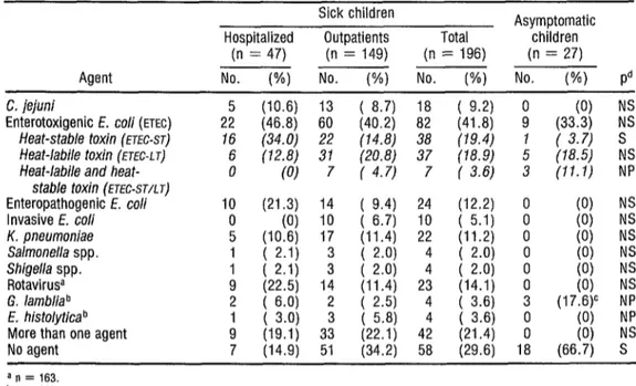
CarnpyZoobacter jejmi was found in 18 (7.2 %) of the sick children but in none of the control children. The enteric disease agent most often isolated was ETEC (from 41.8% of the patients), followed in order by rotavirus (14.1%) enteropathogenic E. cob (12.2%), and KZebsieZZa pnezlmoniae (11.2%). Among the parasites, Giadia ZambZia
(in 3.5 % of the patients) and Ent- amoeba histoZytica (also in 3.5 % ) were both identified. Fifty samples were tested for Ye&z& enterocoditicu and Aeromonas spp., but neither of these microorganisms was detected. In 21.4% of the cases a mixed infection was diag- nosed, but in 29.6% no agents were rec- ognized. A high proportion of the asymptomatic (control) children were found to be infected with ETEC produc- ing heat-stable toxin and with G. lard&a (18.5% and 17.6%, respec- tively) .
A comparison of the hospi- talized patients and outpatients (Table 1) revealed statistically significant dif-
ferences only with respect to heat-stable (ST) ETEC infections.
As Table 2 shows, C. je+zi was not found in association with other etiologic agents in most cases; however, in five of the six cases where it was found in association with another agent, that agent was ETEC. In no instance was C. jejmi found in association with ro-
tavirus .
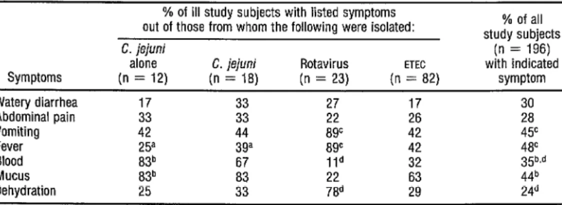
Those 12 subjects from whom C. je&wzi was isolated as the sole etiologic agent exhibited the symptoms indicated in column 1 of Table 3. Ten (83%) had blood in their feces, 10 (83%) had mucus, three (25 %) had fe- ver, five (42%) vomited, and four (33 % ) had abdominal pain. Only three (25 %) exhibited dehydration.
Overall, the symptoms of subjects with C. j,izlni alone were simi-
TABLE 1. Diarrhea1 disease agents isolated from the feces of 196 study children with diarrhea and from 27 asymptomatic (control) children.
Sick children Asymptomatic Hospitalized Outpatients Total children
(n = 47) (n = 149) (n = 196) (n = 27) Agent NO. (%) No. (%) No. (%) No. (%I Pd C. jejuni
Enterotoxigenic E. co/i (ETEC) 225 (46.8) 60
(10.6) 13 ( 8.7) 18 ( 9.2) ;
(40.2) 82 (41.8) (33.3) (0) ;; Heat-stable toxin (ETEC-ST) 16 (34.0) 22 (14.8) 38 (19.4) 1 ( 3.7) s Heat-labile toxin (FJEC-L J) 6 (12.8) 31 (20.8) 37 (78.9)
Heat-labile and heat- 0 (0) 7 ( 4.7) 7 ( 3.6) ; t’8.5) (11.1) :; stable toxin (ETEC-STAT)
Enteropathogenic E. co/i 10 (21.3) 14 ( 9.4) 24 (12.2) 0
Invasive E. co/i 0 (0) 10 ( 6.7) 10 ( 5.1) 0 1:; NNZ K. pneumoniae 5 (10.6) 17 (11.4) 22 (11.2) 0
Salmonella spp. 1 ( 2.1) 3 ( 2.0) I3 NS NS Shigella spp. 1 ( 2.1) 3 ( 2.0) 4 4 ( 2.0) ( 2.0) ii
RotavirW 9 (22.5) 14 (11.4) 23 (14.1) 0 1;; 2 G. lambliab 2 ( 6.0) 2 ( 2.5) 4 ( 3.6) 3 (17.6)c NP E. histolytica b
More than one agent ; [I;:!, 3: ( (22.1) 5.8) 42 4 ( 3J3 (21.4) i 18 NP NS No agent 7 (14.9) 51 (34.2) 58 (29.6) 18 (66.7) S
a n = 163. b n = 112. c n = 17.
TABLE 2. The number of patients from whom Campyktbacter jejuni was the only diarrhea1 disease agent isolated, as com- pared to the number from whom it was isolated with another agent.
Study subjects infected with
C. jejuni Anent(s) isolated No. (%) Campylobacter jejuni 12
C. jejuni and ETEC
C. jejuni and enteropathogenic E. co/i : Total 18
TABLE 3. Initial enteritis symptoms apparently caused by Campylobacter, rotavirus, enterotoxigenic I?. co/i, and other agents.
Symptoms Watery diarrhea Abdominal pain Vomiting Fever Blood Mucus Dehydration
% of ill study subjects with listed symptoms out of those from whom the following were isolated: C. jejuni
alone C. jejuni Rotavirus ETEC
(n = 12) (n = 18) (n = 23) (n = 82) 17 33 17 33 33 is 26 42 44 8gc 42 25a 3ga 8gc 42 83b 67 Ild 32 83b 22
25 786
% of all study subjects
(n = 196) with indicated
symptom
;i 4Y 4P 35b.d
a Difference between the two groups from which Campylobacfer was isolated is statistically significant (pcO.05). ’ Difference between patients from whom Campyhbacter was isolated and all patients is statistically significant (p<O.Oi) c Difference between pabenis from whom rotavirus was isolated and all patients is statistically significant (p<O.OOl). d Difference between patients from wham roiavirus was isolated and all patients is statistically significant (~~0.05).
lar to those of subjects with C. ++uz; in association with another diarrhea1 dis- ease agent. However, a statistically sig- nificant ( p < 0.05) difference was found between the proportion of subjects with fever in the C. $+,vz~ groups, the pro- portion being higher among those with C. ie@nz’ and other pathogens.
Blood and mucus were found more often in the feces of patients with C. ++.G alone than they were in the fe- ces of all subjects (~~0.01). The aver- age duration of the disease for those in-
fected with C. ie&ni alone was 9.1 days, within a range of two to 30 days.
As the Table 4 data show, study subjects O-6 months old were the most likely to yield C. &WZ~ isolates, 72% of the C. iei&zi infections being found in this youngest group. Two of these infected infants were under a month old. One of them, a newborn
TABLE 4. The age groups of the 18 ill study children from whom C. jejuni was isolated.
No. from whom Age group C. jejuni (in months) was isolated /
Cumulative % of No. % subjects from whom tested positive C. jejuni was isolated O-6 13/98 13.3 72.2 7-12 2/45 4.4 83.3 13-24 3153 5.7 100.0
seven days old, had its infection diag- nosed while it was still in the maternity ward. The latter had diarrhea with bloody stools but no fever. Campylo- bacteP was not isolated from the moth- er’s vagina or feces, and it was not possi- ble to determine the origin of the infection. The newborn was treated with erythromycin (40 mg I kg/ day) and stopped excreting C. jejzln; at the end of the seventh day of treatment.
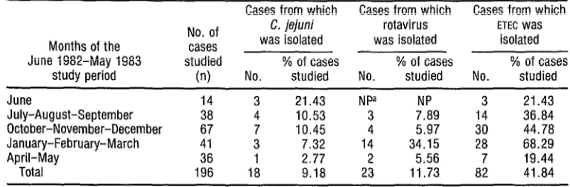
It was also noted that sea- sonal variations in the frequency of C. jejzln; infections differed markedly from
seasonal variations in the frequency of rotavirus infections (Table 5). Specifi-
cally, the highest frequencies of C. je- jzlni infection were found in the hot and
rainy months (June through October), whereas the highest frequencies of ro- tavirus infection were found in the rela- tively cool and dry months (November through March). ETEC was isolated dur- ing all parts of the study period except May and July.
TABLE 5. Seasonal variations in study children testing positive for C. jejuni, rotavirus, and ETEC. k
a Cases from which Cases from which Cases from which
Months of the June 1982-May 1983
study period
June 14 July-August-September 38 October-November-December 67 January-February-March 41 April-May 36 Total 196 No. of cases studied
(n)
C. jejuni rotavirus ETEC Was
was isolated was isolated isolated % of cases % of cases % of cases No. studied No. studied No. studied
3 21.43 NPa NP 4 10.53 3 7.89 3’
10.45
144
5.97 7.32 34.15 1 2.77 2 5.56 18 9.18 23 11.73
D
ISCUSSION
The present study was per- formed to investigate the presence of C. &+z~ in children under two years of age with acute diarrhea. This agent was identified in 9.2 % of the sick children studied, a percentage similar to that found in other Latin American countries (25, 26).
Earlier studies have described the presence of C. &$wz~ in the feces of healthy persons (10, 12-14). In this
study, however, C. &+n; was not iso- lated in any healthy child. This note- worthy finding is consistent with results obtained in industrialized countries, where isolation of CampyZoobacter has always been associated with disease (IO). (In developing countries, especially very poor countries like India and Bangla- desh, the rate of endemicity is high, reaching up to 35 % .)
As in other developing re- gions (11, 12), the pathogen was iso- lated most frequently from children un- der 12 months old. This pattern differed from that found in industrial- ized countries, where the risk of infec- tion is greater in older children (IO, 27).
Moreover, 72% of the infected study
children in Venezuela were under six months old, and two were neonates. One of these neonatal infections mani- fested itself in the maternity ward, as previously described elsewhere (28, 29);
however, it could not be established that transmission had taken place dur- ing delivery. Both infected neonates were afebrile, a circumstance that paral- lels previously reported findings (29).
The symptoms observed among patients with C. &+wz; diarrhea in this study agree essentially with those described by other authors (9, 10). It is
important to note that most of these pa- tients had stools with the dysenteric characteristics of blood or mucus (Table 3). This suggests an invasive pathogenic mechanism associated with production of a cytotoxin (30). However, studies in
Bangladesh (12) have found that a large
proportion of cases with C. ie$ni had watery diarrhea without mucus or blood, a symptomatology that differs from that described in developed coun- tries (10). This watery diarrhea could be
associated with a heat-labile toxin, simi- lar to the heat-labile toxin of Vibnb cho-
krae and that of ETEC described by R&z-Palacios (3 1).
It remains to be determined whether this variation in the reported symptoms of CamPyGobacter infections arises from differences in the host or in the agent strains involved. On the one hand, it is known that not all persons exposed to CampyZoobacter develop the disease (32); however, Klipstein (33) has demonstrated a correlation between dis- ease and toxin production in sympto- matic subjects that contrasts with an ab- sence of toxin in strains isolated from asymptomatic carriers. These observa- tions, and the epidemiologic differences noted between subjects in developed and developing countries, suggest that much remains to be learned about the pathophysiology of C. ie+nz’ and its pathogenic role in acute diarrhea.
ture could be a common factor here, for in general the months when the disease seems most prevalent are the warmest of the year.
Regarding other etiologic agents, ETEC was the one most fre- quently isolated (from 41.8 % of the study subjects). The frequency of ETEC (ST) was found to be similar to that de- scribed by other authors (26, 36). The frequency of ETEC isolates producing heat-labile toxins (LT) was higher than that generally reported elsewhere (26, 35), but the pathogenic significance of this ETEC was reduced by the fact that such strains were also found in 18.5 % of the asymptomatic children. In contrast, ETEC (ST) was found in only 3.7 % of the asymptomatic children. The high fre- quency of ETEC (ST) isolated from the hospitalized children and the low fre- quency detected in healthy children suggests that this agent could be more of a problem in acute diarrhea1 disease in our area than is ETEC (LT).
Rotavirus was the pathogen isolated with the second-greatest fre- quency from the children with diarrhea, This isolation frequency (14.1% ) is sim- ilar to that found in other communities (22, 34), and the seasonal distribution of these isolations resembles that found
by other authors (26, 34, 37). However, the observed 14.1% frequency is low compared to figures indicated by data 2 obtained from other studies in Venezu- Y ela, in which rotavirus was found among
*
52 30-40% of the patients (5, 17, 37). The ? difference could be explained by the or- .$.j igin of the samples studied here (most
u
a were obtained from outpatients), since 4 3
2
rotavirus tends to be more prevalent among hospitalized patients (see
3
Table 1).
The frequency with which K. pnuemoniae was isolated (from 11.2 % of the sick children but none of the 246
asymptomatic children) is noteworthy, since this microorganism has been asso- ciated with production of both heat- labile and heat-stable toxins (26).
G. Zaambdia was found in both sick and asymptomatic children, whereas E. histolytica was found only in sick children. The presence of G. Zamblia in 17.6% of the healthy chil- dren studied indicates that it is neces- sary to study more about this agent’s health implications and epidemiology, since a high prevalence of the parasite in healthy children could present a risk of transmission. In 29.6% of the cases no agent was identified. Part of this per- centage might be accounted for by “new” etiologic agents such as Crypto- spot-i&m, a parasite under study at our laboratory. (In a recent study, Crypto- spori&m oocysts were identified in 10.8% of 120 children under two years old who were suffering from acute diar- rhea-38).
A
CKNOWLEDGMENTS
We wish to extend very spe- cial thanks to Drs. Leonardo Mata and Myron Levine for providing the refer- ence strains of CampyZobacter jejmi used in this study, to Yordy Boher and Gidalia Urbina for their technical assis- tance, and to Yesenia Tedn de Balzano for her secretarial work.
S
UMMARY
The frequency of Campydo- batter jejuni and other enteric disease agents causing diarrheal illness was studied in 196 Venezuelan children un- der two years old who attended two Ca- racas hospitals from June 1982 through May 1983. A group of 27 asymptomatic (control) children was also included in the investigation.
This is the first reported study to examine the prevalence of C. ie&ni infection among this age group in Venezuela. In all, C. jejuni was isolated from 18 (9.2 O/o) of the ill study children but from none of the controls. Entero- toxigenic E. coZi (ETEC) was isolated from 82 (41.8 % ) and rotavirus was iso- lated from 23 (14.1%).
C. jejzmi was most prevalent among the study subjects O-6 months old. (Two of these infected infants were neonates, and one was only seven days old.) Blood and mucus in the stools were very common symptoms of Cam- pyzobacter disease. Also, most of the C. jejzcni-associated cases occurred from
June through October, a period charac- terized by heavy rainfall and warm tem- peratures in Caracas.
These findings suggest a noteworthy prevalence of disease associ- ated with C. jejmi, indicating a need for additional prospective studies to identify transmission mechanisms, ani- mal reservoirs, and asymptomatic infec- tions in poor communities where Cam- pylobacter-associated disease is very
common.
RE
FERENCES
Snyder, I. D., and M. H. Merson. The mag- nitude of the alobal oroblem of acute diar- rhoeal disease; A re%ew of active surveil- lance data. Bd WHO 60:605-613, 1982.
Mata, L. The ChiUren of Santa Ma&a CauquZ: A Prospective FieldStudy of Health
ana’ Growth. MIT Press, Cambridge, 1978.
Martorell, R., J. Habicht, C. Yarbrough, A. Lechtig, R. Klein, and K. Wester. Acute morbidity and physical growth in rural Gua- temalan children. Am] Dis Child 129: 12%
1301,1975.
Ministerio de Sanidad y Asistencia Social. Anuarib de Estadistica y EpidemioLogia. Ca- racas, 1984.
Perez-Schael, I. Estudio etioldgico de la gastroenteritis en Caracas. GEN 36:55- 56,1982.
Kapikian, A. Z., R. H. Yolken, H. B. Green- berg, R. G. Wyatt, A. R. Kalica, R. M. Chanock, and H. W. Kim. Gastroenter- itis Viruses. In: E. H. Lennette and N. J. Schmidt (eds.). Diagnostic Procedures for Vi- ral Riclkettsial, and Chkmydial Infections r’fifi& edition). American Public Health As- &iation, Washington, D.C., 1979, pp. 927-995.
plied to vaccine development. Microbial Rev 47:510-550. 1983.
8 Dekeyser, P., M. Gossuin-Detrain, J. P. Butzler, and J. Sternon. Acute enteritis due to related vibrio: First positive stool cultures. J Infect Dis 125:390-392, 1972.
9 Buttler, J. P., and M. B. Skirrow. Campylo- batter enteritis. Clin Gastroenterol 8:737- 765, 1979.
10 Blaser, M. J,, and L. B. Reller. Campylo- batter enteritis. N Engl J Med 305:1444- 1452, 1981.
11 Richardson, N. J., H. J. Koornhof, V. D. Bokkenheuser, 2. Mayet, and E. U. Rosen. Age-related susceptibility to CampyLobacter jejuni infection in a high-prevalence popula-
tion. Arch Dis Child 58:616-619, 1983. 12
13
14
15
16
17
Glass, R. I., B. J. Stoll, M. I. Huq, M. J. Struelensz M. Blaser, and A. K. M. G. Ki- bri
cl a. Eprdemiologic and clinical features of en emit CampyLobacter jejuni infection in Bangladesh. JZnfect Dis 148:292-296, 1983. Figueroa, G., and M. Araya. Infecciones sin- tomaticas y asintomaticas por Campylobacter jejuni, Rev Chil Pediatr 56~485-489, 1985.
Prasanna-Rajan, D., and V. I. Mathan. Prev- alence of CampyLobacter fetus subsp. lkjuni in healthy o ulations in Southern India. J C/in Miwot!&?15:749-751, 1982.
15:761-768, 1982.
Penner, J. L., and J. N. Hennessy. Passive he- magglutination technique for serotyping Cam
sis o P soluble heat-stable antigens. J Clin Mi- ylobacterfetus subsp. jejuni on the ba- crobiol12:732-738, 1980.
White, L., I. Perez, M. Perez, G. Urbina, H. Greenberg, A. Kapikian, and J. Flores. Rela- tive frequency of rotavirus subgroups 1 and 2 in Venezuelan children with gastroenteritis as assayed by monoclonal antibodies. J C/in Mi- crobiol19:516-520, 1984.
18 Lennette, E. H., A. Balows, W. J. Hausler, Jr., and J. P. Truant (eds.). Manualof Clini- cal Microbiology (thira’ edition). American ~ jociety of Microbiology, Washington, D.C.,
1980, pp. 195-219. 19
20
21
22
23
24
25
26
27
jereny, B. Experimental Shigeila keratocon- unctivitis. Acta Microbial Acad Sci Hung !:293-296, 1955.
Dean, A. G., Y. C. Ching, R. G. Williams, and L. B. Harden. Test for E. coli enterotoxin using infant mice: Application in stud of di- arrhea in children in Honolulu. J In ect Dis P 125:407-411, 1972.
Yolken, R. H., H. B. Greenberg, M. H. Merson. R. B. Sack. and A. 2. Kanikian. Enzyme-linked immunosorbent assay&for de- tection of Escherikhia cofi heat-labile en- terotoxin. J C/in Microbial 6~439-444, 1977. Gracey, M., V. Burke, and J, Robinson. Aeromonas-associated gastroenteritis. Luncet 2:1304-1306, 1982.
Groupe de travail scientifique de I'OMS. In-
fections intestinales dues B Cambdobacter. Yersinia, Sa/mone/Ca et Shigell;. dBull OMi
58:691-711, 1980.
Luechtefeld, N. W., L. L. Wang, M. J. Bla- ser, and L. B. Reller. Campylobacter fetus subsp. jejuni: Background and laboratory di- agnosis. Lab Mea’ 12:481-487, 1981. Mata, L., A. Simhon, R. Padillia, M. M. Gamboa, G. Vargas, F. Hernlndez, E. Mohs, and C. Lizano. Diarrhea associated with ro- taviruses, enterotoxigenic Escherichia co&
Campylobacter, and other a ents in Costa Rican children. Am J Me 2 Hyg 32:146- 153, 1983.
Guerrant, R. L., L. V. Kirchhoff, D. S. Shields, M. K. Nations, J. Leslie, M. A. De- Sousa, J. G. Araujo, L. L. Correira, K. T. Sauer, K. E. McClelland, F. L. Trowbridge, and J. M. Hughes. Prospective study of diar- rhea1 illness in Northeastern Brazil: Patterns of disease, nutritional impact, etiologies, and risk factors. J Infect Dis 148:986-997, 1983. Lassen, J., and G. Kap crud. Epidemiologi- cal aspects of enterms ’ ’ &e to Campylobacter :pp. m810rway. J C/in Microbial 19:153-
1 *
28 Anders, B. J., B. A. Lauer, and J, M. Paisley. Campylobacter gastroenteritis in neonates. Am J Dis Child 135:900-902, 1981. 29 Karmali, M. A., B. Norrish, H. Lior, B.
30
31
32
33
34
Camjy/obacter enterocolitis in a neonatal nursery. J Infect Dir 1491874-877, 1984. Yeen, W. D., S. D. Puthucheary, and T. Pang. Demonstration of cytotoxin from Campylobacter jejzzni. J Gin Pathol 36:1237-1240, 1983.
Ruiz-Palacios, G. M., J. Torres, N. I. Torres, E. Escamilla, B. Ruiz-Palacios, and J. Ta- mayo. Cholera-like enterotoxin produced by CampyZobacter jejtini: Characterization and clinical significance. &met 2:250-252, 1983. Walker, R. I., M. B. Caldwell, E. C. Lee! P. Guerry. T. J. Trust, and G. M. Ruin-Palacros. Patho
Micro 5 hysiology of Campylobacter enter&is. rd Rev 50:81-94, 1986. Klipstein, F. A., R. F. Engert, H. Short, and E. A. Schenk. Pathogenic properties of Cam- pylobacterjejwi: Assay and correlation with clinical manifestations. Infect Zmmun 50:43- 49, 1985.
Georges, M. C., I. K. Wachsmuth, D. M. V. Meunier, N. Nebout, F. Didier: M. R. Siopathis, and A. J. Georges. Parasmc, bacte- rial and viral enteric pathogens associated with diarrhea in the Central African Repub- lic. J C&z Micro&o/ 19:571-575, 1984.
35
36
37
38
Finch, M. J.. and L. W. Riley. Campy/obac- ter infections in the United States: Results of an II-state surveillance. Arch Intern Mea’ 144:1610-1612, 1984.
Black, R. E., K. H. Brown, S. Becker, A. R. M. Abdul Alim, and I. Huq. Longitu- dinal studies of infectious diseases and nhvsi- cal growth of children in rural Banglade~h/II. Incidence of diarrhea and association with known pathogens. Am J Epidemiol 115 : 3 15- 324, 1982.
Viera de Torres, B.. R. Mazzali de Ilja, and J. Es
3 arza. Epidemiological aspects of rotavirus m ection in hospitalized Venezuelan chil- dren with gastroenteritis. Am J Trap Mea’ Hyg 27~567-572, 1978. 2 Perez-Schael, I., Y. Boher, L. Mata, M.
Perez, and F. Venezuelan c I!
Tapia. Cryptosporidiosis in 3 ildren with acute diarrhea.
Am J Trap Med Hyg 34~721-722, 1985. $ 2 a r.r.l