Journal of Dental Science and Therapy
Systemic and Oral Aspects in Celiac Disease for Denistry
Patrícia Maria Magalhães Torres1, Felipe Franco Marçal2, Emerson Dias Ponte2, Renata Asfor Rocha Carvalho Marins2, Fábio Wildson Gurgel Costa3, Crisiane Sá Roriz Fonteles4 and Thyciana Rodrigues Ribeiro3
1 Undergraduate student, Department of Clinical Denistry, Federal University of Ceará
2 DDS, Master´s student, Department of Clinical Denistry, Federal University of Ceará 3 PhD, Adjunct Professor, Department of Clinical Denistry, Federal University of Ceará
4 PhD, Associate Professor, Department of Clinical Denistry, Federal University of Ceará
J. Dent. Sci. Ther 1(1). Page | 12
*Corresponding author: Dr. Thyciana Rodrigues Ribeiro, PhD, Adjunct Professor, Department of Clinical Denistry, Federal University of Ceará, Brazil; Tel/Fax: +55 85 33668232; E mail: [email protected]
Aricle Type: Research, Submission Date: 19 February 2016, Accepted Date:08 March 2016, Published Date:04 May 2016.
Citaion: Patrícia Maria Magalhães Torres, Felipe Franco Marçal, Emerson Dias Ponte, Renata Asfor Rocha Carvalho Marins, Fábio Wildson Gurgel Costa (2016) Systemic and Oral Aspects in Celiac Disease for Denistry. J. Dent. Sci. Ther 1(1): 12-17.
Copyright: © 2016 Thyciana Rodrigues Ribeiro, et al. This is an open-access aricle distributed under the terms of the Creaive
Commons Atribuion License, which permits unrestricted use, distribuion, and reproducion in any medium, provided the original
author and source are credited.
Vol: 1, Issue: 1
Abstract
Celiac disease (CD) is a permanent intolerance to gluten proteins contained in some cereals such as wheat, rye, barley and oats. It is an enteropathy mediated by the immune system and therefore also known as gluten-sensitive enteropathy. It is characterized by total or subtotal villous atrophy of the proximal small intestine, leading consequently to poor absorption of most nutrients. he disease manifests itself mainly in the irst two years of life, bung the small intestine the main afected organ, with clinical manifestations of diarrhea, vomiting and weight loss, but the diagnosis is oten diicult due to the large number of atypical cases of disease. he average incidence of CD is 1 case per 1,000 live births. In Brazil, oicial statistics are unknown. It is estimated that there are 300,000 Brazilians have the disease. Celiac disease is more frequent in women, with a ratio of 2:1, and predominantly afects the white individuals. he objective was to review the literature covering DC and its relation to dentistry. A literature search was conducted in the databases Pubmed, Bireme and Lilacs, being selected scientiic articles published between 1995 and 2015, in Portuguese and English, using the descriptors "Celiac Disease", "dentistry" "Oral Manifestations" and "enamel hypoplasia." In addition to the systemic changes, celiac disease can present oral and dental manifestations, the incidence of oral signs and symptoms in celiac patients shows the importance of the dentist to recognize that these changes may contribute as an aid in the diagnosis of enteropathy.
Keywords: Celiac disease, Dentistry, Oral Manifestations, Enam-el Hypoplasia.
Introducion
Celiac disease (CD) is a chronic intestinal problem caused by a permanent intolerance to gluten proteins contained in some cereals, for instance wheat, rye, barley and oats [1]. In the literature, it can be recognized also as gluten-sensitive enteropathy, non-tropical sprue, idiopathic steatorrhea and celiac sprue. Celiac disease is one of the most common food intolerances, being the
prevalence highly variable between countries, and the worldwide prevalence is estimated in about 1% [2]. he average incidence is 1 case per 1000 live births [3]. According to epidemiological data, the disease is more frequent in women, exhibiting an average ratio of 2:1, and mostly afects the white individuals [4]. his disease resulted from environmental, immunological and genetic factors. Enteric infections and reduced breastfeeding period combined with an excessive intake of processed food containing high gluten concentration can be cited as examples of environmental factors associated to CD [5,6]. he reaction triggered by the immune arsenal will generate a humoral (B cells) or cell (T cells) immune response against the gluten presence. As genetic factors, almost all individuals with celiac disease express human leukocyte antigen (HLA) - DQ2 or (HLA) - DQ8, located on chromosome 6p21. About 5% to 15% of the irst and second degree relatives are at risk of developing disease [3]. he most afected organ is the small intestine [7]. he manifestation occurs in most cases from the second semester of life, which is the period when usually the cereals are introduced in the diet [1]. In addition to the multiple systemic manifestations, this disorder can cause oral and dental injuries. his study aimed to review the literature covering celiac disease and its relation to dentistry based on articles published in the last 20 years.
Eiology
J. Dent. Sci. Ther 1(1). Page | 13 Clinical Features
his disease typically presents four patterns of clinical presenta-tion: classical or typical, non-classical or atypical, asymptomatic or silent and latent form. he classical pattern is diagnosed ear-lier and the most common manifestations are diarrhea, weight loss, anorexia, pallor by iron deiciency anemia, chronic fatigue, frequent vomiting, weakness, irritability, pain and bloating, muscle cramps, stunted growth and gluteal muscles atrophy [10]. he typical pattern is largely characterized by gastrointestinal symptoms that may be cited the triad: chronic diarrhea, bloat-ing and growth retardation [11]. In atypical pattern, digestive symptoms are not sharply expressed, and it is characterized by symptoms such as thyroid dysfunction, epilepsy, short stature, iron deiciency anemia refractory to iron replacement, infertil-ity, constipation and herpetiformis dermatitis [12]. Despite these various possible symptoms, Lahteenoja et al. [13] stated that the oral mucosal lesions or tooth enamel defects may be the only signs present in this pattern. In the silent pattern, patients are asymptomatic or mildly symptomatic, but they have positive se-rology, and exhibit changes in intestinal mucosa. In latent pat-tern, individuals are asymptomatic and they have positive serol-ogy, however, biopsy of the intestinal mucosa may or may not show changes [5]. Celiac crisis can be triggered between the irst and second year of life, which is characterized by severe diarrhea, bleeding, bloating and dehydration. In adolescents, episodes of intestinal infections, anemia and growth retardation can be seen. In females may ultimately result in delayed menarche or stop in the low ater the irst bleeding, while in males delay may occur in the production of spermatozoa [14]. he CD may be associ-ated with some syndromes. In Turner syndrome the prevalence is 4.1 to 8.1% [15]. For Williams syndrome is 8.2% [16], and it is about 12% in individuals with Down syndrome [17]. Celiac disease may be associated with other autoimmune conditions such as Type I Diabetes Mellitus, in which more than 8% of in-dividuals present this disease [18,19] also being associated with eosinophilic esophagitis [20].
Diagnosis
he diagnosis is complex and must be based on some pillars: clinical examination, detailed anamnesis, histopathological analysis of the small intestine and investigation of serum markers [1]. he screening of patients can be done by indirect immunoluorescence method through antibody research. Serological markers are: endomysial antibodies (EmA), anti-tissue transgutaminase antibodies (tTG) anti-gliadin antibodies (AGA) [3]. Intestinal biopsy remains necessary to know the degree of bowel involvement, being the histopathological examination of the small intestine from duodenal-jejunal junction region an indispensable criterion [21]. he conclusion of the diagnosis is made by clinical evaluations and intestinal biopsy by endoscopy [5]. According to Gonçalves [3], endoscopy with biopsy of the small intestine associated with positive serology for CD and clinical improvement due to a gluten- free diet allows the deinitive diagnosis. Genetic testing (HLA typing) can be important to trace the disorder proile within a family environment [22]. According Guerra et al, [6], salivary analysis represents a very important parameter for the diagnosis, since anti-EMA antibodies and anti-tTG have been found in saliva even before clinical signs of disease.
Treatment
Treatment is mainly dietary and done with the institution of gluten-free diet, with the withdrawal of wheat, rye and barley from diet. he exclusion of gluten in the diet leads to remission of symptoms and restoration of normal mucosal morphology. Always at any time of life when these cereals are eaten, it is likely that any tissue amendment will be processed again [14].
Prognosis
here is the possibility of occurrence of various complications when the disease is let untreated, such as stunting, fertility problems, anemia, osteoporosis, neurological disorders, liver disease, herpetiformis dermatitis, diabetes mellitus, selective IgA deiciency, and thyroid diseases. here is an increased risk of developing T cell non-Hodgkin lymphoma, pharyngeal and esophagus carcinoma and adenocarcinoma of the small intestine, being the periodically conducting of abdominal Doppler ultrasound in these patients important in the investigation of the possible complications [1]. Other manifestations include: late menarche, early menopause, episodes of spontaneous abortion, preterm birth and low birth weight [23].
he risk of cancer developing is reduced when there is adherence to a gluten-free diet. Patients, who present severe weight loss, altered bowel habits, as well as the presence of adenopathy, represent alert indication in relation to the possibility of a lymphoma [24].
Denistry consideraions
Celiac disease can present several oral manifestations, being the most cited the higher prevalence of defects in tooth enamel. It can also be noted episodes of recurrent aphthous stomatitis, delayed tooth eruption, decreased salivary low, angular cheilitis and atrophic glossitis [25]. Studies evaluating the presence of recurrent canker sores have been the target of most of the current clinical studies, which show that it is the oral amendment that leads along with the enamel defects, the highest prevalence in celiac subjects.
Between the main oral manifestations that may arise, the enamel hypoplasia is a frequent sign in silent pattern [1] with a prevalence of 1:718 to 1:14,000 [26], which may arise both in the primary and in the permanent dentition [27]. he injury is manifested as a defect in the enamel tissue due to an injury in the ameloblasts. Clinically, the defect is seen as a circle or track with irregularities in the enamel or sot cracks. Teeth can display roughness and sharp accumulation of bacteria plaque, which causes the deposition of extrinsic factors that contribute to a yellow-brown color and a greater susceptibility to dental caries [28]. Hypoplasia has been observed in celiac patients as enamel defects symmetric and chronologically distributed in the four dental hemi-arches. Aine [29] classiied the enamel defects as: grade I with defective enamel color; grade II presenting discrete structural defect with typical horizontal grooves, grade III presenting major structural defects with deep horizontal grooves and large vertical tanks and grade IV with severe structural defect in which the tooth shape can be adjusted [7].
J. Dent. Sci. Ther 1(1). Page | 14
order to prevent the development of caries, periodontal disease or even fracture, since the presence of the enamel surface roughness favors the accumulation of dental plaque. hus, it is important the adoption of health promotion measures by the patient, for instance: greater care in cleaning, use of less abrasive toothpastes and luoride solutions. Dentists may perform topical applications of luoride, and depending on the manifestations severity, rehabilitation with restorative materials of the afected teeth [30].
According to Rauen et al. [7], in celiac patients is noted a higher incidence of recurrent aphthous ulcers (RAU). he etiology may be linked to several factors, such as infections, stress, trauma, systemic diseases, hormonal disorders, hormonal deiciencies and food allergy [6]. Studies show that the majority of cases is related to nutritional deiciency, showing positive reaction for the replacement of folic acid, vitamin B12, or iron. he incidence is signiicantly higher in celiac individuals showing a prevalence of 9.66% to 40.98%, being the average prevalence in the general population indicated by 20% [31]. It is generally presented in the papular or erosive form, surrounded by an erythematous margin located in mucosa, lips, palate or tongue [1].
Protein-energy malnutrition caused by the CD, depending on the period in which this metabolic deicit is established, contributes to the onset of changes in the oral environment, such as delayed tooth eruption, decrease in the size of the teeth, problems in the formation of enamel and salivary gland dysfunction. Other oral manifestations may include: angular cheilitis and atrophic glossitis [7,1].
Methods
his study deals with a literature review of celiac disease and its relation to dentistry. he literature review provides a summarization of results of previous studies bringing general conclusions about the covered subjects. his review was conducted following the steps recommended by Whittemore&Knal [32] to gather and synthesize the knowledge on the subject.
Data collection was characterized by a search on Pubmed, Bireme and Lilacs databases, being selected scientiic articles published between 1995 and 2015, in Portuguese and English, using as descriptors: "Celiac Disease", "Dentistry", " Oral manifestations" and "enamel hypoplasia". he articles were selected, totaling 42 studies.
Results
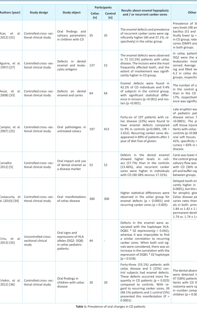
Eight studies selected for this review evaluated enamel defects related to celiac public, where seven of these were controlled and showed that the prevalence of this dental problem is higher in individuals with CD, ranging from 23 to 57.5% between these indices. Most articles notiied incisors and molars as the most frequently afected teeth. One study showed relation between this oral manifestation with the haplotype HLA-DQB1*02. Six articles had as object of study recurrent stomatitis aphthous. Five of these studies were controlled and showed higher levels of this manifestation in public with CD, ranging this prevalence from 37.1 to 48.7%. One study did not ind relation between recurrent canker sores and the haplotype HLA-DQB1*02, but there was correlation of this protein when both signals were used: defect in enamel and recurring canker sores. Other sot tissue lesions were also analyzed by two studies, where one of
these articles did not observe any diference in the prevalence of atrophic glossitis between groups; the other study observed higher prevalence of sot tissue injuries when considered groups (atrophic glossitis, geographic tongue and recurrent aphthous stomatitis, among others).
Dental caries was evaluated by ive studies. One study evaluated statistically and notiied similar rates of caries between celiac and control groups. Among the other four studies, two showed higher rates than the control and the last two lower prevalence. A single study assessed salivary low, pH and bufer efect of saliva, presenting only the salivary low as more prevalent in the celiac group; this last data is controversial to a study that observed higher rates of dry mouth in patients with CD.
Late eruption was the subject of two studies. Both reported higher prevalence in the celiac group than in the control, and the prevalence of the celiac group in these studies were 20 and 26%. Also higher levels were observed regarding to human leukocyte proteins DR7, DQ2, and DR3 as antigen commonly observed in celiac individuals. Moreover, minor salivary levels of streptococci and lactobacilli were observed in the populations of this study. More information about the results of the selected articles can be found in Table 1.
Discussion
he dental enamel hypoplasia is caused by the syndrome of malabsorption associated with micro-lesions in the small intestine that generate hypocalcemia during the enamel formation period [7]. Other factors are also associated with this abnormality, such as malnutrition, deiciency of vitamins A and D and other important nutrients for the tooth germ development [37]. One hypothesis suggests that the occurrence of disturbances in amelogenesis is related to autoimmune mechanisms, such as the action of speciic leukocyte antigens HLA-DQ8 and HLA-DR3 in enamel organ. Hypoplasia of this tissue is a common sign in silent pattern of the disease, especially in untreated children and adolescents [5].
In most cases, enamel defects are symmetric, being located in the four quadrants. he tooth structure might be afected in varying degrees, in which can be veriied the severity of the disease and the periods in which gluten was abolished from the diet. he most afected teeth are the irst molar and permanent incisors because the enamel formation occurs in the period of introduction of gluten in the diet. Deciduous canines and second molars are generally less afected, since its mineralization process begins at one stage when the disease may already be under control. he involvement of permanent canines, premolars and second molars indicates a delay in the diagnosis, since an early diagnosis can be performed to avoid involvement of these dental elements [25].
J. Dent. Sci. Ther 1(1). Page | 15
Authors (year) Study design Study object
Paricipants
Results about enamel hypoplasia
and / or recurrent canker sores Other results Celiac
(n)
Control (n)
Acar, et al.
(2012) [31] Controlled cross- sec-ional clinical study
Oral indings and salivary parameters
in children with CD
35 35
The enamel defects and prevalence
of recurrent canker sores were sig
-niicantly higher (40 and 37.1%, re-specively) in the celiac group.
Prevalence of Streptococcus sali-vary levels (48 and 14%) and Lacto-bacillus (51 and 34%) were stais-ically lower (p = 0.012, p = 0.010) in CD group; rates related to dental caries (DMFS and dfs) were similar in both groups.
Aguirre, et al.
(1997) [27] Controlled cross- sec-ional clinical study
Defects in dental enamel and leuko
-cytes anigens 137 52
The enamel defects were observed in 72 (52.5%) paients with celiac
disease. The incisors were the most
frequently afected teeth, and the extent of involvement was signii-cantly higher in CD group.
In celiac paients, DR7, DR3 and DQ2 were the anigen of human leukocytes most commonly ob-served. Average of decayed, miss-ing and illed teeth were 4.8 and 6.2 in celiac disease and control groups, respecively.
Avsar, et al.
(2008) [33] Controlled cross- sec-ional clinical study Defects on dental enamel and caries 64 64
Enamel defects were found in 42.2% of CD individuals and 9.4% of subjects in the control group, with signiicant staisical difer-ence in incisors (p <0.001) and mo-lars (p <0.001).
The number of caries-free paients
in the control group was higher
than in the CD group (38% and 17%, respecively) and the difer-ence was signiicant (p<0.001).
Campisi, et al.
(2007) [25] Controlled cross- sec-ional clinical study Oral pathologies in untreated celiacs 197 413
Forty-six of 197 paients with ce-liac disease (23%) were found to have enamel defects compared to 9% in controls (p<0.0001, OR = 2.652). Recurring canker sores dis-appeared in 89% of paients ater 1 year of diet free of gluten.
Late erupion was observed in 26% of pediatric paients with celiac disease versus 7% of controls (p <0.0001). The prevalence of oral sot issue injuries was 42% in pa-ients with celiac disease and 2% in controls (p <0.0001). Lesions of the oral sot issues have sensiivity = 42%, speciicity = 98% and test ac-curacy = 83% in diagnosis of celiac
disease.
Carvalho
(2012) [5] Controlled cross- sec-ional clinical study
Oral impact and use
of dental enamel as a disease marker
52 52
Defects in the dental enamel
showed higher levels in celi-acs (57.7%) than in the controls (13.46%), also recurrent canker sores were higher in individuals with CD (40.38% vesrsus 17.31%).
Carie was lower in CD group than in
the control group (2.11 versus 3.9), salivary low was higher in paients with CD (36% versus 12%), while pH and bufer capacity were similar between groups.
Costacurta, et
al. (2010) [34] Controlled cross- sec-ional clinical study Oral manifestaions of celiac disease 300 300
Higher staisical diferences were observed in the celiac group for enamel defects (p = 0.0001) and recurring canker sores (p = 0.005).
Delayed tooth erupion was signii-cantly higher in celiac group (p = 0.0001), but this was not signiicant for atrophic glossiis (p = 0.664). Celiac disease paients had higher caries rates than healthy individu-als in both: primary (DMFT 2.31 ± 1.84 vs 1.42 ± 1.13; p = 0.021) and permanent deniion (DMFT 2.97 ± 1.74 vs. 1.74 ± 1.64; p = 0.0001).
Erriu, et al. (2013) [35]
Uncontrolled cross- secional clinical study
Oral signs and expressions of HLA alleles (DQ2- DQ8)
in celiac pediatric
paients.
44
-Defects in the enamel were as
-sociated with the haplotype HLA-DQB1 * 02 expression(p = 0.042), whereas it was impossible to ind a similar correlaion to recurring canker sores. When both oral
sig-nals were considered, there was an
increase in the correlaion with the expression of DQB1 * 02 haplotype (p = 0.018).
Ertekin, et al.
(2012) [36] Controlled cross- sec-ional clinical study
Oral indings in
children with celiac disease
20 20
Forty-three (53.1%) paients with celiac disease and 5 (25%) con-trol subjects had enamel defects.
These defects occurred more fre
-quently in CD paients (p = 0.025)
compared to controls. With re -gard to recurring canker sores, 39
(48.1%) paients and 1 control (5%) presented this manifestaion (P = 0.0001).
The dental absence and xerostomia were detected in 11 (13.6%) and 47 (58%) paients, respecively. Pa-ients with CD that presented xe-rostomia were signiicantly greater in number compared with healthy children (p = 0.008).
J. Dent. Sci. Ther 1(1). Page | 16
It is suggested that the occurrence of idiopathic aphthous lesions, particularly in patients with defects in the formation of tooth enamel, is a relevant criterion for the investigation of CD [5]. It is not yet established if the aphthous ulcers are a direct manifestation of CD or if they occur due to indirect efects of malabsorption on the mucous basal cell, that are in process of division already susceptible to irritation by a pre-existing disease. Burning tongue, with papillary atrophy is related to deiciencies in vitamin B, folic acid and iron, which have their absorption afected by the efects of CD in the small intestine [1].
he speciic oral manifestations in children have been studied by comparing a group of children sufering from CD and a control group. he control group was composed in equivalent number of patients. he prevalence of enamel defects was 40% in the celiac group, and of these, 85.7% had defects grade I and II; and 14.3% had grade II defects. he control group did not exhibit this event [31]. In a study that examined the prevalence and distribution of enamel defects comparing CD patients to a control group, a prevalence of enamel defects was found in 52.5% in the celiac group and 42.3% in the control group. In a group of 137 patients with CD, 32 exhibited defect grade I, 16 patients with defect grade II, 3 patients with defect grade III, and 1 patient had defect grade IV [27].
In an investigation about the presence and distribution of enamel defects and caries prevalence, comparing children with CD and a control group, the prevalence of symmetrical enamel defects was signiicantly higher (42.2%) in CD group compared to those without disease (9.4%). his study also found an association between enamel defects and dental caries. he number of individuals free of caries in the control group was higher (38%) compared to the celiac group (17%) [33].
In relation to the frequency of oral lesions in CD compared to healthy individuals, a study found that from 197 patients with CD, 46 showed enamel defects (23%). he control group showed a prevalence of 9%. Regarding recurrent aphthous stomatitis, 19% of a sample of patients with CD had RAU, and 1% of the control group had this condition. Delayed tooth eruption was noted in 26% of celiac patients, and in 7% of the control group [25]. In a study where enamel defects were analyzed, there was a prevalence of 57.7% versus 13.46% in celiac patients and control group, respectively. Aphthous stomatitis was exhibited 40.38% versus 17.31% in individuals with and without CD, respectively. Caries prevalence was observed in 2.11% of celiac patients and in 3.9% of the control group. Decreased salivary low was observed in 36% of celiac patients versus 12% in group without this disease [5]. In an evaluation of the prevalence of oral manifestations in CD compared to a group of healthy subjects, performed in 300 patients, it was found that in celiac this prevalence was 33%, and in control group this value was 11% [34]. In a research with the aim to correlate the DC and the appearance of oral signs, it was found, in 44 pediatric patients with CD, a prevalence of 38.6% of defects in tooth enamel [35].
In a group of 81 patients with CD compared to a group with 20 healthy subjects, without signiicant diferences in age, gender and blood calcium levels, enamel defects were found in 53.1% of the irst group versus 25% of the control group. Of patients in the celiac group, 56 exhibited the classic pattern of the disease and
25 the atypical pattern. he permanent irst molar was the most afected tooth among carriers of the disease [36].
Regarding the most afected dentition between primary, mixed and permanent regarding to enamel defects, it was concluded that the mixed and permanent dentition showed higher prevalence (51%), having the primary dentition an average prevalence of 9.6%. hese indings conirm the hypothesis of the environmental component as inluential factor in the manifestations of CD, because the development of permanent teeth occurs early in life. Regarding the deciduous dentition, the hypothesis that the immunological and genetic components may be responsible for dental enamel defects can be reinforced [37].
In a study that aimed to evaluate the chronology of tooth eruption, comparing celiac patients to a group of healthy subjects, it was found a delay in tooth eruption in 56% of cases in a group of patients with CD individuals [39].
In research conducted in order to compare the hypoplastic enamel of patients with and without CD, hypoplastic enamel from celiac patients showed changes in the distribution of prisms and less interprismatic substance [40]. However, in a study of enamel chemical composition, comparing a group of celiac a group of individuals without the disease, it was detected a minor proportion of calcium/phosphorus in the dental enamel in the teeth group of patients with celiac disease [5].
Regarding the prevalence of dental caries, the results are conlicting. It can be related to greater fragility of the enamel due to the efects caused by CD [5]. But other study conirms that a lower incidence of this disease in celiac can be justiied by the more controlled diet of these individuals, resulting from a strictly free gluten food, protein present in many cariogenic foods such as breads, porridges and starches [33].
In relation to the salivary low changes and their efects in the oral cavity of patients with CD, some authors reported that celiac patients have a higher predisposition to xerostomia [41]. However, some authors have reported that CD does not afect the salivary low, but afects its composition, because the albumin values, IgA, IgM, amylase and myeloperoxidase were altered [42].
Conclusion
Celiac Disease is a chronic intestinal injury caused by permanent intolerance to gluten proteins contained in some cereals. he primary oral manifestations associated with this disease are the dental enamel defects. It is important that the dental professional is able to recognize changes in the oral cavity present in patients with clinical symptoms of celiac disease, since they can even contribute in the diagnosis of this enteropathy, especially in atypical pattern. A very detailed anamnesis is essential. he patient should be referred for medical evaluation immediately ater the veriication of these symptoms. he involvement of a multidisciplinary team is critical to guide the patient in order to provide a better quality of life.
References
1. Silva PC, Almeida PDV, Azevedo LR, Grécio AMT, Machado MAN, Lima AAS. Celiac disease: a review. ClinPesqOdontol. 2006; 2(5/6):401-406.
J. Dent. Sci. Ther 1(1). Page | 17
3. Gonçalves FJA. Estudo da Incidência de DoençaCelíacanaRegiãoAu-tónoma da Madeira. Portugal. Dissertação [MestradoemBioquími-caAplicada] - Universidade da Madeira; 2012.
4. Heap GA, Van Heel DA. Geneics and pathogenesis of celiac
disease. SeminImmunol. 2009; 21(6):346-354. doi: 10.1016/j. smim.2009.04.001.
5. Carvalho FK. DoençaCelíaca: Repercussõesbucais e estado do esmalte dental comomarcador da doença. RibeirãoPreto (SP). Tese [doutorado] - Universidde de São Paulo; 2012.
6. Guerra FA, Garbin Junior EA, Griza GL, Érnica NM, Marins ACM, Conci RA, et al. Manifestaçõesorais da doençacelíaca - Revista da Literatura. Rev. Odontologia. 2015; 15(2):117-149.
7. Rauen MS, Back JCV, Moreira EAM. Doençacelíaca: suarelação com a saúdebucal. Rev Nutr. 2005; 18 (2):271-276.
8. Roriz, J V. Manifestaçõesbucaisempacientes com doençasgastro-intesinaisinlamatórias. Brasília. Dissertação [MestradoemCiência da Saúde]-Universidade de Brasília; 2008.
9. Sollid LM. Molecular basis of celiac disease. Annual Rev. Immun.
2000;18:53-81.
10. Sdepanian VL, De Morais MB, Fagundes NETO U. Celiac disease: evoluion in knowledge since its original centennial descripion up to the presnt day. ArqGastroenterol. 1999; 36(4):244-257.
11. Teixeira NFG. DoençaCelíacaAtualizada. Covilhão. Dissertação [MestradoemMedicina] – Universidade da Beira Interior; 2012. 12. Modelli ICS. Prevalência da doençacelíacaemcriançasdesnutridas,
nafaixaetária de 12 a 36 meses. Brasília. Tese [doutorado] - Universidade de Brasília; 2009.
13. Lähteenoja H, Toivaner A, Viander M, Maki M, Irjala K, Raiha I, et al.
Oral mucosal changes in coeliac paients on a gluten-free diet. Eur J Oral Sci. 1998; 106(5):899-906.
14. Kotze LMS, Uiyama SRR, Nisihara RM, Mocelin V, Carvalho RFA, Zeni MPB, et al. Comparação dos anicorpos ani-reiculina e an-iendomísioclasse IgA paradiagnósico e controle da dietanadoen-çacelíaca. ArqGastroenterol. 1999; 36:177- 184.
15. Bonamico M, Pasquino AM, Mariani P, Danesi HM, Culasso F, Mazzani L, et al. Prevalence and clinical picture of celiac disease in Turner syndrome. J ClinEndocrinol Metab. 2002; 87(12):5495-5498.
16. Giannoi A, Tiberio G, Castro M, Virgilli F, Colistro F, Ferrei F, et al. Coeliac disease in Wiliams syndrome. J Med Genet. 2001; 38(11):767-768.
17. Zachor DA, Mroczek-Musulman E, Brow P. Prevalence of celiac disease in Down syndrome in the United States. J PediatrGastroenterolNutr. 2000; 31(3): 275-279.
18. Bapista ML, Koda YK, Mitsunori R, Nisihara, Iosshii SO. Prevalence of celiac disease in Brazilian children and adolescents with type 1 diabetes mellitus. J PediatrGastroenterolNutr. 2005 ; 41 (5):621-624. 19. Barera G, Bonfani R, Viscardi M, Bazzigaluppi E, Calori G, Meschi F, et
al. Occurrence of celiac disease ater onset of type 1 diabetes: a 6-year prospecive longitudinal study. Pediatrics. 2002; 109 (5):833-838.
20. Fey A, Kotze LM, Serapião M. Esofagiteeosinoílica: revisão de literature. ACM arqcatarin Med. 2012; 41(2).
21. Green PH, Jabri B. Celiac disease. Annu Rev Med. 2006; 57:207-221.
22. Megiorni F, Pizzui A. HLA-DQA1 and HLA-DQB1 in Celiac disease
predisposiion: Pracical implicaions of the HLA molecular typing. J Biomed Sci. 2012; 19:88. doi: 10.1186/1423-0127-19-88.
23. Scanlon SA, Murray JA. Update on celiac disease – eiology,
diferenial diagnosis, drug targets, and management advances. ClinExpGastroenterol. 2011; 4:297-311. doi: 10.2147/CEG.S8315.
24. Bucckley O, Brien JO, Ward E, Doody O, Govender P, Torreegiane
WC. The imaging of coeliac disease and its complicaions. Eur J Radiol. 2008; 65(3):483-490.
25. Campisi G, Di Liberto C, Iacono G, Compilato D, Di Prima L, Calvino F, et al. Oral pathology in untreated coelic disease. Aliment. Pharmac. Ther. 2007; 26(11-12):1529-1536.
26. Ferreira, RAMH. Possíveiscausas e tratamentosparapacientespor-tadores de amelogênese. Belo Horizonte. Monograia [Especializa-ção] - Universidade Federal de Minas Gerais; 2011.
27. Aguirre JM, Rodriguez R,Oribe D, Vitoria JC. Dental enamel defects in celiac paients. Oral Surg Oral Med Oral Path Oral Rad Endod. 1997; 84(6):646-650.
28. Moura MM. Soluçõesestéicasparaalterações da odontogênese. Belo Horizonte. Tese [doutorado] - Universidade Federal de Minas Gerais; 2012.
29. Aine L. Coeliac-type permanente-tooth enamel defects. Ann Med.
1996; 28(1):9-12.
30. Passos IA, Costa JDMC, Melo JM, Forte FDS, Sampaio FC. Defeitos do esmalte: eiologia, caracterísicasclínicas e diagnósicodiferencial. Rev InstCiêncSaúde. 2007; 25(2):187-192.
31. Acar S, Yetkiner AA, Ersin N, Oncag O, Aydogdu S, Arikan C. Oral
indings and salivary parameters in children with celiac disease: a preliminary study. Med PrincPract. 2012; 21(2):129–133. doi: 10.1159/000331794.
32. Whitemore R, Knal K. The integraive review: updated
methodology. J AdvNurs. 2005; 52(5):546–553.
33. Avsar A, Kalayci AG. The presence and distribuion of dental enamel defects and caries in children with celiac disease. Turk J Pediatr.
2008; 50(1):45-50.
34. Costacurta M, Maturo P, Bartolino M, Docimo R. Oral manifestaions
of coeliac disease: A clinical-staisic study. Oral Implantol(Rome). 2010;3(1):12-19.
35. Erriu M, Abbate GM,Pili FMG, Novara F, Orrù G, Montaldo C, et al. Oral Signs and HLA-DQB1*02 Haplotypes in the Celiac Paediatric Paient: A Preliminary Study. Autoim. Diseas. 2013; 2013.
36. Ertekin V, SümbüllüMA, Tosun MS, Selimoglu MA, Kara M, Kiliç N. Oral indings in children with celiac disease. Turk J Med Sci. 2012; 42(4):613-617.
37. Pastore L, Carroccio A, Compilato D, Panzarella V, Serpico R, Lo Muzio L. Oral manifestaions of celiac disease. J ClinGastroenterol. 2008; 42(3):224-232. doi: 10.1097/MCG.0b013e318074dd98.
38. Goud A, Deshpande S. Prosthodonic rehabilitaion of denino-genesis imperfect. ContempClin Dent. 2011; 2(2):138-141. doi: 10.4103/0976-237X.83072.
39. Costacurta M, Condò R, Sicuro L, Perugia C, Docimo R. Cervical
vertebral maturaion and dental age in coeliac paients. Oral Implantol (Rome). 2011; 4(3-4):11–17.
40. Bossu M, Bartoli A, Orsini G, Luppino E, Polimeni A. Enamel hypoplasia in coeliac children: a potenial clinical marker of early diagnosis. Eur J Paediatr Dent. 2007;8(1):31-37.
41. Painen P, Aine L, Collin P, Hieanen J, Korpela M, Enckell G, et al.
Oral indings in coeliac disease and Sjogren´s syndrome. Oral Dis. 2004; 10(6):330-334.
42. Lenander-Lumikari M, Ihalin R, Lahteenoja H. Changes in whole