Original Article
Artigo Original
Janaina Patricio de Lima1 Irineu Tadeu Velasco1 Denise Frediane Barbeiro1 Eliane Schochat1
Descritores
Audiometria de Resposta Evocada Audição Isquemia Encefálica Sepse Tronco Encefálico
Keywords
Audiometry, Evoked Response Hearing Brain ischemia Sepsis Brain Stem
Correspondence address:
Janaina Patricio de Lima
Rua José Benedito Salinas, 68, apartamento 152B, Jardim Itapeva, São Paulo (SP), Brasil, CEP: 04674-200.
E-mail: [email protected]
Received: 09/08/2014
Study carried out at the Laboratory of Clinical Emergencies, School of Medicine, Universidade de São Paulo – USP – São Paulo (SP), Brazil.
(1) Universidade de São Paulo – USP – São Paulo (SP), Brazil.
Conlict of interests: nothing to declare.
Auditory brainstem response in gerbils
submitted to ischemia and sepsis
Potencial evocado auditivo de tronco encefálico
em gerbils submetidos à isquemia e sepse
ABSTRACT
Introduction: An ischemic stroke is a clinical condition that affects thousands of people worldwide. As a result of this injury, neuronal death can be observed, and in the natural course of recovery, the individual may develop sepsis. Sepsis is a systemic inlammatory response that can lead the patient to death. To assess the clinical condition of a patient with this condition, the Auditory Brainstem Response (ABR) can be useful, since it is not an invasive procedure, it is a fast technique and it can be done at the bedside. Purpose: To assess auditory brainstem response (ABR) latency values in gerbils subjected to ischemia and sepsis. Methods: ABR values were collected from 72 adult male gerbils, which were divided into six groups: control, sepsis, ischemia, sham, ischemia with sepsis, and sham with sepsis. For the induction of sepsis, lipopolysaccharide (LPS) was applied intraperitoneally in gerbils. The animals were anesthetized with a ketamine/xylazine combination before collection; their ABR were collected before any procedure (base collection), after ischemia, and 24 hours after the application of LPS. The absolute latency of wave IV was evaluated, and the values were compared between groups. Results: There were signiicant differences in the groups submitted to sepsis in the latency value of wave IV in relation with the other groups. Conclusion: ABR was sensitive to sepsis with the increase in latency of wave IV during the development of the disease in the experimental model used.
RESUMO
INTRODUCTION
Clinical use of the auditory brainstem response (ABR) in assessing the integrity of auditory pathways and in the inves-tigation of the electrophysiological threshold is already rou-tine(1,2). However, using this potential as a prognostic index in various diseases is still object of study in the ield of research. Studies aimed at characterizing the ABR in these situations are of great value, because, for clinical use in situations where the patient lies in bed, as in the case of patients who have suffered a stroke or were victims of sepsis, well-deined parameters are needed. With this approach, experimental studies play an impor-tant role, as it is possible to reduce variables in the laboratory, and thus better understand the mechanisms and the results(3).
Ischemic stroke and sepsis are two diseases that affect a large number of patients. Ischemia can be classiied as either global or focal, that is, when there is total or partial lack of oxygen delivery; and as either complete or incomplete, when there is no reperfusion, that is, the return of the vascular blood low(4,5). The lack of oxygen in neurons leads to neuronal death. In addition, during ischemia and reperfusion, there is exces-sive neuron excitation, a phenomenon known as excitotoxicity, which is caused by excess glutamate release by oxygen-deprived neurons. This increase in glutamate release is responsible for the death of postsynaptic neurons, because this substance in excessive concentration is toxic to neurons(6,7).
Many patients are left with postischemic stroke sequelae. In addition, in the natural course of recovery, the individ-ual may develop sepsis(8), which is a systemic, exacerbated proinlammatory response that has a complex development, which, if not controlled, affects many organs and may even cause death(9).
These patients are often kept in intensive care units, and transporting them for laboratory tests is dificult and risky. In this perspective, the ABR becomes a valuable tool, as it is a nonin-vasive, fast test and can be performed at the bedside(10), which may indicate some level of prognosis for the patient’s condition.
The objective of this study was to investigate the ABR latency values of wave IV in adult gerbils in pre- and postisch-emia, pre- and postsepsis conditions, compared with a control group and a sham group.
METHODS
This study was approved by the Ethics Committee for Analysis of Research Projects of the Clinical Board of Hospital das Clínicas and of the School of Medicine of Universidade de São Paulo (Protocol No. 0456/09). The research was conducted at the Laboratory of Medical Research on Clinical Emergencies, Universidade de São Paulo.
A total of 72 male gerbils (Meriones unguiculatus), weigh-ing between 56 and 79 g, aged 3 to 5 months, kept in controlled environmental conditions with light/dark cycle (12/12 hours), temperature in the range of 22 to 27°C, humidity of 45 to 65%, and sanitized environment, receiving common gerbil food and water ad libitum, from the vivarium of the Department of Clinical Medicine, School of Medicine, Universidade de São Paulo.
The use of these rodents is justiied by the fact that they show a failure in the formation of the circle of Willis, which provides a single irrigation for 80% of the brain parenchyma. Thus, the surgical preparation of the experimental model is lim-ited to the temporary carotid occlusion, eliminating unneces-sary complications and producing far more consistent results. The animals were divided into six experimental groups, and each group consisted of two animals, namely:
1. sham (surgery without carotid occlusion); 2. ischemia (surgery with carotid occlusion); 3. sepsis;
4. sham with sepsis; 5. ischemia with sepsis; and
6. control (without any type of procedure).
For the ischemia procedure, the gerbils were anesthetized with a 3% halothane in a gas mixture of 30% oxygen and 70% nitrous oxide and subjected to global cerebral ischemia for 7 minutes with bilateral carotid occlusion. During the isch-emic period (7 minutes), followed by the reperfusion period (30 minutes), the animals were monitored and maintained at a controlled temperature of 36.0±0.2°C. Then, the animals were maintained on food and water ad libitum.
The ischemia and surgery procedure without occlusion of the carotid (sham group) was performed on the irst day of the experiment, after the irst ABR collection (base collection).
Regarding the groups with sepsis, during the second day of the experiment, that is, 24 hours after ischemia, they were injected with 1 mg/kg lipopolysaccharide (LPS) from Escherichia coli serotype 026: B6 (Sigma) intraperitoneally, thus inducing inlammation (sepsis) in the animal.
For the electrophysiological assessment, the equipment used was the Navigator model by Biologic. The stimuli were presented through 3A insert phones, positioned in the animals’ left ear, with their right ear not occluded. The stimulation hap-pened solely on the left ear.
Before all records were made, the animals were anes-thetized with a peritoneal ketamine injection (Parke-Davis, São Paulo, Brazil) at a concentration of 100 mg/kg of body weight and Rompun, whose active principle is xylazine (Bayer HealthCare, Sao Paulo, Brazil) at a concentration of 4 mg/kg of body weight. The ABR was recorded by the use of three electrodes positioned in Cz, left mastoid M1, according to the international 10-20 system(11), and a ground electrode. The left mastoid was used as reference, and the animal’s paw was used for the ground electrode. The animals remained in a passive listening situation. To capture the ABR, 2,000 click-type stimuli were used with thin polarity and presentation rate of 13 stimuli per second, intensity of 80 dBnHL, and insert earphones coupled to the animal’s auricullar pavillion. Filters used were 100 Hz pass/low and 1,500 Hz pass/high ilters. The recording window was of 10.66 ms after the stimulation. All responses were reproduced, thus ensuring the reproducibility of the acquired potential.
held on the irst day of the experiment). The ABR was col-lected again on the second day in all animals after the ischemic procedure performed in those who belonged to the applicable group (collection A); and a third sample was collected in all animals 24 hours after the LPS injection in animals belonging to the infected group (B collection). Only wave IV of ABR was analyzed, owing to a visualization problem with the other waves(12). The professional who examined the waves did not know to which group the animals belonged, neither at which step of the study they were.
The results were compared in pre- and postimplementation of the ischemic event and application of LPS.
For the statistical analysis, ANOVA and Tukey’s tests(13) were used. Values lower than 0.05 (p<0.05) were consid-ered signiicant.
RESULTS
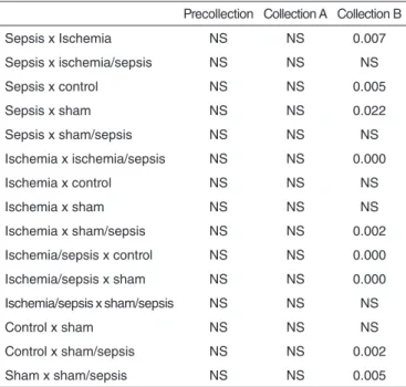
Table 1 shows the mean and standard deviation of the latency values of wave IV of ABR per group at all moments of the study. With the data, we can see a signiicant difference in the irst collection (pregathering) and inal collection (B) val-ues (Table 2) in all groups that were induced to sepsis (sepsis, sham with sepsis, and ischemia with sepsis). The opposite is observed in groups in which sepsis was not induced (control, ischemia, and sham).
DISCUSSION
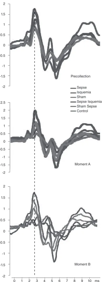
The data in Tables 1 and 2 and Figure 1 show the values of wave IV of ABR in the different groups studied and their signii-cant difference. One can see the difference between the induced sepsis groups (sepsis, ischemia/sepsis, and sepsis/sham) from those not induced to the inlammatory process (sham, ischemic, and control). On the other hand, in collection B, there was no difference between the ischemic and nonischemic groups at any point of the search. Thus, we have an increase in wave IV latency in the development of sepsis (collection B, 24 hours after the application of LPS), which is not observed in the other groups. This shows that the ABR was able to identify the ani-mal with sepsis but not the aniani-mal with ischemia.
Sepsis is a disease that can lead to poor brain function, owing to the increase of cytokines that can cause apoptosis and alterations in synaptic transmission. It is a process that involves local and systemic alterations and is not yet fully understood. It is known, however, that there is an excess inlammation of
the organism, and, through positive feedback and synergistic responses of various systems, the inlammation is potentiated without control. Among the main events observed with exces-sive production of pro- and anti-inlammatory cytokines, there is the activation of the adhesion molecules, increased arachidonic acid, oxygen-free radicals, nitric oxide, and platelet activating factor. This lack of control of these substance causes damage to vital organs, and on the central nervous system, alterations in cytokine levels in sepsis can impair brain function, resulting in a condition known as septic encephalopathy(14,15).
No studies were found involving ABR and sepsis, but only studies with other evoked potentials. In one study(16), the authors veriied the cortical (N20-N70) and subcortical (N13-N20) somatosensory potentials to assess the incidence of septic encephalopathy in patients with severe sepsis and septic shock. The conclusion of the group was that these potentials are sen-sitive to this type of evaluation, and their loss was associated with disease severity.
The groups subjected only to ischemia did not differ sig-niicantly from those without ischemia and without sepsis. The value of absolute latency of wave IV did not change between
Table 1. Descriptive analysis (mean and standard deviation) of latency (ms) of wave IV of the auditory brainstem response in moments of the precollection and collections A and B, according to the evaluated groups
Collection Control
(n=12)
Sepsis (n=12)
Ischemia (n=12)
Sham (n=12)
Ischemia/sepsis (n=12)
Sepsis/sham (n=12)
Precollection 3.03±0.18 3.03±0.20 2.99±0.12 2.99±0.23 3.03±0.18 3.01±0.19
A 3.00±0.13 3.08±0.19 3.04±0.16 2.97±0.15 3.15±0.33 3.07±0.14
B 2.98±0.23 3.99±1.03 3.02±0.21 3.10±0.30 4.42±0.93 4.52±0.61
Table 2. Statistical analysis comparing the latency of wave IV of the auditory brainstem response between the groups evaluated
Precollection Collection A Collection B
Sepsis x Ischemia NS NS 0.007
Sepsis x ischemia/sepsis NS NS NS
Sepsis x control NS NS 0.005
Sepsis x sham NS NS 0.022
Sepsis x sham/sepsis NS NS NS
Ischemia x ischemia/sepsis NS NS 0.000
Ischemia x control NS NS NS
Ischemia x sham NS NS NS
Ischemia x sham/sepsis NS NS 0.002
Ischemia/sepsis x control NS NS 0.000
Ischemia/sepsis x sham NS NS 0.000
Ischemia/sepsis x sham/sepsis NS NS NS
Control x sham NS NS NS
Control x sham/sepsis NS NS 0.002
Sham x sham/sepsis NS NS 0.005
the precollection and collection B. In this study, the animals underwent carotid occlusion on the i rst day of the experiment, and the ABR was collected 24 hours after the procedure. The analysis was also performed just for wave IV.
In a study made with cats subjected to ischemia by occlu-sion of the common carotid, the authors(17) found the pres-ence of wave I and abspres-ence of the other waves in the ABR. In this study, the ABR was evaluated immediately after the surgical procedure.
In a later study, the authors found the latencies of ABR waves in Wistar rats subjected to occlusion of the anterior cerebellar artery. They divided the study into two groups according to col-lection: ipsilateral to the occlusion (group A) and contralateral to the occlusion (collection B). The authors observed different types of waves in group A, from total absence of ABR waves to waves I, II, III and IV, with an increase in interpeak latencies I-IV and II-IV. In relation to group B (whose collection was contralateral to the occlusion), the authors observed the pres-ence of waves I, II, III and IV, with an increase in interpeak latencies I-IV and II-IV. All samples were taken within 60 min-utes after occlusion. The authors justii ed the response variabil-ity in group A because of possible variations in vessels in the anterior cerebellar artery. According to them, when there is a signii cant contralateral movement, there may be blood supply to the cochlea, which maintains the presence of ABR waves.
In our study, we believe that the ABR values held before and after ischemia can be justii ed, because the potential assesses the brainstem region. It is known that ischemic damage are related mainly to the cortical regions (CA1 area of the cortex)19). In addition, the ABR in this study was collected 24 hours after the ischemia procedure; thus, there may have been injuries during the lack of blood supply, as cited by Inui et al.(18), but the body itself may have recovered from event. In this study, the ABR was not sensitive to ischemia.
CONCLUSION
This study found a strong correlation between the increases in latency of wave IV of ABR in animals induced to sepsis. Thus, the evaluation of this potential can serve as a prognostic factor for identifying the damage to the brainstem in gerbils with sepsis. However, the same effect was not observed in animals subjected to ischemia, whose wave IV value did not change.
*JPL conducted the tests in gerbils and drafted the manuscript; ITV and ES corrected the manuscript; DFB was responsible for the process of inducing ischemia and sepsis in the animal studied.
REFERENCES
1. Casali RL, Santos MFC. Potencial evocado auditivo de tronco encefálico: padrão de respostas de lactentes termos e prematuros. Braz J Otorhinolaryngol. 2010;76(6):729-38.
2. Pedriali IVG, Kozlowski L. Potenciais evocados auditivos de tronco encefálico na detecção do neurinoma do acústico. Arquivos Int Otorrinolaringol. 2005;9(1):303-6.
Figure 1. Layout of the average of the potential of the auditory brainstem response in each study group in all the moments (precollection, A, and B)
2
1.5
1
0.5
0
-0.5
-1
-1.5
-2
2
1.5
1
0.5
0
-0.5
-1
-1.5
-2 2.5
2
1.5
1
0.5
0
-0.5
-1
-1.5
-2
Precollection
Sepse Isquemia Sham
Sepse Isquemia Sham Sepse Control
Moment A
Moment B
3. Seelig VC. Questões atuais relacionadas ao uso de modelos animais em pesquisa cientíica [monograia]. Porto Alegre: Universidade Federal do Rio Grande do Sul; 2007.
4. Lenzi GL, Frackowiak RS, Jones T. Cerebral oxygen metabolism and blood low in human cerebral ischemic infarction. J Cereb Blood Flow Metab. 1982;2(3):321-35.
5. Joaquim MAS, Patriota GC, Bianco AM. Isquemia encefálica, cascatas vasodilatadoras e alterações bioquímicas. Arq Bras Neurocir. 2010;29(2):58-63. 6. Zipfel GJ, Lee JM, Choi DW. Reducing calcium overload in the ischemic
brain. N Engl J Med. 1999;341(20):1543-4.
7. Stehno-Bittel L. Neuroplasticidade. In: Lundy-Ekman L. Neurociência: fundamentos para reabilitação. Rio de Janeiro: Elsevier; 2008. p. 61-70. 8. Basile-Filho A, Suen VMM, Martins MA, Coletto FA, Marson F.
Monitorização da resposta orgânica ao trauma e à sepse. Medicina (Ribeirão Preto). 2001;34:5-17.
9. Juncal VR, Britto Neto LA, Camelier AA, Messeder OHC, Farias AMC. Impacto clínico do diagnóstico de sepse à admissão em UTI de um hospital privado em Salvador, Bahia. J Bras Pneumol. 2011;37(1):85-92. 10. Jardim M, Person OC, Rapoport PB. Potencial evocado auditivo de
tronco encefálico como auxílio diagnóstico de morte encefálica. Pró-Fono R Atual Cient. 2008;20(2):123-8.
11. Jasper HH. The ten-twenty electrode system of the International Federation. Electroenceph Clin Neurophysiol. 1958;10:371-5.
12. Smith DI, Kraus N. Postnatal development of the auditory brainstem response (ABR) in the unanesthetized gerbil. Hear Res. 1987;27(2):157-64.
13. Field A. Descobrindo a estatística usando o SPSS. 2ª edição. São Paulo: Artmed; 2009.
14. Hotchkiss RS, Tinsley KW, Swanson PE, Karl IE. Endothelial cell apoptosis in sepsis. Crit Care Med. 2002;30(5 Suppl):S225-8.
15. Silva FP. Nomenclatura e Epidemiologia. In: Silva FP, Velasco IT. Sepse. Barueri: Manole; 2007. p. 12-8.
16. Zauner C, Gendo A, Kramer L, Funk GC, Bauer E, Schenk P, et al. Impaired subcortical and cortical sensory evoked potential pathways in septic patients. Crit Care Med. 2002;30(5):1136-9.
17. Sohmer H, Gafni M, Havatselet G. Persistence of auditory nerve response and absence of brain-stem response in severe cerebral ischaemia. Electroencephalogr Clin Neurophysiol. 1984;58(1):65-72.
18. Inui H, Murai T, Matsunaga T. Auditory brainstem response indings in brainstem ischemia following selective occlusion of the anterior inferior cerebellar artery in the rat. Eur Arch Otorhinolaryngol. 1995;252(3):181-5.