* Corresponding author.
E-mail: [email protected] (A. Zille).
0102-695X/$ - see front matter © 2014 Sociedade Brasileira de Farmacognosia. Published by Elsevier Editora Ltda. All rights reserved. http://dx.doi.org/10.1016/j.bjp.2014.11.007
Original article
Properties and controlled release of chitosan microencapsulated
limonene oil
Jefferson M. Souza
a, Artemísia L. Caldas
a, Shafagh D. Tohidi
b, Javier Molina
c,
António P. Souto
b, Raul Fangueiro
b, Andrea Zille
b,*
aDepartamento de Moda, Design e Estilismo, Universidade Federal do Piauí, Campus Ministro Petronio Portela, Teresina, PI, Brazil bCentro de Ciência e Tecnologia Têxtil, Universidade do Minho, Campus de Azurém, Guimarães, Portugal
cDepartamento de Ingeniería Textil y Papelera, EPS de Alcoy, Universitat Politècnica de València, Alcoy, Spain
A RT I C L E I N F O
Article history:
Received 26 September 2014 Accepted 2 November 2014
Keywords: Cellulose Chitosan
Controlled release Essential oil Microcapsule Limonene
A B S T R A C T
Chitosan microcapsules containing limonene essential oil as active ingredient were pre-pared by coacervation using three different concentrations of NaOH (0.50, 1.00, 1.45 wt%) and fixed concentrations of chitosan and surfactant of 0.50 wt%. The produced microcap-sules were fully characterized in their morphology and chemical composition, and the ki-netic release analysis of the active ingredient was evaluated after deposition in a non-wo-ven cellulose fabric. The concentration of 1.00 and 1.45 wt% clearly show the best results in terms of dimension and shape of the microcapsules as well as in the volatility results. However, at the concentration of 1 wt% a higher number of microcapsules were produced as confirmed by FTIR and EDS analysis. Free microcapsules are spherical in size with disperse diameters between 2 and 12 μm. Immobilized microcapsules showed sizes from 4 to 7 μm, a rough surface and loss of spherical shape with pore formation in the chitosan walls. SEM analysis confirms that at higher NaOH concentrations, the larger the size of the microcap-sules. This technique shows that by tuning NaOH concentration it is possible to efficiently control the release rate of encapsulated active agents demonstrating great potential as in-sect repellent for textiles.
© 2014 Sociedade Brasileira de Farmacognosia. Published by Elsevier Editora Ltda. All rights reserved.
Introduction
The microencapsulation of substances has as principle the preparation of an emulsion, which involves the compound to be encapsulated (a solid, liquid or gaseous product) in order to protect it and to preserve its potential (Mahdavi et al., 2014). The encapsulation process also involves merger, absorption or dispersion of the combinations of solid, liquid or gaseous encapsulated bioactive
improve the efficiency of encapsulated materials or to create new applications, including functions in textile products, thus allowing them repellent, odorous, moisturizing or antimicrobial properties, among others. They can be used in a wide range of clothing, such as pants, socks, underwear and gloves (Nelson, 2002; Nazzaro et al., 2012). Flavours have a large range of applications in the industry. However, some of them are very sensitive to environmental or industrial process conditions. The flavour loss may reach values of 90% in free form due to their extreme volatility and reactivity with other components. The release kinetics of the active elements within the microcapsules depends directly on the processes and formulation parameters. They are specifically designed to release components when subjected to certain parameters. The active ingredient can be released through two methods: forced and controlled release. The forced release is obtained by rupturing the microcapsule membrane under thermal and/or mechanical conditions, such as friction. The controlled release is based on the diffusion of the encapsulated active element through the membrane or its degradation (Jamekhorshid et al., 2014). With respect to the materials used in the production of microcapsules, polysaccharides, such as alginate, starch and cellulose; and proteins as collagen and gelatin are widely used due to their ability to bind to flavour compounds, plus their biodegradability and low cost. They are used for the production of those materials in the food and pharmaceutical area (Can Karaca et al., 2013; Soliman, 2013). Researchers have been recently exploring the use of chitosan as an encapsulating agent (Peng et al., 2010a; 2010b; Estevinho et al., 2013a; Nuisin et al., 2013). Chitosan, the N-deacetylated derivative of chitin, is a cationic polyelectrolyte due to the presence of amino groups, one of the few occurring in nature. This gives chitosan singular chemical and biological characteristics, such as: biocompatibility, antibacterial properties, heavy metal ion chelation ability, gel-forming properties and hydrophilicity (Santos et al., 2013). Due to its chemical configuration and to features like abundance, low toxicity, hydrophobicity, biodegradability, biocompatibility and antimicrobial activity, chitosan is employed for the preparation of films, gels, microspheres and microcapsules. It has been used in various areas such as biotechnology, cosmetics, food and pharmaceuticals, as a way to release active compounds, among others (Kong et al., 2010; Cruz-Romero et al., 2013). The use of chitosan in protein and drug delivery systems is being actively researched and reported in the literature (Chen et al., 2013). Chitosan has one important advantage over other encapsulating agents, which is the possibility to establish covalent or ionic bonds with the crosslinking agents, building a network of sorts, in which the active substance is retained. In consequence, these chemical bonds carry advantages in terms of controlled release (Estevinho et al., 2013b).
There is a variety of techniques used for encapsulating drugs, foods and cosmetics, such as: spray-drying, complex coacervation, atomization and liposomes (Gharsallaoui et al., 2007; Gou et al., 2013). When choosing the material to be used for this process, a number of factors must be taken into consideration, such as: physical and chemical properties of the core, porosity and solubility of the wall, viscosity, mechanical properties, film-forming ability and vitreous transition, compatibility of the core with the wall, and the wall material used must be insoluble and non-reactive with the core (Gouin, 2004).
The encapsulation of essential oils allows optimization of its functionality, a factor that enables a more prolonged action of its active principle, since essential oils are characterized by their high volatility. Essential oils can be extracted from different parts of the plants, such as: roots, leaves, rose petals, stems and fruits, as well as condiments. They have properties ranging from antimicrobial, healing, and odorants, among others (Silva et al., 2010). The essential oils with antimicrobial activity by its anti-quorum-sensing activity become an important mechanism for the reduction of virulence and pathogenicity of bacteria. Among the essential oils with antibacterial activity against the bacteria E. coli and S. aureus, the lemon essential oil (Citrus limon L.), with limonene, β-pinene, γ-terpinene and citral (neral and geranial) as main compounds showed interesting antimicrobial features (Kim and Morr, 1996; Rodrigues et al., 2008; Donsì et al., 2011).
In this study, chitosan microcapsules containing limonene essential oil (EO) as active ingredient were prepared by coacervation process by NaOH dripping technique. The produced microcapsules were fully characterized in their morphology and chemical composition by Attenuated Total Reflectance-Fourier Transform Infrared Spectroscopy (ATR-FTIR), Energy Dispersive X-ray Spectroscopy (EDS), optical and scanning electron microscopy (SEM). Finally, the kinetic release analysis of the active ingredient was evaluated after deposition in a cellulose non-woven fabric for its potential as insect repellent.
Materials and methods
Materials
Chitosan (ChitoClear hq95-43000) was purchased from Primex (Iceland). The non-ionic surfactant was Lutensol ON 30 (BASF). All the other materials were purchased from Sigma-Aldrich and used without further purification.
Preparation of microcapsules
The process for the manufacturing of the microcapsules was as follows: 2 ml of limonene essential oil was added, in 20 ml of 0.5 wt% chitosan solution along with 0.5 wt% of surfactant. The solution was stirred at 700 rpm for 10 min. Then, the emulsion containing the essential oil was dripped into three different 100 ml solutions containing 0.5, 1 and 1.45 wt% of NaOH (0.12, 0.25, 0.36 M) stirred at 100 rpm. The solution was left on slow agitation for a period of 30 min after dripping. After the resting period, 1 ml of each microcapsule sample was removed for observation under an optical microscope. The solution was then filtered, washed three times with distilled water and dried at 30°C for a period of 15 h, in order to evaporate any remaining water from its surface.
Optical microscopy
Scanning Electron Microscopy (SEM) and Energy Dispersive X-ray Spectroscopy (EDS)
Morphological analyses of microcapsules were carried out with an Ultra-high resolution Field Emission Gun Scanning Electron Microscopy (FEG-SEM), NOVA 200 Nano SEM, FEI Company. Secondary electron images were performed with an acceleration voltage at 5 kV. Backscattering Electron Images were realized with an acceleration voltage of 15 kV. Samples were covered with a film of Au-Pd (80-20 weight %) in a high-resolution sputter coater, 208HR Cressington Company, coupled to a MTM-20 Cressington High Resolution Thickness Controller. The atomic composition of the microcapsules were examined with the energy dispersive spectroscopy (EDS) capability of the SEM equipment using an EDAX Si(Li) detector and an acceleration voltage of 5 kV.
Impregnation of microcapsules onto cellulose
Commercial 100% bleached cellulose non-woven fabric was used in this study. The samples were pre-washed with a solution of 1% non-ionic detergent at 30ºC for 30 min and then rinsed with water for another 15 min. Microcapsules were applied to non-woven cellulose fabric at a laboratory scale, reproducing the industrial application conditions. The fabric was padded at a speed of 2.5 m min-1 with the pressure of 4 bars to remove excess solution. After padding, the fabric was air-dried and then rinsed with deionized water and air-dried again.
Attenuated Total Reflectance-Fourier Transform Infrared Spectroscopy (ATR-FTIR)
The chemical structure of microcapsules deposited on a cotton fabric was characterized using an Attenuated Total Reflectance Fourier Transform (ATR-FTIR) spectrophotometer (Perkin Elmer, Spectrum 100). Each spectrum was acquired in transmittance mode on a ZnSe ATR crystal cell by accumulation of 256 scans with a resolution of 4 cm-1 and a wave number range of 4000-600 cm-1.
Release kinetics
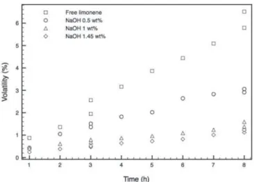
With the aim of implementation on textiles as an insect repellent, 2 ml of free essential oil and samples of microcapsules produced with 0.5, 1 and 1.45 wt% of NaOH were weighed and placed in an oven. The oven was calibrated at 35°C, the estimated temperature of the human body surface, to measure the rate of volatility. The results related to weight loss were collected every 60 min for 8 h. The data was then processed in Matlab software, and the percentage of weight loss was estimated.
Results and discussion
Various researchers have proposed different models for the release of active agents from chitosan microcapsules. The selection of the most appropriate method to determine the
active ingredient behavior depends on many parameters such as particle size requirement, molecular weight of chitosan, thermal and chemical stability of the active substance, type and concentration of cross-linker, reproducibility of the release kinetic and several other variables like stirring speed, additives etc. One of the most employed techniques is the emulsion cross-linking. In this method, a water-in-oil emulsion is prepared by emulsifying a chitosan aqueous solution in the oil phase. However, the system variables are difficult to control since the size of the microcapsules depends on the extent of cross-linking agent and by stirring speed (Agnihotri et al., 2004). Another versatile method is the coacervation/precipitation. This method is based on the chitosan property to precipitate in contact with high pH solutions since it is insoluble in an alkaline medium. Microparticles are produced by blowing a chitosan solution into an alkali medium like sodium hydroxide, sodium hydroxide (NaOH) or ethanediamine, to form coacervate drops. The advantage of this method is its intrinsic simplicity with the advantage to control the diameter, porosity and strength of the droplets only varying the concentration and ratio of the chitosan and alkali solutions (Ravi Kumar, 2000). However, observing the available literature regarding the preparation of chitosan microcapsules by the coarcervation method using NaOH, it can be noted that the alkali concentration is rarely studied as a variable of the system (Table 1). Moreover, despite the effect of chitosan and acetic acid concentrations are widely studied, the obtained microcapsule diameters are often excessive in average sometimes reaching the size of a millimeter. In this work, we demonstrate that simply by varying the NaOH concentration it is possible to obtain small particle diameters and tune the size of the microcapsules and the release kinetics. Moreover, these results were achieved using the lowest concentrations of surfactant and chitosan (according to the literature), which will contribute to save steps, reagents, and to reduce the cost of the process.
applying too much NaOH can cause excessively high viscosity of the entire emulsification system and produce microcapsules in a bulky group formation (Hsieh et al., 2006). However, this is not the case.
The images obtained by SEM were used to observe the external morphology and the average size of the microcapsules after deposition of the microcapsules in the cellulose fabric (Fig. 2). The mean size of the microcapsules based on volume distribution is 10 μm. However, it is clear that increasing the amount of NaOH, the microcapsule diameter also increases ranging between 2 and 12 μm. The microcapsules showed a rough surface and loss of the spherical shape. For the three samples, we can see that they have shrunk due to the loss of encapsulated EO. It also shows pore formation in the chitosan walls, allowing the controlled release of the encapsulated EO. The microcapsules are distributed individually without excessive agglomeration, and no visible oil residues on the surface of the microcapsules can be seen.
To confirm the microcapsule structure, an EDS analysis was performed. The elemental composition of the microcapsule is
shown in Table 2. Chitosan contains nitrogen in its amine, and acetylamine groups on its backbone. The nitrogen amount was an average 0.9 At%, in close agreement with the theoretical nitrogen composition, indicating that chitosan was present in the microcapsule walls (Santos et al., 2013). An interesting fact is also the low amount of P and Ca in the microcapsules A (0.5 wt% of NaOH). This could be attributed to the thicker wall of the microcapsules incorporating fewer ions during the spherification process.
After deposition of the microcapsules in the cellulose fabric, the ATR-FTIR spectra (Fig. 3) of the chitosan microcapsules incorporating limonene showed the dominant absorption peaks at 3360, 2920, 1430 and 1010 cm-1 respectively attributed to the v (O-H), vs (CH2), δ (CH-O-H) and
v (C-O) of pure cellulose (Chung et al., 2004). The intensities of methylene peaks at 2920 and 2850 cm−1 are attributed to asymmetric and symmetric CH2 stretch, and in the case of pure cellulose indicating the amount of waxes remaining on the fabric, but in the case of the treated fabric it can be related to the amount of chitosan microcapsules. The peak increase Figure 1 – Microphotographs of chitosan microcapsules (40 × 65). The microcapsules were prepared with chitosan concentration of 0.5 wt% and 0.5 wt% (A), 1.0 wt% (B) and 1.45 wt% (C) NaOH concentrations at 700 rpm stirring rate.
Microcapsule type
Chitosan (% w/v)
NaOH (M)
Active ingredient
Average Diameter (μm)
Encapsulation
efficiency (%) Reference
Chitosan 0.5 0.25 Fish oil 1-14 83 (Klaypradit and Huang, 2008)
Chitosan 0.5 0.5 Citronella oil 225 98 (Hsieh et al., 2006)
Chitosan 2 1 - 224 - (Liu et al., 2011)
Chitosan 1 0.25 Celecoxib 2-15 68 (Cheng et al., 2010)
Chitosan/Lecithin 2 2 Rosemary oil - - (Magdassi et al., 1997)
Chitosan 1 0.25 Miconazole 2-4 96 (Yuen et al., 2012)
Chitosan/Alginate 0.5 1 Guaifenesin 500 - (Lee et al., 1997)
Chitosan/Alginate 1 0.1 Shark liver oil 1-2 87 (Peniche et al., 2004)
Chitosan/Casein 0.5 0.5 Diltiazem 890 42 (Bayomi et al., 1998)
Chitosan/Silk fibroin 0.8 5 n-Eicosane 23 64 (Deveci and Basal, 2009)
Chitosan/PEG 1.3 1 Isonazid - 93 (Gupta and Ravi Kumar, 2001)
Chitosan/Gelatin 2 0.1 Limonella oil - 60 (Maji and Hussain, 2008)
Chitosan/PAA 4 2 Aspirin 1000 94 (Nascimento, 2001)
Table 1
Figure 3 – ATR-FTIR spectra of pure cellulose fabric, and fab-rics with chitosan microcapsule synthetized with different percentages of NaOH (0.5, 1 and 1.45 wt%).
Figure 2 – SEM Microphotographs (×20000) of Chitosan microcapsules synthetized with different percentage (w/w) of chitosan: 0.5% (A); 1% (B) and 1,45% (C).
Element 0.5 wt% (At%) 1 wt% (At%) 1.45 wt% (At%)
C 77.71 73.03 75.25
O 12.86 16.53 12.97
N 0.89 1.09 0.91
P 0.64 1.19 1.21
Pd 1.06 1.64 1.59
Ca 1.31 2.92 2.75
Au 5.52 3.59 3.71
Cl 0 0 0.75
K 0 0 0.87
Table 2
Variation of atomic percentages (At%) of C, O, N, P and Ca atoms in the microcapsules.
between 990-1100 cm-1 after microcapsule deposition may be attributed to the C-O stretching of free and condensed C-OH groups (Goodarzi et al., 2013). The significant intensification of the characteristic IR peaks for -CH3 (1360 cm-1) and the appearance of the peak for CH in-plane bend (1430 cm-1) suggests the formation of hydrocarbon fragments on the fabric surface due to the presence of limonene (Zheng et al., 2010). Attributed to the adsorbed water molecules in pure cellulose, the intensification of the band at 1640 cm-1 in the microcapsule treated fabrics can be related to the carbonyl stretching of the secondary amide band (amide I) of the pure chitosan (Yan et al., 2013). The band around 1580 cm-1 can be assigned to C-N stretching vibration and refers to the amide group because of the NH2 bending vibration (Monllor et al., 2007). This last band is therefore not observed in the control cellulose fabric spectra and it can be clearly assigned to the chitosan microcapsules. Moreover, the spectra of the chitosan microcapsules, especially those obtained with 1% of NaOH, exhibited an intense peak at 880 cm-1, characteristic of the saccharide structure of chitosan (Wan et al., 2006).
Conclusions
The microcapsules produced at the concentration of 1.45 wt% of NaOH clearly show the best results in terms of dimension and shape of the microcapsules as well as in the volatility. However, the microcapsules produced at the concentration of 1 wt% of NaOH are produced in higher numbers and showed very similar results in volatility. The dispersion can be considered satisfactory for all the samples, which indicates that the used surfactant and chitosan concentrations (0.5 wt%) were optimal. The optical microscope analysis showed microcapsules with spherical size with dispersion diameters between 2 and 12 μm. The SEM analysis of the immobilized microcapsules showed rough surface and loss of spherical shape, with pore formation in the chitosan walls. However, these differences on surface morphology of the microcapsules wall are favourable to the slow release of limonene on the non-woven fabric, thus increasing the durability of the fragrance. The average diameter of immobilized chitosan microcapsules was enlarged from 4 to 7 μm when the amount of the NaOH concentration increased from 0.5 to 1.45 wt%. FTIT-ATR and EDS analyses confirmed the presence of a high concentration of chitosan microcapsules on the non-woven surface. Moreover, it was confirmed that the microcapsules synthetized from 1 wt% of NaOH were the highest in number. The results indicate that the chitosan microcapsules synthetized from 1 wt% of NaOH solution obtained the best results in terms of number of microcapsules and volatility, achieving a more prolonged effect on the non-woven fabric. This technique shows great potential as a functionalization method for the controlled release of insect repellent on textiles.
Authors’ contributions
JMS and ALC for running the laboratory work analyzed and drafted the data, and writing of the manuscript. JM, APS and
RF supervised the laboratory work and contributed to critical reading of the manuscript. AZ designed the study, analyzed and drafted the data, contributed to the analysis of the spectroscopic data, supervised the laboratory work, contributed to critical reading of the manuscript and revised the final manuscript. All the authors have read the final manuscript and approved the submission.
Conflicts of interest
The authors declare no conflicts of interest.
Acknowledgments
JMS and ALC acknowledge CAPES Foundation, the Ministry of Education of Brazil, Proc. nº 8976/13-9 e Proc. Nº 1071/13-0, respectively, and the Department of Textile Engineering of the University of Minho, Portugal. J. Molina is grateful to the Conselleria d’Educació, Formació i Ocupació (Generalitat Valenciana) for the Programa VALi+D Postdoctoral Fellowship. AZ (C2011- UMINHO-2C2T-01) acknowledges funding from Programa Compromisso para a Ciência 2008, Portugal. Shafagh Dinparast Tohidi would like to thank the Portuguese Foundation of Science and Technology for providing the PhD grant SFRH/ BD/94759/2013.
R E F E R E N C E S
Agnihotri, S.A., Mallikarjuna, N.N., Aminabhavi, T.M., 2004. Recent advances on chitosan-based micro- and nanoparticles in drug delivery. J. Controll. Release 100, 5-28.
Bayomi, M.A., Al-Suwayeh, S.A., El-Helw, A.M., Mesnad, A.F., 1998. Preparation of casein-chitosan microspheres containing diltiazem hydrochloride by an aqueous coacervation technique. Pharm. Acta Helv. 73, 187-192.
Can Karaca, A., Low, N., Nickerson, M., 2013. Encapsulation of flaxseed oil using a benchtop spray dryer for legume protein-maltodextrin microcapsule preparation. J. Agr. Food Chem. 61, 5148-5155.
Chen, M.-C., Mi, F.-L., Liao, Z.-X., Hsiao, C.-W., Sonaje, K., Chung, M.-F., Hsu, L.-W., Sung, H.-W., 2013. Recent advances in chitosan-based nanoparticles for oral delivery of macromolecules. Adv. Drug Deliver. Rev. 65, 865-879.
Cheng, S.-Y., Yuen, M.C.-W., Lam, P.-L., Gambari, R., Wong, R.S.-M., Cheng, G.Y.-M., Lai, P.B.-S., Tong, S.-W., Chan, K.-W., Lau, F.-Y., Kok, S.H.-L., Lam, K.-H., Chui, C.-H., 2010. Synthesis, characterization and preliminary analysis of in vivo biological activity of chitosan/celecoxib microcapsules. Bioorg. Med. Chem. Lett. 20, 4147-4151.
Chung, C., Lee, M., Choe, E.K., 2004. Characterization of cotton fabric scouring by FT-IR ATR spectroscopy. Carbohyd. Polym. 58, 417-420.
Cruz-Romero, M.C., Murphy, T., Morris, M., Cummins, E., Kerry, J.P., 2013. Antimicrobial activity of chitosan, organic acids and nano-sized solubilisates for potential use in smart antimicrobially-active packaging for potential food applications. Food Control 34, 393-397.
Deveci, S.S., Basal, G., 2009. Preparation of PCM microcapsules by complex coacervation of silk fibroin and chitosan. Colloid Polym. Sci. 287, 1455-1467.
Donsì, F., Annunziata, M., Sessa, M., Ferrari, G., 2011. Nanoencapsulation of essential oils to enhance their antimicrobial activity in foods. LWT - Food Sci. Technol. 44, 1908-1914.
Estevinho, B.M.A.N., Rocha, F.A.N., Santos, L.M.D.S., Alves, M.A.C., 2013a. Using water-soluble chitosan for flavour microencapsulation in food industry. J. Microencapsul. 30, 571-579.
Estevinho, B.N., Rocha, F., Santos, L., Alves, A., 2013b. Microencapsulation with chitosan by spray drying for industry applications A review. Trends Food Sci. Tech. 31, 138-155.
Gharsallaoui, A., Roudaut, G., Chambin, O., Voilley, A., Saurel, R., 2007. Applications of spray-drying in microencapsulation of food ingredients: An overview. Food Res. Int. 40, 1107-1121. Goodarzi, V., Jafari, S.H., Khonakdar, H.A., Ghalei, B., Mortazavi,
M., 2013. Assessment of role of morphology in gas permselectivity of membranes based on polypropylene/ ethylene vinyl acetate/clay nanocomposite. J. Membrane Sci. 445, 76-87.
Gou, G.J., Dong, L.E., Bao, F.J., Wang, Z.Y., Jiao, L., Huang, J., Sun, Y., Xue, B., 2013. A review on research of the sustained release drug delivery system based on magnesium aluminate layered double hydroxide. Appl. Mech. Mater. 320, 495-504.
Gouin, S., 2004. Microencapsulation. Trends Food Sci. Tech. 15, 330-347.
Greay, S.J., Hammer, K.A., 2011. Recent developments in the bioactivity of mono- and diterpenes: anticancer and antimicrobial activity. Phytochem. Rev. DOI: 10.1007/s11101-011-9212-6.
Gupta, K.C., Ravi Kumar, M.N.V., 2001. pH dependent hydrolysis and drug release behavior of chitosan/poly(ethylene glycol) polymer network microspheres. J. Mater. Sci.-Mater. Med. 12, 753-759.
Hsieh, W.-C., Chang, C.-P., Gao, Y.-L., 2006. Controlled release properties of chitosan encapsulated volatile Citronella oil microcapsules by thermal treatments. Colloid. Surfaces B 53, 209-214.
Jamekhorshid, A., Sadrameli, S.M., Farid, M., 2014. A review of microencapsulation methods of phase change materials (PCMs) as a thermal energy storage (TES) medium. Renew. Sust. Energ. Rev. 31, 531-542.
Kim, Y.D., Morr, C.V., 1996. Microencapsulation properties of gum arabic and several food proteins: spray-dried orange oil emulsion particles. J. Agr. Food Chem. 44, 1314-1320. Klaypradit, W., Huang, Y.-W., 2008. Fish oil encapsulation with
chitosan using ultrasonic atomizer. LWT - Food Sci. Technol. 41, 1133-1139.
Kong, M., Chen, X.G., Xing, K., Park, H.J., 2010. Antimicrobial properties of chitosan and mode of action: A state of the art review. Int. J. Food Microbiol. 144, 51-63.
Lee, K.Y., Park, W.H., Ha, W.S., 1997. Polyelectrolyte complexes of sodium alginate with chitosan or its derivatives for microcapsules. J. Appl. Polym. Sci. 63, 425-432.
Liu, L., Yang, J.-P., Ju, X.-J., Xie, R., Liu, Y.-M., Wang, W., Zhang, J.-J., Niu, C.H., Chu, L.-Y., 2011. Monodisperse core-shell chitosan microcapsules for pH-responsive burst release of hydrophobic drugs. Soft Matter 7, 4821-4827.
Magdassi, S., Bach, U., Mumcuoglu, K.Y., 1997. Formation of positively charged microcapsules based on chitosan-lecithin interactions. J. Microencapsul. 14, 189-195.
Mahdavi, S.A., Jafari, S.M., Ghorbani, M., Assadpoor, E., 2014. Spray-drying microencapsulation of anthocyanins by natural biopolymers: a review. Dry Technol. 32, 509-518.
Maji, T.K., Hussain, M.R., 2008. Microencapsulation of Zanthoxylum limonella oil (ZLO) in genipin crosslinked chitosan-gelatin complex for mosquito repellent application. J. Appl. Polym. Sci., 111, 779-785.
Monllor, P., Bonet, M.A., Cases, F., 2007. Characterization of the behaviour of flavour microcapsules in cotton fabrics. Eur. Polym. J. 43, 2481-2490.
Nascimento, A.M.C.M.L., 2001. Impregnation and release of aspirin from chitosan/poly(acrylic acid) graft copolymer microspheres. J. Microencapsul. 18, 679-684.
Nazzaro, F., Orlando, P., Fratianni, F., Coppola, R., 2012.
Microencapsulation in food science and biotechnology. Curr. Opin. Biotech. 23, 182-186.
Nelson, G., 2002. Application of microencapsulation in textiles. Int. J. Pharm. 242, 55-62.
Nuisin, R., Krongsin, J., Noppakundilograt, S., Kiatkamjornwong, S., 2013. Microencapsulation of menthol by crosslinked chitosan via porous glass membrane emulsification technique and their controlled release properties. J. Microencapsul. 30, 498-509.
Peng, H., Xiong, H., Li, J., Chen, L., Zhao, Q., 2010a. Methoxy poly(ethylene glycol)-grafted-chitosan based microcapsules: Synthesis, characterization and properties as a potential hydrophilic wall material for stabilization and controlled release of algal oil. J. Food Eng. 101, 113-119.
Peng, H., Xiong, H., Li, J., Xie, M., Liu, Y., Bai, C., Chen, L., 2010b. Vanillin cross-linked chitosan microspheres for controlled release of resveratrol. Food Chem. 121, 23-28.
Peniche, C., Howland, I., Carrillo, O., Zaldıvar, C., Argüelles-Monal, W., 2004. Formation and stability of shark liver oil loaded chitosan/calcium alginate capsules. Food Hydrocolloid. 18, 865-871.
Pirvu, C.D., Hlevca, C., Ortan, A., Prisada, R., 2010. Elastic vesicles as drugs carriers through the skin. Farmacia 58, 128-135. Ravi Kumar, M.N.V., 2000. A review of chitin and chitosan
applications. React. Funct. Polym. 46, 1-27.
Rodrigues, S.N., Fernandes, I., Martins, I.M., Mata, V.G., Barreiro, F., Rodrigues, A.E., 2008. Microencapsulation of limonene for textile application. Ind. Eng. Chem. Res. 47, 4142-4147. Santos, C., Silva, C.J., Guimaraes, R., Buttel, Z., Tamagnini, P.,
Zille, A., 2013. Fabrication and characterization of PVA, PVA/chitosan, and PVA/cyanobacterial exopolysaccharide nanofibrous composite nanofiltration membranes prepared by electrospinning. Abstr. Pap. Am. Chem. S 245.
Shateri-Khalilabad, M., Yazdanshenas, M.E., 2013. Fabrication of superhydrophobic, antibacterial, and ultraviolet-blocking cotton fabric. J. Text. I 104, 861-869.
Silva, P.S., Viccini, L.F., Singulani, J.L., de Siqueira, E.P., Zani, C.L., Alves, T.M.A., 2010. Chemical composition of the essential oil and hexanic fraction of Lippia and Lantana species. Rev. Bras. Farmacogn. 20, 843-849.
Soliman, E.A., 2013. Microencapsulation of essential oils within alginate: formulation and in vitro evaluation of antifungal activity. J. Encapsulation Adsorption Sci. 3, 48-55.
Wan, Y., Wu, H., Yu, A., Wen, D., 2006. Biodegradable polylactide/ chitosan blend membranes. biomacromolecules 7, 1362-1372. Yan, E., Fan, S., Li, X., Wang, C., Sun, Z., Ni, L., Zhang, D., 2013.
Electrospun polyvinyl alcohol/chitosan composite nanofibers involving Au nanoparticles and their in vitro release
Yang, Z., Song, B., Li, Q., Fan, H., Ouyang, F., 2004. Effects of surfactant and acid type on preparation of chitosan microcapsules. China Particuology 2, 70-75.
Yuen, C.-W.M., Yip, J., Liu, L., Cheuk, K., Kan, C.-W., Cheung, H.-C., Cheng, S.-Y., 2012. Chitosan microcapsules loaded with either miconazole nitrate or clotrimazole, prepared via emulsion technique. Carbohyd. Polym. 89, 795-801.
Zheng, Y., Liu, H.Y., Gurgel, P.V., Carbonell, R.G., 2010.