ABSTRACT
INTRODUCTION
Implant stability is one of the crucial factors for a long-term success of osseointegration.There are different methods of measuring implant stability, such as percussion, radiograph, Periotest® (Siemens
AG, Modautal, Germany), Dental Fine Tester®
(Kyocera, Kyoto, Japan), thread cutting force and the reverse torque test. However, they have been criticized for lack of resolution, poor sensitivity and
susceptibility to being inluenced by the operator.
Resonance frequency analysis (RFA) offers a clinical, non-invasive measure of stability and presumed osseointegration of implants12-13,18.
Implant primary stability can be obtained by choosing an implant that matches bone quality and by applying an appropriate surgical technique according to the bone quality3. Sennerby, Thomsen
and ericson20 (1992) analyzed the healing process
in the early stage of implantation by performing a research on reaction of bone tissue in rabbit
Comparison of implant stability after different
implant surface treatments in dog bone
Sun-Jong KIM1, Myung-Rae KIM 2, Jae-Suk RIM3, Sung-Min CHUNG4, Sang-Wan SHIN5
1- DDS, MSD, PhD, Oral and Maxillofacial Surgery, School of Medicine, Ewha Womans University, Seoul, Korea. 2- DDS, MSD, PhD, Oral and Maxillofacial Surgery, School of Medicine, Ewha Womans University, Seoul, Korea. 3- DDS, MSD, PhD, Oral and Maxillofacial Surgery, College of Medicine, Korea University, Seoul, Korea. 4- DDS, MSD, PhD, Well Dental Clinic, Seoul, Korea.
5- DDS, MPH, PhD, Advanced Prosthodontics, Institute for Clinical Dental Research, Graduate School of Clinical Dentistry, Korea University , Seoul, Korea.
Corresponding address: Sun-Jong Kim - 911-1 Mok-dong - Yangcheon-gu - Seoul - Korea - Phone: 82-2-2650-5196 - Fax: 82-2-2650-5764 - e-mail:
Received: January 2, 2009 - Modiication: September 21, 2009 - Accepted: September 28, 2009
O
bjectives: The purpose of this investigation was to evaluate the effects of different implant surface treatments on implant stability in dog mandibles. Material and Methods: A total of 30 implants (Dentium Co, Seoul, Korea) were placed in 5 dog mandibles. Bone quality was assessed at each site. Implant stability was evaluated using 2 different methods. An Osstell™ resonance frequency analyzer (RFA) was used to determine the stability at baseline (day 1), and 3, 6 and 10 weeks after surgery. Animals were euthanized 10 weeks after implant installation. Specimens were obtained and submitted to the laboratory processing. Sections were stained with hematoxylin and eosin for histologic and histomorphometric analyses. All implantation sites in dog mandibles demonstrated bone types II and III. Results and Conclusions: All implants showed good primary stability at baseline in terms of insertion torque. The results of this study suggest that surface treatment may have signiicant effects on biological stability 3 weeks after implant placement. Further studies are needed to conirm these initial observations in poor quality bone.Key words: Implant surface. Primary stability. Bone quality. Histomorphometry. Resonance frequency analysis.
cortical and cancellous bone. In this study, those authors emphasized the importance of cortical
bone ixation. Although it is relatively easy to
obtain implant primary stability in cortical bone,
it is somewhat dificult to achieve implant primary
stability in areas such as the maxillary molar area where severe bone resorption, poor bone quality and lack of bone quantity are present. Some implant researchers who have been interested in soft bone have attempted to overcome this limitation by implant design and surface treatment. Glauser, et al.5 (2001)havereported that implant design and surface treatment have a signiicant inluence on
soft bone.
The aim of this study was to evaluate the effects of surface treatment on implant primary stability using RFA and histomorphometric analysis.
MATERIAL AND METhODS
kg were used in this study. Animal selection/ management and surgery protocol followed the routines approved by the Animal Care and Use Committee, Korea University, Seoul, Korea. Animals had access to a standard laboratory diet and water until the beginning of the study. A total of 30
cylindrical implants (Implantium®, Dentium Co.,
Seoul, Korea, 3.4 mm x 6 mm) were used in this study (Figure 1).
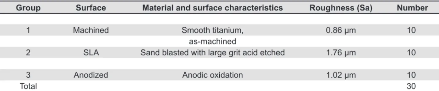
Implants were divided into 3 groups: those which were machined and did not undergo any surface treatment (group 1), those which were treated by SLA (group 2) and those which were anodized by oxidized electricity using pulse power (Autoelectric Co., Seoul, Korea) (group 3). The surface roughness of all groups was measured at the top of the thread using a confocal laser scanning profilometer (TopScan3D®; UBM Messtechnik GmbH, Germany)(Table 1).
Animals were preanesthetized by subcutaneous injection of buprenorphine HCl (Hanlim Co., Seoul, Korea, 0.02 mg/kg)/acepromazine (0.1 mg/kg)/ atropine (0.02 mg/kg). They were then sedated with methohexital (5 mg/kg) and maintained on gas
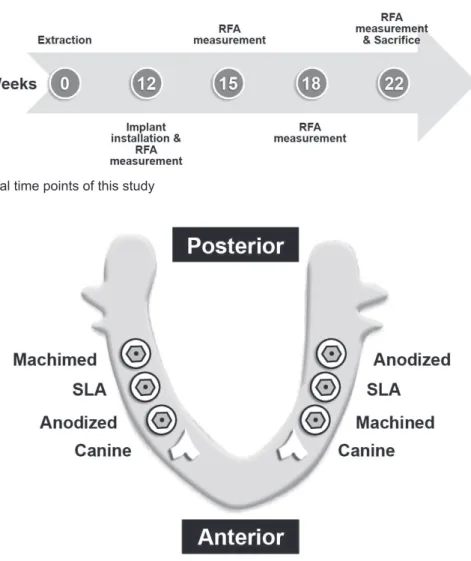
anesthesia (2% isolurane/O2). After scrubbing the surgical site with potadine, 1 mL of 2% lidocaine (Yu-Han Co., Seoul, Korea; 1:100,000) was injected into each surgical site for local anesthesia. In order to create an edentulous alveolar ridge, 4 mandibular premolar teeth were extracted from both sides of the lower jaw. After 3 months of healing period, implants were installed under general anesthesia. This experiment was undertaken as follows (Figure
2). For each implant, the insertion torque (IT), which represents the cutting resistance of bone when its rotation is stopped, was registered in Ncm (INTRAsurg 300, KaVo, Bieberach, Germany).
The implants were placed at the bone level. After stable installation of implants, cover screws were connected to them (Figure 3)
All implantation sites tested in dog mandibles were demonstrated to be bone type II or III. The surgical wound was sutured with 3-0 non-absorbable sutures (Mersilk®, ethicon Co., U.K.).
In order to prevent infections, kanamycin (50 mg/kg, Dong-A Co. Seoul, Korea) was injected intramuscularly for 7 days after surgery. One week after the operation, the sutures were removed, and a soft diet was provided for 2 weeks after surgery. A regular chow diet was supplied after 2 weeks. Implant Stability Quotient (ISQ) was recorded using an Osstell mentorTM (Integration Diagnostics
AB, Gothenburg, Sweden) at baseline (the day of surgery), and 3, 6 and 10 weeks after implant installation. All items measured in the experiment were recorded with an Osstell mentor Data Manager (OmDM). The program also calculated the mean and standard deviations. Generalized Linear Model in Statistical Package for the Social Science (SPSS) for Windows (ver. 11.0, SPSS Inc.) was used for comparative analysis among the groups.
Animals were sacriiced 10 weeks after implant
installation, and histomorphometric analysis was performed in order to measure the degree of osseointegration. Specimens including implant (N=30) were prepared for histomorphometric analysis. The specimens were stained with hematoxylin and eosin for light microscopy. Histomorphometric analysis was performed by attaching a Kappa Dx30 digital camera (Opto-electronics, Gleichen, Germany) on the light microscope (Olympus BX51, Olympus Co., Tokyo, Japan) and transferring digital images to a computer monitor. Then, a quantitative analysis was performed using Kappa image base metro (Kappa Opto-electronics, Germany) as an image-analysis software: A. The bone-to-implant direct contact ratio (BIC) was measured at the thread using a x40
magniication; B. The mineralized bone ratio was
calculated by measuring the total surface area of
Group Surface Material and surface characteristics Roughness (Sa) Number
1 Machined Smooth titanium, 0.86 μm 10
as-machined
2 SLA Sand blasted with large grit acid etched 1.76 μm 10
3 Anodized Anodic oxidation 1.02 μm 10
Total 30
Table 1- Implants used in this study (3 groups)
all threads (bone density: trabeculae, bone volume/ total volume).
RESULTS
All implants showed a mean insertion torque value of 18.12±6.53 Ncm as compared to a set torque of 40 Ncm and higher initial stability at baseline with an ISQ valuethan recommended ISQ (Implant Stability Quetient) value 70. No statistically
signiicant differences were found among the 3
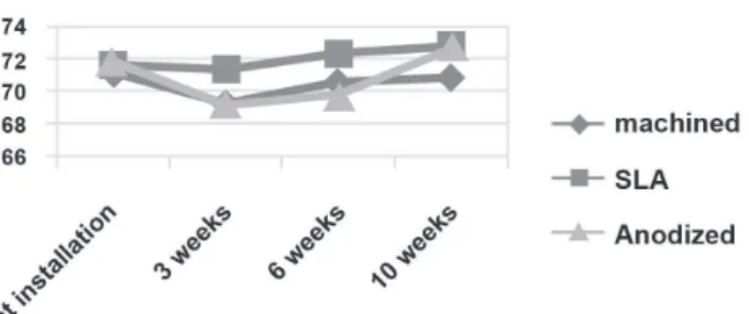
groups immediately after implantation (P>0.05). The 3 groups showed similar ISQ changing patterns 0, 3, 6 and 10weeks after implant installation,
with a decreasing pattern during the irst 3 weeks
and increasing 3 weeks later (P<0.05) (Table 2) (Figure 4). The bone-to-implant contact ratios of
groups 1, 2 and 3 in all threads were 60.8%, 69.6%
and 73.6%, respectively. There was a signiicant
difference in the contact ratio between groups 1 and 3 (P<0.05). The bone-to-implant contact ratio of groups 1, 2 and 3 in the 3 best continuous threads with abundant bone quantity showed values of 68.6%, 81.2% and 83.2%, respectively. There
was a signiicant difference in the contact ratio
between groups 1 and 2 and between groups 1 and 3 (P<0.05) (Figure 5). For all threads, bone density was 65.4% in group 1, 72.5% in group 2 and 71.1%
in group 3. There was no signiicant difference in
bone density between the 3 groups (P>0.05). For the 3 best continuous threads with abundant bone quantity, the bone density was 66.3% in group 1, 76.5% in group 2 and 75.7% in group 3 (Figure 6). Surface at installation 3rd weeks 6th weeks 10th weeks
Machined 71.33±2.42 69.33±3.14 70.67±2.58 70.83±3.31
SLA 71.67±3.33 71.36±3.72 72.33±1.63 72.83±1.94
Anodized 71.83±2.48 69.17±5.91 69.83±5.04 72.67±1.75
Table 2- ISQ during healing period
Figure 2- Experimental time points of this study
DISCUSSION
Dental implant surface has shown rapid evolution during the past few years6,17,23. This is because implant surface can have a signiicant effect on
long-term implant maintenance. Direct bone contact with metallic substrate can be achieved in
thin-ilm coated implants16.
It has been demonstrated that implant stability depends on the contact patterns between bone and implant7. Stable initial ixation may have an overt
effect on long-term implant stability3. Several1,21,22
reports indicate cortical bone, not bone in general to be mainly responsible for implant stability.
Meredith, et al.12,13 (1996, 1997) developed a
reversible experimental method to measure implant stability quantitatively. This reversible method allowed the measurement of osseointegration depth according to elapsed time15. Friberg, et
al.3 (1999) have indicated that in compact bone,
stability decreases as time passes despite excellent bone adhesion and increased contact between the implant and bone, whereas in soft bone, stability increases as time passes. Those authors have also proposed that the initial implant stability decreases as a result of bone compression caused by mechanical bone relaxation, biological changes during the primary bone recovery stage, and initiation of marginal bone resorption. Concerning the changes in stability according to elapsed time, Glauser, Portmann and Ruhstaller5 (2001) installed
20 TiUnite and 27 machined implants in the molar area of 9 and 15 patients, respectively. They performed RFA for 6 months after implantation,
which showed a decreasing pattern for the irst 3
weeks and then an increasing pattern thereafter. In this study, ISQ values at baseline, 3, 6 and 10
weeks after implant installation were signiicantly
different among 3 groups. There was a changing pattern of ISQ values that slightly decreased from implantation to 3 weeks post-implantation and increased thereafter in all groups.
SLA treatment, a combination of blasting and acid treatment, is performed by sand blasting the
implant with 25-50 μm Al2O3 particles and then etching with HCl/H2SO4 mixed solution. Cochran et al.2 observed an increase in alkaline phosphatase
activity, DNA absorption in 3H chimicin's and collagenase by biochemically testing the condition of cultured cells in the SLA-treated titanium phase. When the anodizing method is used, a thick
porous oxide ilm is formed. This ilm can increase
frictional force between implant and bone, and bone quantity and bone quality changes depending on the pore size10. Also, an improved surface
accelerates recovery in the early stage by protein
absorption, platelet accumulation and activity, ibrin
maintenance, and augmentation of the surrounding bone tissues9. When bone quality is poor, the
contact rate between bone and implant decreases up to below 25%, hindering implant primary stability which is an important factor in successful osseointegration. In this study, the 3 groups did not
show any signiicant difference in primary stability
because the experimental dogs had type II and III bone quality mandibles and because the implants
were completely ixated in all groups. This is in agreement with the indings of O'Sullivan, et al.14
(2004), who reported that implant primary stability
did not show any signiicant difference between type II and IV bone. They also found a signiicant
difference in the mean maximum insertion torque between type II and IV bone and between type
III and IV bone, whereas no signiicant difference
was noted between types II and III bone. Several reports have demonstrated the relationship between surface treatment and bone quality. The Figure 4- Changing pattern of implant stability quotient
during the healing period following implant installation in different types of implants (P<0.05)
Figure 5- The bone-to-implant contact (BIC) ratio in different types of implant
authors have suggested that factors related to bone density and implant diameter/length may affect the level of implant primary stability. Furthermore, greater stability was observed in male patients than in female patients. High implant primary stability was achieved in all jaw regions, although the use of thinner drills and/or tapered implants cannot fully compensate for the effect of soft bone16.In
contrast, Ganeles, et al.4 (2008) reported that
even in poor bone quality, SLActive surface were safe and predictable when used in immediate and early loading procedures. The survival rate was comparable with that of conventional loading. The mean bone-level change was not deemed to be
clinically signiicant and corresponded well with
typical bone resorption observed in conventional implant loading. Lazzara, et al.11 (1999)reported
that the osseointegration rate of a dual-acid-etched implant was twice as high as that of a machined implant (73% versus 34%). In this study, the rate was higher in implants with anodized surfaces, followed by those with SLA surfaces and those with machined surfaces. In other words, surface-treated implants showed a higher osseointegration rate (69%-76%) than machined implants (60%) (P<0.05). Johansson, et al.8 (1998) installed
pure titanium and titanium-aluminum-vanadium in rabbit bone and measured bone density 1, 6 and 12 months after implantation. Bone density did not show significant differences around different implant surfaces. In the present study, bone density was higher in SLA-treated implants, followed by anodized and machined implants, but
the differences were not statistically signiicant
(P>0.05). In SLA-treated implants, there was a significant difference in bone density between cancellous and compact bone. This result suggests
that there is a signiicant difference in the bone
response according to bone quality in textured surface. It is well known that implants with rough surfaces increase the contact surface area between implant and bone and thus improve the success rate of implants. Implants with rough surfaces show a high success rate and an excellent clinical outcome when used in poor quality bone20. Wennerberg, et
al.24 (1993) stated that the bone-to-implant contact
ratio was higher in titanium implants with surface
roughness (Sa) of about 1.4 μm than in smoother implants (Sa=0.7-1.2 μm) or rougher implants (2.2 μm). In the present study, the bone-to-implant
contact ratio was higher in anodized surfaces with
a roughness of 1.02 μm (73.6%±14.4%) and SLA-treated surfaces with a roughness of 1.76 μm
(69.6%±12.5%) than in machined surfaces with a
roughness of 0.86 μm (60.82%±13.11%).
In the present study, a good condition was created for implant primary stability by providing microthreads and connecting them to a compact
bone area. Marginal bone resorption depending on elapsed time was minimized. It is considered that
there were no signiicant differences among the 3
groups because of the good mechanical stability and the microthread design, which reduced marginal bone resorption.
CONCLUSION
The surface treatment had insigniicant effects
and did not affect implant stability in a compact bone (dog mandible). Further studies are needed
to conirm the effects of microthreads on implant
stability in bone.
ACKNOWLEDgEMENTS
The authors acknowledge Dentium (Seoul, Korea) for donating the Implantium® ixtures for
this study.
REFERENCES
1- Albrekttson T, Brånemark PI, Hansson HA, Lindström J. Osseointegrated titanium implants. Requirements for ensuring a long-lasting, direct bone-to-implant anchorage in man. Acta Orthop Scand. 1981;52(12):155-70. 2- Cochran DL, Buser D, ten Bruggenkate CM, Weingart D, Taylor TM, Bernard JP, et al. The use of reduced healing times on ITI implants with a sandblasted and acid-etched (SLA) surface: early results from clinical trials on ITI SLA implants. Clin Oral Implants Res. 2002;13(2):144-53. 3- Friberg B, Sennerby L, Meredith N, Lekholm U. A comparison between cutting torque and resonance frequency measurements of maxillary implants. A 20-month study. Int J Oral Maxillofac Surg. 1999;28(4):297-303. 4- Ganeles J, Zöllner A, Jackowski J, ten Bruggenkate C, Beagle J, Guerra F. Immediate and early loading of Straumann implants with a chemically
modiied surface (SLActive) in the posterior mandible and maxilla: 1-year
results from a prospective multicenter study. Clin Oral Implants Res. 2008;19(11):1119-28.
5- Glauser R, Portmann M, Ruhstaller P. Initial implant stability using different implant designs and surgical techniques. Appl Osseointeg Res. 2001;2(1):6-8.
6- Gottlander M, Albrektsson T. Histomorphometric studies of hydroxylapatite-coated and unhydroxylapatite-coated CP titanium threaded implants in bone. Int J Oral Maxillofac Implants. 1991;6(4):399-404.
7- Jafin RA, Berman CL. The excessive loss of Brånemark ixtures in type
IV bone: a 5-year analysis. J Periodontol. 1991;62(1):2-4.
8- Johansson CB, Han CH, Wennerberg A, Albrektsson T. A quantitative comparison of machined commercially pure titanium and titanium-aluminum-vanadium implants in rabbit bone. Int J Oral and Maxillofac Implants. 1998;13(3):315-21.
9- Larsson C, Emanuelsson L, Thomsen P, Ericson LE, Aronsson BO,
Kasemo B, et al. Bone response to surface modiied titanium implants:
studies on the tissue response after 1 year to machined and electropolished implants with different oxide thicknesses. J Mater Sci Mater Med. 1997;8(12):721-9.
10- Larsson C, Thomsen P, Aronsson BO, Rodahl M, Lausmaa J, Kasemo
B, et al. Bone response to surface modiied titanium implants: studies on
the early tissue response to machined and electropolished implants with different oxide thickness. Biomaterials. 1996;17(6):605-16.
13- Meredith N, Book K, Friberg, Jemt T, Sennerby L. Resonance frequency measurements of implant stability in vivo. A cross-sectional and longitudinal study of resonance frequency measurements on implants in the edentulous and partially dentate maxilla. Clin Oral Impl Res. 1997;8(3):226-33. 14- O’Sullivan D, Sennerby L, Jagger D, Meredith N. A comparison of two methods of enhancing implant primary stability. Clin Implant Dent Relat Res. 2004;6(1):48-57.
15- Ostman PO, Hellman M, Wendelhaq I, Sennerby L. Resonance frequency analysis measurements of implants at placement surgery. Int J Prosthodont. 2006;19(1):77-83
16- Coelho PG, Suzuki, M. Evaluation of an IBAD thin-ilm process as
an alternative method for surface incorporation of bioceramics on dental implants: a study in dogs. J Appl Oral Sci. 2005;13(1):87-92
17- Rompen E, DaSilva D, Lundgren AK. Stability measurements of a double-threaded titanium implant design with turned or oxidized surface. Appl Osseointegration Res. 2000;2(1):18-20.
18- Sennerby L, Meredith N. Implant stability measurements using resonance frequency analysis: biological and biomechanical aspects and clinical implications. Periodontol 2000. 2008;47:51-66
19- Sennerby L, Thomsen P, Ericson LE. A morphometric and biomechanic comparison of titanium implants inserted in rabbit cortical and cancellous bone. Int J Oral and Maxillofac Impl. 1992;7(1):62-71.
20- Tufekci E, Brantley WA, Mitchell JC, Foreman DW, Georgette FS. Crystallographic characteristics of plasma-sprayed calcium phosphate coatings on Ti-6Al-4V. Int J Oral and Maxillofac Implants. 1999;14(5):661-72.
21- Turkyilmaz I, McGlumphy EA. Inluence of bone density on implant
stability parameters and implant success: a retrospective clinical study. BMC Oral Health. 2008;8:32.
22- Turkyilmaz I, McGlumphy EA. Is there a lower threshold value of bone density for early loading protocols of dental implants? J Oral Rehabil. 2008;35(10):775-81.
23- Wennerberg A, Albrektsson T, Andersson B. Design and surface characteristics of 13 commercially available oral implant systems. Int J Oral and Maxillofac Implants. 1993;8(6):622-33.