* Corresponding author.
E-mail: saude@ef.ufop.br (D.A.S. Guimarães). A R T I C L E I N F O
Article history:
Received 10 October 2013 Accepted 16 December 2013
Keywords:
Campomanesia velutina Myricitrin
Anti-inflammatory Antinociceptive Inflammatory mediators
A B S T R A C T
Campomanesia velutina (Cambess) O. Berg, Myrtaceae, popularly known as “gabiroba” or “guavira”, is used in traditional Brazilian medicine to treat several diseases, including inflammation and rheumatism. Extraction and isolation from leaves of the plant afforded the active compound myricetin 3-O-rhamnoside, also known as myricitrin. The ethanolic extract of leaves of C. velutina and its ethyl acetate and methanolic fractions were evaluated in inflammation (carrageenan-induced paw oedema) and analgesic models (acetic acid-induced abdominal writhing and hot plate test). Moreover, the ethanolic extract, its fractions and the isolated compound were also in vitro evaluated for their ability to modulate NO, TNF-α and IL-10 production from J774A.1 macrophages stimulated by LPS/IFN-γ.In vivo assays showed remarkable anti-inflammatory activity of ethanolic extract, ethyl acetate and methanolic fractions. The antinociceptive activity of ethanolic extract and A was demonstrated in acetic acid-induced abdominal writhing test. In vitro assays demonstrated that ethyl acetate and methanolic fractions fraction and myricitrin inhibited NO production from macrophages J774A.1. Also Myricitrin induced production of IL-10 anti-inflammatory cytokine. None of the samples was able to inhibited TNF-α production. The results demonstrated for the first time the anti-inflammatory and antinociceptive activity of C. velutina.
© 2013 Brazilian Society of Pharmacognosy. Published by Elsevier Editora Ltda. All rights reserved.
Original Article
Extracts from the leaves of
Campomanesia velutina
inhibits
production of LPS/INF-
γ
induced inflammatory mediators
in J774A.1 cells and exerts anti-inflammatory and
antinociceptive effects
in vivo
Marcela C.P. Michel
a, Andrea Grabe Guimarães
b, Carmem A. Paula
a, Simone A. Rezende
c,
Marcos E.G. Sobral
d, Dênia A. Saúde Guimarães
a,*aLaboratório de Plantas Medicinais, Escola de Farmácia, Universidade Federal de Ouro Preto (UFOP), Ouro Preto, MG, Brazil
bLaboratório de Farmacologia Experimental, Escola de Farmácia, Universidade Federal de Ouro Preto (UFOP), Ouro Preto, MG, Brazil cLaboratório de Imunoparasitologia, Núcleo de Pesquisas em Ciências Biológicas, Universidade Federal de Ouro Preto (UFOP), Ouro
Preto, MG, Brazil
dDepartamento de Ciências Naturais, Universidade Federal de São João Del-Rei (UFSJ), São João Del-Rei, MG, Brazil
Introduction
Inflammation is a natural process that protects the body against infection and injury and is characterized by the activation of
histamine and bradykinin (Sobolewski et al., 2010). NO produced by macrophages plays a crucial role in antimicrobial, antiviral and antitumor activities. However, overproduction of NO is related to septic shock, vascular collapse and death (Chi et al., 2003). Tumor necrosis factor-α (TNF-α) is one of the most important pro-inflammatory cytokines, playing a crucial role in inflammation, stimulating the production of other cytokines and pro-inflammatory mediators (Baugh and Bucala, 2001). However, the excessive production of TNF-α is related to the exacerbation of the inflammatory process and the development of autoimmune diseases (Williams et al., 2007). The inflammatory process can be limited by the production of immunomodulatory cytokines like interleukin-10 (IL-10). This cytokine acts as a potent inhibitor of macrophage function by blocking the synthesis of pro-inflammatory cytokines like TNF-α, IL-6 and IL-12 and by reducing the harmful effects of excessive activation of this cell during inflammation (Conti et al., 2003).
Campomanesia, Myrtaceae, can be found from northern Argentina to the island of Trinidad and from the Brazilian coast to the Andes, Peru, Ecuador and Colombia (Landrum, 1986). They are popularly known as “gabiroba” or “guavira”, and have been widely been used to treat various diseases. The bark and leaves of Campomanesia species, prepared by infusion, are used in folk medicine as depurative, anti-diarrhea, anti-rheumatic, anti-inflammatory, antipyretic, antiseptic of the urinary tract, in the treatment of diabetes, stomach problems and reduction of blood cholesterol (Alice et al., 1995).
Campomanesia velutina (Cambess) O. Berg is endemic in Brazil, found in the Brazilian cerrado bioma, savanna and Brazil´s Atlantic Forest (Landrum, 1986). Although the traditional use of Campomanesia velutina as anti-inflammatory agent, there is a lack of investigations on the pharmacological properties of this plant. Thus, the aim of this study is to in vivo
evaluate the antinociceptive and anti-inflammatory activity of ethanolic extract from Campomanesia velutina leaves and its fractions. In order to establish themselves the possible mechanism of action, the ethanolic extract, its fractions and the isolated flavonoid were also tested in vitro for their ability to modulate the production of NO, TNF-α and IL-10 from J774A.1 macrophages stimulated by LPS/IFN-γ.
Materials and methods
General experimental procedures
Melting points were recorded on Buchi M-560 melting point apparatus. NMR spectra were obtained on a Bruker Avance Drx-400 MHz spectrometer. Tetramethylsilane (TMS) was used as internal reference and deuterated methanol (MeOD) as solvent. HPLC analysis was performed on Waters Liquid Chromatography (model Alliance 2695), equipped with vacuum degasser, a quaternary pump, an auto sampler, a diode array detector - DAD (Waters 2996) and reversed phase C18 column Shimadzu ODS (250 mm × 4,6 mm, 5 µm). HPLC grade reagents were purchase from Tedia. Water was purified by Milli-Q system from Millipore. Absorbance was measured on Molecular Devices microplate reader. Thin layer chromatograph (TLC) was
carried out on analytical plates coated with silica gel (Merck 60G). Spots were visualized by spraying with anisaldehyd/ sulfuric acid or by exposition to UV light. Sephadex LH-20 was obtained from Sigma and Poliamida-6 from Fluka. Carrageenan and indomethacin were purchase from Sigma Aldrich. Acetic acid and morphine hydrochloride were purchased from Merck Inc. (Brazil). Lipopolysaccharide (LPS) was obtained from Sigma and interferon-γ (IFN-γ) from RD Systems. ELISA kits were purchase from PeproTech (Brazil). Analytical grade reagents were from Merck.
Plant material
The leaves of Campomanesia velutina (Cambess) O. Berg, Myrtaceae, were collected in Lagoa Santa, Minas Gerais, Brazil, in October 2007, with permission of Instituto Chico Mendes de Conservação da Biodiversidade/Sistema de Autorização e Informação em Biodiversidade (license no 17021-5). The plant botanical identification was realized by Dr. Marcos E. Guerra Sobral, Departamento de Ciências Naturais, Universidade Federal de São João Del-Rei, Minas Gerais, Brazil. A voucher specimen was deposited in the Herbarium of UFSJ, reference number HUFSJ 4637.
Preparation of plant extract and fractions
The leaves of the plant materials were air-dried, powdered to a fine grade (410 g) and extracted by percolation with ethanol, at room temperature, for 27 days. The solvent was eliminated by evaporation under reduced pressure, resulting in the dried crude ethanolic extract (101 g). Part of the ethanolic extract (EE, 60 g) was submitted to filtration column chromatography on silica gel, eluted with hexane, ethyl acetate and methanol to yield the hexanic (H, 0.31 g), ethyl acetate (A, 8.30 g) and methanolic (M, 25.55 g) fractions, respectively.
Isolation
Initially were added 22 ml of acetone part of methanolic fraction (15 g) to afford a soluble portion and an insoluble portion. To the soluble portion (1.44 g) was added distilled water until a precipitate was formed. This was separated by filtration. The filtrate obtained (0.44 g) was partitioned with a mixture of n-buthanol/water (1:1, v/v) to yield an n-buthanolic and an aqueous fraction.
Fingerprints chromatograms by HPLC
EE, A, M and myricitrin were solubilized in methanol to yield a concentration of 5 mg/ml (EE, A and M) and 1 mg/ml (myricitrin), and then filtered through a 0,45 µm Millex syringe filters. The volume injected was 25 μl. The elution gradient was performed in a system with 20% methanol and 80% water taking 55 min to reach 100% methanol and 5 min to return to the initial condition. The flow rate was kept constant at 0.8 ml/ min and the separation temperature was 25°C. The UV-DAD detector was set to record between 200 and 600 nm and UV chromatograms were recorded at 254 nm.
Animals
All experiments were performed with male Swiss albino mice (30 ± 5 g) supplied by Animal House of Universidade Federal de Ouro Preto. Animals were maintained in a temperature-controlled room (22 ± 2°C) with 12 h light/dark cycles and free access to food and water. The animals were limited to a water-only diet 12 h prior to each experiment in order to avoid interference with food substance absorption. The experimental protocols were approved, under the number 2010/060, by UFOP Ethical Committee of Animal Experimentation. The protocols were in accordance to the Guide for the Care and Use of Laboratory Animals, published by the US National Institute of Health (NIH Publication, revised in 1985).
Preparation of test samples and drugs
Indomethacin and morphine were dissolved in distilled water and Tween-80 (95:5). Acetic acid and carrageenan were dissolved in distilled water just before use. EE, A and M were solubilized in Tween-80, DMSO and distilled water (1:1:8) and 0.2 ml were administered by oral gavage at doses of 100 and 300 mg/kg. Morphine (10 mg/kg) and indomethacin (10 mg/ kg) were administered orally by gavage also (0.2 ml) and the reference drugs used. To the control group was given a vehicle (Tween-80, DMSO and distilled water 1:1:8).
Carrageenan-induced paw edema assay
The in vivo anti-inflammatory activity was determined by the carrageenan-induced paw edema method in mice, according to previously described by Winter et al. (1962) with modifications. The animals were divided into eight groups and the respective groups were treated with vehicle, indomethacin and with both doses (100 and 300 mg/kg) of EE, A and M. Half an hour after administration of the various agents, edema was induced by injection of carrageenan (0.02 ml, 0.1%, w/v) into the sub-plantar tissue of the right hind paw. Only performed needle introduction was into the left paw, corresponding to activity induced by mechanical perforation (control). To measure variation edema, a digital caliper rule (Starret) was used. The paws were measured before and 1, 2, 3, 4, 5 and 6 h after carrageenan administration, with or without treatments. The paw edema was expressed in millimeters (mm) and was calculated as the percentage of variation between the time zero and 1, 2, 3, 4, 5 and 6 h after carrageenan.
Acetic acid-induced abdominal writhing
The test was performed as described by Koster et al. (1959). The animals were divided into eight groups and the respective groups were treated with vehicle, indomethacin and with both doses (100 and 300 mg/kg) of EE, A and M 30 min before intraperitoneal (i.p.) injection of acetic acid 0.8 % v/v, 0.1 ml/10 g body weight. The number of abdominal writhing was counted cumulatively over a period of 30 min immediately after the acetic acid injection. The antinociceptive activity was expressed as percentage of inhibition of abdominal writhing according to the following formula:
“Inhibition (%)=” (Number of Writhes [Control] – Number of Writhes [Treatment]) × 100 (Number of Writhes [Control])
Hot plate test
The hot-plate test was used to measure response latencies according to the method described by Eddy and Leimbach (1953). The animals were divided into eight groups and placed on a metal plate heated to a temperature of 56 ± 1°C and the time between placement of the mice on the platform and shaking or licking of the hind paws or jumping was recorded as the hot-plate latency (seconds). A cut-off period of 60 s was imposed to avoid tissue damage to the paws. Then, animals were treated with vehicle, morphine and with both doses (100 and 300 mg/kg) of EE, A and M. The reaction time of animals was recorded again 30, 60 and 120 min after oral administration. The significant increase in latency time compared to the initial observation was considered positive antinociceptive response.
Cell line and culture conditions
The J774A.1 (ATCC® TIB-67™) murine macrophage cell line were cultured in RPMI supplemented with L-glutamine (2 mM), sodium pyruvate (1 mM), penicillin (100 Units/ml) and fetal bovine serum (10%). Cells were incubated at 37°C in a humidified 5% CO2 atmosphere.
Cell viability
NO assay
J774A.1 cells were seeded onto a 48-well culture plate at density 3.5 × 10⁵ cells per well and incubated for 2 h at 37°C in a 95% air and 5% CO2 atmosphere adheres to. The medium was carefully discarded and 200 μl of fresh medium was added. Cells were treated with different non-cytotoxic concentrations of EE, A, M and myricitrin dissolved in DMSO for 1 h before stimulation with LPS and IFN-γ (25 ng/ml and 25 UI/ml, respectively) for 24 h. The presence of nitrite, a table oxidized product of NO, was measured in cell culture medium using Griess reagent (Green et al., 1982). Briefly, 50 μl of supernatant was transferred to a 96-well plate and incubated with 100 μl of Griess Reagent for 10 min at room temperature. The absorbance was measured at 570 nm using a microplate reader. NO concentration was determined using comparison with a sodium nitrite standard curve. The end concentration of DMSO was adjusted to less than 0.3% for all treatments. Dexamethasone was used as a reference standard.
Measurements of TNF-α and IL-10 levels
J774A.1 cells were seeded onto a 48-well culture plate at density 3.5 × 10⁵ cells per well and incubated for 2 h at 37°C in a 95% air and 5% CO2 atmosphere adheres to. The medium was carefully discarded and 200 μl of fresh medium was added. Cells were treated with different non-cytotoxic concentrations of EE, A, M and myricitrin dissolved in DMSO for 1 h before stimulation with LPS and IFN-γ (25 ng/ml and 25 UI/ml, respectively) for 24 h. After incubation period, the supernatant was collected and used to estimate the levels of TNF-α and IL-10 by specific ELISA kits according to the manufacturer´s instruction. The end concentration of DMSO was adjusted to less than 0.3% for all treatments. Dexamethasone was used as a reference standard.
Statistical analysis
In vivo results presented were the means ± SEM from experiments performed with eight animals per group. In vitro
results were obtained from three independent experiments in duplicate and were presented as mean ± SEM. Results were submitted to analysis of variance (One-way ANOVA) followed by Dunnett’s test for analysis of test groups compared to control group and the analysis of variance (One-way ANOVA) followed by Bonferroni’s test for analysis between groups. The differences were considered significant when p-values were less than 0.05 (p < 0.05). Statistical analysis was performed using the PRISM software (GraphPad Software, Inc., San Diego, CA, version 5.01).
Results and discussion
Fingerprints from EE, A and M were obtained order to establish themselves in the chromatographic pattern and level of complexity of samples. The HPLC profile of EE, A and M at 254 nm were presented in Fig. 1. At first sight, fingerprint chromatogram off EE (Fig. 1A) did not reveal the complex metabolic profile. However, integration of the chromatogram showed the presence of 58 peaks with the main peaks at
retention time until 30 min. The A fraction showed a complex metabolomic profile (Fig. 1B) with a concentration of peaks in retention time between 30 and 45 min. M fraction also showed the chromatographic profile not too complex and the integration revealed the presence of 46 peaks with the main peaks at retention time until 25 min (Fig. 1C). The HPLC profile demonstrated that from the fractionation of EE were obtained two fractions with different profiles metabolomics, which fulfilled the objective of obtaining samples of different composition. Myricitrin was recognized in EE chromatogram by comparing retention time and UV spectra with purified substance. The HPLC chromatogram of the purified substance was shown in Fig. 1D.
The anti-inflammatory effect of the crude ethanolic extract of C. velutina and its fractions were evaluated in carrageenan-induced paw edema, an animal model widely used for the
screening of anti-inflammatory compounds and has been used to assess often the anti-oedematogenic effect of natural products, exhibiting a high degree of reproducibility (Morris, 2003). The edema produced by carrageenan injection remains located in the area of administration (Cicala et al., 2007) and is characterized by biphasic response which involves the release of several mediators, cell migration and plasma exudation (Loram et al., 2007). The first phase (0-2.5 h) is associated with the release of serotonin and histamine in the first 90 min, and bradykinin in an intermediate stage (90-150 min) (Moncada and Higgs, 1993). On the second phase (3-4 h) the edema is maintained by the production of PG’s after the induction of COX-2 and NO release after iNOS expression in activated leukocytes infiltrated the edema (Salvemeni et al., 1996). At this time the swelling reaches its largest volume.
The group was treated with vehicle was considered as maximum of inflammation and all others were treatments compared to this group. Those shown in Table 1, the effect
of indomethacin began in the second hour after carrageenan injection and remained throughout the experiment. Oral treatment with EE at major dose (300 mg/kg) was effective in reducing the edematogenic response from first hour after carrageenan injection and also remained throughout the experiment. Moreover, the anti-inflammatory activity of EE was similar to indomethacin during the entire experiment. These results suggest that EE acts at different phases of carrageenan-induced inflammatory process, probably by inhibiting the release and/or actions of several inflammatory response mediators involved at, like vasoactive amines, kinins, PG and NO. Fractions A and M significantly inhibited the edema from third hour after carrageenan injection. Thus, the activity of these fractions only in the second phase of the inflammatory process is an indication that the mechanism of action of substances presents in A and M fractions are related to PG and NO.
In order to elucidate the possible mechanism of anti-inflammatory action of EE, A and M, they were evaluated for their
Treatment Swelling thickness (%)
(After carrageenan) 1 h 2 h 3 h 4 h 5 h 6 h
Control conditiona 1,88 ± 1,10d 0,27 ± 3,32d 4,14 ± 2,81d 4,27 ± 3,12d 3,01 ± 1,95d 1,05 ± 1,56c
Vehicle 14,22 ± 1,84 22,00 ± 2,84 30,22 ± 3,10 25,58 ± 3,99 18,36 ± 4,19 12,32 ± 2,97
Indomethacin 6,62 ± 1,93 11,04 ± 1,93b 15,25 ± 2,79c 10,65 ± 2,25c 5,80 ± 2,61c 0,72 ± 2,18c EE 100 mg/kg 13,77 ± 2,89 17,97 ± 3,10 18,26 ± 3,64b 13,24 ± 3,87b 8,10 ± 2,59b 1,51 ± 1,90c EE 300 mg/kg 4,86 ± 2,60b 11,47 ± 2,67b 14,40 ± 2,98c 11,20 ± 2,93c 6,92 ± 1,86b 1,53 ± 1,38c A 100 mg/kg 12,24± 2,09 15,74 ± 2,57 10,50 ± 3,62d 15,43± 3,69 22,09 ± 2,66 2,00 ± 2,33c A 300 mg/kg 12,86 ± 1,64 15,01 ± 1,69 4,70 ± 2,39d 15,14 ± 1,59 18,70 ± 2,33 2,55 ± 2,49c
M 100 mg/kg 11,96 ± 2,01 13,67 ± 1,61 19,27 ± 2,26 11,67 ± 1,60b 5,85 ± 1,65b 2,36 ± 1,37c
M 300 mg/kg 11,78 ± 1,89 13,15 ± 1,30 7,99 ± 1,86d 8,20 ± 3,17# 7,41 ± 1,32b 2,54 ± 1,32c
Results represent the mean ± S.E.M. variation of the percentage of full length between the dorsal and ventral faces of the paws before and 1, 2, 3, 4, 5 and 6 h after carrageenan.
aWithout carrageenan. bp < 0.05.
cp < 0.01.
dp < 0.001, when compared to the group treated with vehicle (ANOVA followed Dunnett’s test).
Table 1
Effect of oral administration of ethanolic Campomanesia velutina extract (EE) and its ethyl acetate (A) and methanolic (M) fractions on carrageenan-induced paw edema.
ability to modulate NO, TNF-α and IL-10 production from J774A.1 macrophages stimulated by LPS/IFN-γ.
First of all, the viability of the J774A.1 macrophages in the presence of different concentrations of EE, A, M and myricitrin were evaluated. The cytotoxicity on J774A.1 cells was negligible and did not show statistic difference when compared to the control. The exception occurred for the A fraction, since the concentration of 640 and 1280 µg/ml showed cell viability lower than 90%. Thus, EE, A and M fractions were assayed in vitro using concentrations lower than 640 µg/ml. Myricitrin was in vitro assayed using concentrations lower than 20 µg/ml.
Inhibition of NO production was evaluated in supernatants of J774A.1 cells stimulated by LPS (25 µg/ml) and IFN-γ (25 UI/ml) in the presence or absence of the EE, A and M. The NO is produced from
absence of samples, unstimulated J774A.1 cells secreted basal levels of NO, while LPS/IFN-γ stimulation resulted in NO production increase (Fig. 2). EE and M were not able to reduce NO production from J774A.1 macrophages (Fig. 2A and C). However, the fraction significantly inhibited NO release from J774A.1 macrophages in all concentrations evaluated (Fig. 2B), this might be demonstrated that one of the possible mechanism of action of this fraction.
The production of TNF-α and other pro-inflammatory interleukins is a crucial part of the immune response to many inflammatory stimuli. The local effect of TNF-α can be considered
beneficial to the host. However, overproduction leads to severe systemic toxicity and even death (Herath et al., 2003). J774A.1 cells in the absence of any stimulus secreted basal levels of TNF-α, while LPS/IFN-γ stimulation resulted in an appreciable increase of TNF-α level (Fig. 3). EE was able to inhibit the TNF-α production, but this reduction was not significant compared to control (Fig. 3A). A and M did not produce significant inhibition in any evaluated concentration (Fig. 3B and C). The results obtained suggest that the mechanism of action of anti-inflammatory activity observed for C. velutina does not involve inhibition of this cytokine.
Figure 2 - A, effect of the ethanolic extract (EE); B, effect of ethyl acetate fraction (A); C, effect of methanolic fraction (M) on NO production by LPS/IFN-γ stimulated J774A.1 macrophages. The cells were treated with EE, A and M dissolved in DMSO at different non-cytotoxic concentrations for 1 h and then stimulated with LPS (25 µg/ml) and IFN-γ (25 UI/ml) for 24 h. Supernatants were collected and nitrite (NO) concentration was determined by Griess reagent. Data are represented as mean ± S.E.M. *p < 0.05.
**p < 0.01.
***p < 0.001 compared with LPS/IFN-γ-stimulated macrophages alone (ANOVA followed Dunnett’s test).
Figure 3 - A, effect of the ethanolic extract (EE); B, effect of ethyl acetate fraction (A); C, effect of methanolic fraction (M) on TNF-α production by LPS/IFN-γ stimulated J774A.1 macrophages. The cells were treated with EE, A and M dissolved in DMSO at different non-cytotoxic concentrations for 1 h and then stimulated with LPS (25 µg/ml) and IFN-γ
(25 UI/ml) for 24 h. Supernatants were collected and TNF-α concentration was determined by specific ELISA kit. Data are represented as mean ± SEM.
*p < 0.05. **p < 0.01.
Treatment Number of writhes Inhibition (%)
Vehicle 58.62 ± 7.09
-Indomethacin 18.37 ± 5.44 68.66b
EE 100 mg/kg 34.37 ± 7.53 41.37
EE 300 mg/kg 9.37 ± 4.59 84.01c
A 100 mg/kg 20.25 ± 7.09 65.45a
A 300 mg/kg 16.75 ± 8.19 71.43
M 100 mg/kg 33.00 ± 13.93 43.71
M 300 mg/kg 38.71 ± 10.08 33.96
Results represent the mean ± SEM (n = 8) of the number of abdominal writhing after i.p. acetic acid administration.
ap < 0.05 bp < 0.01.
cp < 0.001, when compared to the group treated with vehicle (ANOVA
followed Dunnett’s test). Table 2
Effect of oral administration of ethanolic Campomanesia velutina extract (EE) and its ethyl acetate (A) and methanolic (M) fractions in the acetic acid-induced abdominal writhing test.
It is well established that inflammatory response may be resolved through the clearance of the inflammatory cells or through the release of endogenous anti-inflammatory mediators such as IL-10. IL-10 is a late cytokine produced by several cell types that has a special physiological significance in limiting and preventing an excessive immune response and in limiting collateral damage (Sabat et al., 2010). Thus, the effect of EE, A and M in IL-10 production from J774A.1 macrophages stimulated by LPS/IFN-γ was evaluated. In the absence of samples, cells secreted appreciable amounts of IL-10 after stimulation with LPS/IFN-γ, while unstimulated J774A.1 cells secreted smaller significant levels of IL-10 (Fig. 4). Unfortunately, EE, A and M induced a significant inhibition of IL-10 production at all concentrations tested (Fig. 4A, B and C). Therefore, the anti-inflammatory activity of EE, A and M are not related to increased production of IL-10.
The cytokines network is very complicated and until now isn´t completely understood (Bony et al., 2012). Despite the results obtained on in vitro tests, EE, A and
M demonstrated a strong anti-inflammatory activity, which suggests a general inhibitory effect on macrophage activation, probably acting on different targets.
The antinociceptive potential of EE and its A and M fractions was evaluated by both chemical and thermal nociception induced models. Acetic acid i.p. injection produced 58.62 ± 7.09 writhes in the group treated with vehicle (control group). Indomethacin significantly reduced the number of writhes with 68.66% of inhibition. EE (300 mg/kg) and A (100 and 300 mg/kg) produced significant reduction in the number of abdominal writhing with 84.01, 65.45 and 71.43% of inhibition, respectively. The antinociceptive activity of these groups was similar to indomethacin (Table 2) and there is no statistic difference between indomethacin and these groups. Notice that EE at the dose of 300 mg/kg was more active than A and M. These findings suggest that the highest activity of EE can be attributed to the synergism between the various molecules present in the extract. Thus, the fractionation of the crude extract resulted in loss of synergistic effects, leading to reduction of antinociceptive activity to A fraction and absence of antinociceptive activity to M fraction.
The acetic acid-induced writhing abdominal visceral pain model is employed as a screening tool for the assessment of antinociceptive or anti-inflammatory activity of new
Figure 4 - A, Effect of the ethanolic extract (EE); B, effect of ethyl acetate fraction (A); C, effect of methanolic fraction (M) on IL-10 production by LPS/IFN-γ stimulated J774.A1 macrophages. The cells were treated with EE, A and M dissolved in DMSO at different non-cytotoxic concentrations for 1 h and then stimulated with LPS (25 µg/ml) and IFN-γ
(25 UI/ml) for 24 h. Supernatants were collected and IL-10 concentration was determined by specific ELISA kit. Data are represented as mean ± SEM.
*p < 0.05. **p < 0.01.
analgesic agents (Souza et al., 2009). However, despite good sensitivity of this test, it is considered nonspecific because the nociceptive action of acetic acid appears to involve different mechanisms (Le Bars et al., 2001) such as the release of endogenous mediators by the sympathetic system (Duarte et al., 1988), opioid mechanisms (Collier et al., 1968) and the increase in the level of COX and LOX products as well as the release of many inflammatory mediators like bradykinin, substance p and cytokines (TNF-α, IL-1β and IL-8) by peritoneal macrophages and mast cells (Ikeda et al., 2001). Thus, in order to avoid misinterpretation, the hot plate test was employed. This model of nociception is a sensitive and specific test used to show the involvement of central mechanisms (Le Bars et al., 2001). The oral administration of EE, A and M did not produce a significant prolongation of latency times, indicating that analgesic effects does not involve central mechanisms of pain inhibition. Thus, the antinociceptive effect of EE and its A fraction is peripherally mediated and may be related to inhibition of the production and/or action of mediators involved in inflammation such as bradykinin, substance P, TNF-α, IL-1β, IL-8, NO, which can interfere with the transduction mechanism signal of primary afferent nociceptors (Ong et al., 2011). In vitro results showed that EE and A did not reduce TNF-α production on J774A.1 cells. However, unlike EE, the fraction was able to reduce NO production, indicating an involvement of NO pathway on antinociceptive activity of this fraction.
Myricitrin was isolated from M fraction obtained from
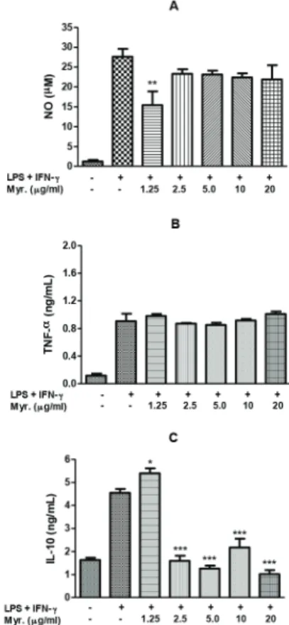
C. velutina ethanolic extract and was the first flavonoid isolated on this specie. Previous studies demonstrated the anti-inflammatory and antinociceptive activity of this flavonol (Meotti et al., 2006) and its ability to inhibit NO production from RAW 264.7 macrophages stimulated by LPS (Chen et al., 2000) and TNF-α production from RAW 264.7 cells (Shimosaki et al., 2011). Thus, the contribution of myricitrin for the observed anti-inflammatory and analgesic activity was also evaluated on in vitro tests. In this study, was not capable to myricitrin inhibited TNF-α production from J774A.1 macrophages stimulated by LPS and IFN-γ on concentrations tested (Fig. 5B). However, myricitrin significantly inhibited NO production (Fig. 5A) and increased IL-10 secretion in macrophages stimulated by LPS/IFN-γ (Fig. 5C). It is interesting to note the effect over NO and IL-10 production vanishes at higher concentrations. This may be attributed to post-transcriptional effects of this flavonoid, since it is well established that IL-10 can be regulated by post-transcriptional mechanisms in macrophages (Németh et al., 2005). Also it is possible that at concentrations above 1.25 mg/ml myricitrin is acting as a pro-oxidative molecule (Comalada et al., 2006). These results indicate that myricitrin may contribute for the anti-inflammatory and antinociceptive activity of the species.
The anti-inflammatory and antinociceptive activity of
C. velutina observed after oral administration of EE, A and M suggests that active compounds are well absorbed by the gastrointestinal tract. Moreover, activities of C. velutina could be attributed to the action of different classes of substances, probably acting on different targets of inflammation, since
extracts with different composition showed anti-inflammatory and antinoceptive activity.
Campomanesia velutina is used in traditional Brazilian medicine for the treatment of painful and inflammatory conditions. The results of the present study demonstrate, for the first time, that
Campomanesia velutina anti-inflammatory and antinociceptive activity presents. Although the complete mechanisms underlying these actions remain to be elucidated, the anti-inflammatory activity seems to involve vasoactive amines, kinins, prostaglandins, NO and IL-10 pathway. The antinociceptive activity is mediated by peripheral mechanisms. The isolated compound myricitrin seems to contribute to anti-inflammatory and antinociceptive activity, since it was able to modulate NO and IL-10 production. However, further studies are necessary to corroborate its use in humans.
Figure 5 - Effect of myricitrin on NO (A), TNF-α (B) and IL-10 (C) production by LPS/IFN-γ stimulated J774.A1 macrophages. The cells were treated with myricitrin dissolved in DMSO at different non-cytotoxic concentrations for 1 h and then stimulated with LPS (25 µg/ml) and IFN-γ (25 UI/ml) for 24 h. Supernatants were collected and nitrite (NO) concentration was determined by Griess reagent, TNF-α and IL-10
concentration was determined by specific ELISA kit. Data are represented as mean ± S.E.M.
*p < 0.05. **p < 0.01.
Authors’ contributions
MCPM (PhD student) contributed in running the laboratory work, analysis of the data and drafted the paper. MEGS contributed in plant identification and herbarium confection. AGG and SAR contributed to biological studies in vivo and
in vitro, respectively. CAP contributed to critical reading of the manuscript. DASG designed the study, supervised the laboratory work and contributed to critical reading of the manuscript. All the authors have read the final manuscript and approved the submission.
Acknowledgments
The authors would like to thank FAPEMIG (Projeto CDS-APQ-01426-11), Rede TOXIFAR/FAPEMIG, CAPES and Universidade Federal de Ouro Preto for financial support. The authors would like to thank also Leidiane Cristina Ferreira and Nayara Almeida de Assis for collaboration and Dr. Luis Carlos Crocco Afonso for supplying J774A.1 macrophages.
R E F E R E N C E S
Alice, C.B., Siqueira, N.C.S., Mentz, L.A., Silva, G.A.A.B., José, K.F.D., 1995. Medicinal Plants of Use People: Atlas Pharmacognosy. Canoas: Ulbra.
Baugh, J.A., Bucala, R., 2001. Mechanisms for modulation TNF-α in immune and inflammatory disease. Curr. Opin. Drug Discov. Devel. 4, 635-650.
Bony, E., Boudard, F., Dussossoy, E., Portet, K., Brat, P., Giaimis, J., Michel, A. 2012. Chemical composition and
anti-inflammatory properties of the unsaponifiable fraction from Awara (Astrocaryum vulgare M.) pulp oil in activated J774 macrophages and in a mice model of endotoxic shock. Plant Food Hum. Nutr. 67, 384-392.
Cicala, C., Morello, S., Alfieri, A., Velleco, V., Marzocco, S., Autore, G., 2007. Haemostatic imbalance following carrageenan-induced rat paw oedema. Eur. J. Pharmacol. 577, 156-161. Chen, Y.C., Yang, L.L., Lee, T.J.F., 2000. Oroxylin A inhibition of
lipopolysaccharide-induced iNOS and COX-2 gene expression via suppression of nuclear factor-kB activation. Biochem. Pharmacol. 59, 1445-1457.
Chi, D.S., Qui, M., Krishnaswamy, G., Li, C., Stone, W., 2003. Regulation of nitric oxide production from macrophages by lipopolysaccharide and cathecolamines. Nitric Oxide 8, 127-132. Collier, H.O.J., Dinneen, J.C., Schneider, C., 1968. The abdominal
constriction response and its suppression by analgesic drugs in the mouse. Br. J. Pharmacol. Chemother. 32, 295-310. Comalada, M., Ballester, I., Bailón, E., Sierra, S., Xaus, J., Gálvez,
J., Medina, F.S., Zarzuelo, A., 2006. Inhibition of pro-inflammatory markers in primary bone marrow-derived mouse macrophages by naturally occurring flavonoid: analysis of the structure activity relationship. Biochem. Pharmacol. 72, 1010-1021.
Conti, P., Kempuraj, D., Kandere, K., Di Gioacchino, M., Barbacane, R.C., Castellani, M.L., Felaco, M., Boucher, W., Letourneau, R., Theoharides, T.C., 2003. IL-10, an
inflammatory/inhibitory cytokine, but not always. Immunol. Lett. 86, 123-129.
Duarte, J.D.G., Nakamura, M., Ferreira, S.H., 1988. Participation of the sympathetic system in acetic acid induced writhing in mice. Braz. J. Med. Biol. Res. 21, 341-343.
Eddy, N.B., Leimbach, D., 1953. Synthetic analgesics. II. Dithienylbutenyl and dithienylbutylamines. J. Pharmacol. Exp. Ther. 107, 385-393.
Green, L.C., Wagner, D.A., Glogowski, J., Skipper, P.L., Wishnok, J.J., Tannebaum, S.R., 1982. Analysis of nitrate, nitrite and (15N) nitrate in biological fluids. Anal. Biochem. 126, 131-138. Herath, H.M.T., Takano-Ishikawa, Y., Yamaki, K., 2003.
Inhibitory effect of some flavonoids on tumor necrosis factor-α production in lipopolysaccharide stimulated mouse macrophage cell line J774.1. J. Med. Food. 6, 365-370. Ikeda, Y., Ueno, A., Naraba, H., Oh-ishi, S., 2001. Involvement of
vanilloid receptor VR1 and prostanoids in the acid-induced writhing responses of mice. Life Sci. 69, 2911-2919.
Koster, R., Anderson, M., Beer, J. 1959. Acetic acid for analgesic screening. Fed. Proc, 18, 412-417.
Landrum, L.R., 1986. Campomanesia, Pimenta, Blepharocalyx, Legrandia, Acca, Myrrhinium and Luma (Myrtaceae). New York: Flora Neotropica New York Botanical Garden.
Le Bars, D., Gozariu, M., Cadden, S.W., 2001. Animal models of nociception. Pharmacol. Rev. 53, 597-652.
Loram, L.C., Fuller, A., Fick, L.G., Cartmell, T., Poole, S., Mitchell, D., 2007. Cytokines profiles during carrageenan-induced inflammatory hyperalgesia in rat muscle and hind paw. J. Pain 8, 127-136.
Meotti, F.C., Missau, F.C., Ferreira, J., Pizzolatti, M.G., Mizuzaky, C., Nogueira, C.W., Santos, A.R.S., 2006. Anti-allodynic property of flavonoid myricitrin in models of persistent inflammatory and neuropathic pain in mice. Biochem. Pharmacol. 72, 1707-1713.
Moncada, S., Higgs, A., 1993. The L-arginine-nitric oxide pathway. N. Engl. J. Med. 329, 2002-2012.
Morris, C.J., 2003. Carrageenan-induced paw edema in the rat and mouse. Methods Mol. Biol. 225, 115-121.
Németh, Z.H., Lutz, C.S., Csóka, B., Deitch, E.A., Leibovich, S.J., Gause, W.C., Tone, M., Pacher, P., Vizi, E.S., Haskó, G., 2005. Adenosine augments IL-10 production by macrophages through an A2B receptor-mediated post transcriptional mechanism. J. Immunol. 175, 8260-8270.
Ong, H.M., Mohamad, A.S., Makhtar, N., Khalid, M.H., Khalid, S., Perimal, E.K., Mastuki, S.N., Zakaria, Z.A., Lagis, N., Israf, D.A., Sulaiman, M.R., 2011. Antinociceptive activity of methanolic extract of Acmella uliginosa (Sw.) Cass. J. Ethnopharmacol. 133, 227-233.
Punturee, K., Wild, C.P,, Vinitketkumneun, U., 2004. Thai medicinal plants modulate nitric oxide and tumor necrosis factor-α in J774.2 mouse macrophages. J. Ethnopharmacol. 95, 183-189.
Sabat, R., Grütz, G., Warszawska, K., Kirsch, S., Witte, E., Wolk, K., Geginat, J., 2010. Biology of interleukin-10. Cytokine Growth Factor Rev. 21, 331-344.
Salvemini, D., Wang, Z.Q., Bourdon, D.M., Stern, M,K., Currie, M.G., Manning, P.T., 1996. Evidence of peroxynitrite involvement in the Carr-induced rat paw edema. Eur. J. Pharmacol. 303, 217-220.
Sobolewski, C., Legrand, N., Morceau, F., Diederich, M., 2010. Inflammation: Novel arrows for an ancient target. Biochem. Pharmacol. 80, 1769-1770.
Souza, M.M., Pereira, M.A., Ardenghi, J.V., Mora, T.C., Bresciani, L.F., Yunes, R.A., Monache, F.D., Cechinel-Filho, V., 2009. Filicene obtained from Adiantum cuneatum interacts with the cholinergic, dopaminergic, glutamatergic, GABAergic, and tachykinergic systems to exert antinociceptive effect in mice. Pharmacol. Biochem. Behav. 93, 40-46.
Williams, R.O., Paleolog, E., Feldmann, M. 2007. Cytokine inhibitors in rheumatoid arthritis and other autoimmune disease. Curr. Opin. Pharmacol. 7, 412-417.